![First Aids For The Orthopedics Board PDF [PDF]](https://pdfs.asia/img/200x200/first-aids-for-the-orthopedics-board-pdf.jpg)
7 0 11 MB
FIRST AID
FOR® THE
ORTHOPAEDIC BOARDS Se co nd Editio n
Ro b e rt A. Ma lin za k, MD Ma rk J. Alb ritto n , MD Tre vo r R. Picke rin g, MD
New York / Chicago / San Francisco / Lisbon / London / Madrid / Mexico City Milan / New Delhi / San Juan / Seoul / Singapore / Sydney / Toronto
First Aid fo r the ® Orth o p a e d ic Bo a rd s, Se co nd Editio n Copyright © 2009, 2006 by The McGraw-Hill Companies, Inc. All rights reserved. Printed in the United States of America. Except as permitted under the United States Copyright Act of 1976, no part of this publication may be reproduced or distributed in any form or by any means, or stored in a data base or retrieval system, without the prior written permission of the publisher. First Aid for the ® is a registered trademark of The McGraw-Hill Companies, Inc.
1 2 3 4 5 6 7 8 9 0 QPD/ QPD 12 11 10 9 8
ISBN 978-0-07-159894-1 MHID 0-07-159894-4 ISSN 1554-3196
NOTICE Medicine is an ever-changing science. As new research and clinical experience broaden our knowledge, changes in treatment and drug therapy are required. The authors and the publisher of this work have checked with sources believed to be reliable in their efforts to provide information that is complete and generally in accord with the standards accepted at the time of publication. However, in view of the possibility of human error or changes in medical sciences, neither the authors nor the publisher nor any other party who has been involved in the preparation or publication of this work warrants that the information contained herein is in every respect accurate or complete, and they disclaim all responsibility for any errors or omissions or for the results obtained from use of the information contained in this work. Readers are encouraged to confirm the product information sheet included in the package of each drug they plan to administer to be certain that the information contained in this work is accurate and that changes have not been made in the recommended dose or in the contraindications for administration. This recommendation is of particular importance in connection with new or infrequently used drugs.
This book was set in Electra LH by International Typesetting and Composition, Inc. The editors were Catherine A. Johnson and Peter J. Boyle. The production supervisor was Sherri Souffrance. Project management was provided by Preeti Longia Sinha, International Typesetting and Composition, Inc. Quebecor World Dubuque was printer and binder. This book is printed on acid-free paper.
CONTENTS
Pre face
v
Acknowle dgm e nts
vii
Introduction
ix
Ch a p te r 1
Basic Scie nce
Ch a p te r 2
Orthopaedic Anatom y
17
Ch a p te r 3
Orthopaedic Traum a
31
Ch a p te r 4
Joint Arthroplasty
73
Ch a p te r 5
Sports Medicine
105
Ch a p te r 6
The Hand
125
Ch a p te r 7
Foot and Ankle
149
Ch a p te r 8
Orthopaedic Tum ors
167
Ch a p te r 9
Pediatric Orthopaedics
193
Ch a p te r 10
The Spine
217
Ch a p te r 11
Miscellaneous Facts
231
Inde x
1
247
iii
PREFACE
The first edition of First Aid for the Orthopaedic Boards was five years in the making. Over the past eighteen months, we have revised the material for this edition to reflect the changing emphasis of the Board exam and to incorporate new data relevant to those seeking Board certification or recertification. Our aim––to provide orthopaedic exam takers with a concise resource of the most important testable orthopaedic facts––has not changed. The topics cover the high-yield subject matter on the Boards, presented in bulletpoint format to facilitate easy and rapid appropriation of facts needed to answer frequently tested questions. Any minutia presented has been important on previous exams and is included to help maximize your scores. Overall, the facts presented have been collected and organized in the best manner we know possible. We invite any and all advice, criticism, and insight to help make this resource even better for you and all future test takers. We certainly have a vested interest in improving this text, as we will be preparing for recertification in just a few years ourselves! Best of luck on examination day and it is our sincere wish that First Aid for the Orthopaedic Boards has enabled you to overshoot your examination goals. Robert A. Malinzak, MD Mark J. Albritton, MD Trevor R. Pickering, MD
v
ACKNOWLEDGMENTS
Rob, Mark, and Trevor would like to thank the residents, faculty, and staff of the outstanding Duke Orthopaedic Residency Program who assisted in their orthopaedic journey. We strongly feel Duke Orthopaedics is one of the outstanding institutions in the country and are very thankful for having been a part of something so strong. Rob thanks his lovely and brilliant wife, Dr. Sandy Moreira, for strongly standing by his side and remaining the most remarkable person he knows. Also, he gives thanks to his wonderful children, Mackenzie and Jacob, as they provide the ultimate strength for his days. And he is thankful to Mom, Dad, Dave, and Ann as they provided the structure and vision for him to arrive at this day. Mark thanks his amazing wife, Laura, for her patience and encouragement during this process; he truly cherishes her. He also thanks his two great children, Dylan and Drake, for being the greatest inspiration in his life, and he is indebted to his parents for all of their love and support. Trevor thanks Cris and Nora for their love and support. He thanks the Center for Hip and Knee Surgery for a wonderful opportunity. Finally, he wishes to thank Oxford Orthopaedics and Sports Medicine in Oxford, Mississippi, for a terrific partnership.
vii
INTRODUCTION
Congratulations on continuing your journey toward orthopaedic board credentialing. The trek began with SATs, MCATs, and USMLEs only now to include the very relevant OITE, our Step I Orthopaedic Board Examination and the 10-year Recertification Examination. No matter which portion of the journey you are currently traversing, First Aid for the Orthopaedic Boards will prepare you for your current exam by offering the detailed high-yield facts popular on such exams. We have not prepared a thorough orthopaedic encyclopedia, as there are several excellent resources that provide this amount of orthopaedic detail. The thrust of our composition is to provide medical professionals, medical students, orthopaedic residents, and orthopaedic surgeons with the relevant key, testable orthopaedic information. This concise compilation of orthopaedic facts will certainly benefit your understanding of orthopaedics, not only for the exam, but also for your medical practice. We have scoured the popular examinations and are presenting the most recent frequently tested facts required for passing such examinations. Also included are mnemonics, tables, charts, and figures that we found helpful in keeping certain obtuse, yet important facts in the rapid recall portion of our cranium.
BACKGROUND
The Orthopaedic In-Training Examination (OITE) is given each November and is an excellent measuring stick of your progress through residency. This examination is administered at your local training program and is an untimed examination. Different institutions vary the test-day format, but the overall purpose of this annual examination is for the resident to take note of his/her orthopaedic progress. The quick rule is that if you score in the lower third of your class’s scale, you have a high chance of failing the real board examination. On the other hand, if you score in the top third, you are almost assured of passing the real examination. Those in the middle third have more variable chances. Overall, the better prepared you are throughout residency and the OITE, the more likely you will be able to perform very well on the day of your Step I examination. After completing residency you will be expected to take and pass Step I of the orthopaedic certification process. Step I is administered each July in Chicago. Registration needs to be completed by March of your testing year. The American Board of Orthopaedic Surgery (ABOS) has an excellent Web site (www.abos.org) for you to check dates and progress, as well as hotel and travel arrangements. We recommend making your hotel and travel plans well in advance. Everyone has heard of examinees traveling and arriving the night before the test in ix
a frenzy as their flight, car, or other travel plans ran amok. If possible, arrive 2 days in advance to alleviate that portion of possible stress and get the lay of the land. Step I of the orthopaedic board exam is a one-day written examination including 320 questions administered in two 3.5-hour sessions. An hour is allocated for lunch. The board offers that there is no curve and no predetermined failure rate. Approximately 30–35% of the questions are known as they have been given on prior tests, and the Board knows how these questions should play out. A score of 70% correct answers (or 224 out of 320) should get you through the passing door. First-time test takers from the United States and Canada have a pass rate of around 93% and repeat takers a 40% pass rate (but that was before this book was available!). After successfully taking the Step I examination, you go home and await your scores, which arrive in about 8 long weeks. Step II of the orthopaedic board certification process is the oral examination, which occurs on completion of your second full year in practice. You will provide to the Board at your expense a complete list of every surgical case you performed from July through December. Then you will be expected to bring with you to the exam all the relevant information on 10 of the 12 cases the Board asks you to discuss. This list includes clinic notes, operative reports, x-rays (pre- and postop), and follow-up notes. Thorough documentation is crucial, and prompt organization is vital for your oral board examination. Recertification is now required every 10 years for orthopaedic surgeons. Although there are several courses to quickly relearn the minutia required for the exam, First Aid for the Orthopaedic Boards will enable you to have the most important facts at your fingertips. You can study in between cases during room turnover, in clinic, and in any other few minutes you find. In the end, your preparedness for the examination will pay huge dividends. First Aid for the Orthopaedic Boards will enable you to feel secure in your ability to handle the key testable orthopaedic information. Remember that guessing cannot hurt you on the few questions that you are not sure of and that most would offer that your first choice is usually the better of your responses. We hope to have provided you with a resource that allows you to improve your orthopaedic knowledge in a way that best uses what little “free” time you may have. We wish you the very best on your voyage toward the examination. Our goal is for every reader of First Aid for the Orthopaedic Boards to not only pass the examination but to do significantly better than his/her initial expectation. We trust that the facts and information provided here are true to the best of our ability. Any mistakes are our sole responsibility and are absolutely unintended. We appreciate any and all constructive criticism, noted omissions, future mnemonics, or other ideas that can make First Aid for the Orthopaedic Boards a stronger text for future Step I and recertification candidates.
CHAPTER 1
Basic Science
Bone
2
BONE MATRIX
2
BONE METABOLISM
3
BONE DISORDERS
3
FRACTURE
5
BONE GRAFTS
5
P HYSEAL ZONES (RPHM)
6
Cartilage
6
ARTICULAR CARTILAGE LAYERS
6
COLLAGEN TYPES
7
Muscle
7
Ligaments
8
Nerves
8
ENCAPSULATED SKIN RECEPTORS
8
NONENCAPSULATED SKIN RECEPTORS
9
Genetics, Immunology, Molecular Biology
9
MICROBIOLOGY, INFECTIONS, ANTIBIOTICS, AND OTHER MEDICATIONS
11
ANTIBIOTIC MECHANISMS OF ACTION
12
Coagulation BIOMECHANICS
13 14
1
BASIC SCIENC
BONE
Bon e Ma trix ■ ■ ■
■
■ ■ ■
40% organic: Primarily collagen I, gives tensile strength. 60% inorganic: Primarily hydroxyapatite, gives compressive strength. Proteoglycans: Compressive strength, made of glycosaminoglycans (GAG) complexes. Osteocalcin: Made by osteoblasts; attract osteoclasts, inhibited by parathyroid hormone (PTH), stimulated by 1,25-dihydroxy vitamin D (1,25[OH 2]D). Regulates bone density. Osteonectin: Secreted by platelets and osteoblasts, matrix mineralization. Osteopontin: Cell binding protein. Important cytokines and growth factors for bone: ■ Transforming growth factor-β (TGF-β): Causes mesenchymal cells to differentiate into osteoblasts; present in fracture callus. ■ Insulin-like growth factor (IGF): Stimulates type I collagen, involved in cartilage matrix synthesis. ■ Interleukin-1 (IL-1): Stimulates osteoclasts. ■ Bone morphogenic protein (BMP).
Osteoblasts produce type I collagen; high alkaline phosphatase activity. ■
■
■ ■
From undifferentiated mesenchymal cells (controlled by Cbfa, plateletderived growth factor [PDGF], and insulin-derived growth factor [IDGF]). Downregulated directly by PTH, glucocorticoids, prostaglandins (PGEs), and leptin. Upregulated by estrogens. Produce osteocalcin when stimulated by 1,25-(OH 2)D, which in turn upregulates osteoblasts.
Osteoclasts resorb bone and are irregularly shaped giant cells. ■ ■ ■ ■ ■ ■ ■
Ruffled boarders increase surface area. Binds to bone via integrins. IL-1 is a potent stimulator. Produce tartrate-resistant acid phosphatase. Receptors for calcitonin which inhibit bone resorption. Pamidronate inhibits osteoclasts. Resorbs bone in multiple myeloma and metastatic disease.
RANKL: ■ ■ ■ ■ ■ ■ ■
■
2
Released by osteoblasts. Binds to RANK receptor on osteoclast. Activates osteoclast. Leads to bone resorption. Important in bone metabolism and tumors—especially multiple myeloma. Osteoprotegerin (OPG) is a decoy inhibitor and stops osteoclast activation. Upregulate RANKL and downregulate OPG (increase bone resorption/ destruction): ■ PTH ■ 1,25-(OH 2)D ■ IL-1β ■ Tumor necrosis factor-α (TNF-α ) ■ PGE2 Downregulate RANKL and upregulate OPG (decrease bone resorption/ destruction):
■ ■
BASIC SCIENCE
■
IL-4 TNF-β Interferon N gamma
Bon e Me ta b olism ■
■ ■ ■
■
■
Bone is resorbed by
Vitamin D strongly stimulates absorption of calcium (Ca) and phosphateinositol (PO 4) from intestines. 1-α hydroxylase activates vit D (25[OH]-VitD to 1,25[OH 2]-VitD). PTH stimulates production of 1,25-(OH 2) vitamin D, Ca elevated, Pi lowered. Calcitonin from clear cells of the thyroid gland: ■ Decreases osteoclast activity and number while decreasing the tubular resorption of Ca and PO 4. ■ Lowers serum Ca levels. ■ Osteoclasts have receptors for calcitonin. ■ Inhibits osteoclast bone resorption (i.e., lowers Ca). Corticosteroids cause low bone formation and high resorption, but retain normal trabecular osteoid volume. ■ Increased Ca loss, reduced Ca absorption. Estrogen reduces femoral neck fracture rate and increases bone density in the femoral neck. ■ Limits bone loss by limiting bone resorption. ■ Ca and PO 4 metabolism involves skin, liver, kidney, intestines, thyroid, parathyroid glands. ■ Ca requirements: ■ Lactating women, 2000 mg/d. ■ Pregnant women, 1500 mg/d. ■ Postmenopausal woman with healing fracture, 1500 mg/d.
osteoclasts not only in bone homeostasis but bonedestroying tumors.
RANKL is the primary mediator of osteoclast activation.
Bon e Disord e rs ■
■ ■
■
■
■
■
■ ■
■
■
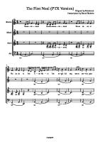
Osteomalacia is due to an inability to maintain serum Ca and phosphate levels. Most patients will have normal hemoglobin and low Ca and PO 4. Osteomalacia in renal osteodystrophy is most often the result of aluminum-containing phosphate-binding antacids. Osteopetrosis: Failure of osteoclastic and chondroclastic resorption. ■ Autosomal dominant. ■ Lack of marrow-derived osteoclast precursor cells (Albers-Schönberg disease). Osteoporosis has normal bone mineralization and abnormal osteoclast function. World Health Organization (WHO) definition is > 2.5 standard deviations (SD) below peak bone mass of a 25-year-old. T score below -2 SD from “normal” young female, initiate treatment. ■ If -1.5 and risk factors, start treatment. ■ Z score is versus own age-matched controls. Menopause associated with 2%–3% per year bone loss for 6–10 years. Type I postmenopausal: Trabecular bone; distal radius and vertebral fractures. Type II senile: Trabecular and cortical bone; hip and pelvic fractures. ■ Senile osteoporosis is the uncoupling of bone formation and resorption. Idiopathic transient osteoporosis (ITOH) (see Figure 1-1): ■ Unknown etiology affecting pregnant females or late-middle-aged males.
3
1-α hydroxylase activates vit D (25[OH]-VitD to 1,25[OH]2-VitD)
BASIC SCIENC
Id io p a th ic tra n sie n t o ste o p o ro sis o f th e h ip (ITOH). Notice the de cre ase d signal in the affected left hip. FIGURE 1-1 .
Presents with a painful hip and magnetic resonance imaging (MRI) evidence of diffuse osteoporosis. ■ Treat with protected weight bearing. ■ Some advocate calcitonin to alleviate the pain. ■ Resolves with time. Rickets: Failure in zone of provisional calcification (i.e., a part of the hypertrophic zone). Primary hyperparathyroidism: parathyroid adenoma: ■ Increased: Serum Ca, PTH, 1,25-VitD, urine Ca, Alk Phos (or normal) ■ Decreased: Serum Phos ■ Normal: 25 VitD Hypoparathyroidism: ■ Increased: Serum Phos ■ Decreased: Serum Ca, PTH, 1,25-Vit D, urine Ca ■ Normal: Alk Phos Pseudohypoparathyroidism (genetic disorder, resistance to PTH): ■ Increased: PTH (or normal), Serum Phos ■ Decreased: Serum Ca, 1,25-VitD, urine Ca ■ Normal: PTH (or increased), Alk Phos, 25 VitD Vit D deficiency rickets: ■ Increased: PTH, Alk Phos ■ Decreased: Serum Ca (or normal), Serum Phos, 25 VitD, 1,25-VitD, urine Ca Hypophosphatemic rickets (“phosphate diabetes”) ■ X-linked dominant ■ Most common form of rickets ■ Decreased: Serum Phos ■
■
■
Osteoporosis is a quantitative problem, whereas
■
osteomalacia is a qualitative problem. ■
■
■
4
■
BASIC SCIENCE
■
Normal: Serum Ca, PTH, 25 VitD, 1,25-VitD, urine Ca Treatment: Phosphate and vit D (to offset effect of phosphate supplementation)
Fra cture ■ ■ ■
■ ■
■ ■ ■ ■
■
■ ■ ■
■ ■ ■
Fracture repair: Inflammation, soft callus, hard callus, then remodeling. Initial response: Decreased blood flow. Maximum vascularization at the fracture occurs around 2 weeks. ■ Blood flow normal at 3–5 months. ■ Inflammation 24–72 hours. ■ Primary callus repair happens within 2 weeks. ■ Soft callus is converted to hard callus by enchondral ossification. ■ Amount of callus indirectly proportional to amount the fracture is immobilized. ■ Remodeling up to 7 years. Unstable fracture repair has type II collagen early, then type I collagen later. Bone is weaker in tension than compression, thus get transverse fracture with three-point bending. Bending forces result in transverse with butterfly fracture. Torsion causes spiral fracture. Cast: Periosteal bridging callus, enchondral ossification. Rigid compression plate: Primary bone healing, cutting cone remodeling. ■ Near cortex: Direct Haversian remodeling, inhibits callus formation. ■ Far cortex: Often, there is a gap. Gap healing: Lamellar bone is 90 degrees to longitudinal axis of bone. IM nail: ■ Early: Periosteal callus ■ Late: Medullary callus Enchondral ossification. Ex-fix: If rigid: Primary healing; less rigid: Periosteal callus. Hypertrophic nonunion: Failed endochondral ossification, no osteoid, mostly type II cartilage. Smoking decreases the eventual strength of the healed fracture. Blood flow is the main determinant in fracture healing. Medial clavicle ossifies at 25 years old.
Bon e Gra fts ■ ■
■ ■
■
Osteoinduction: Signals local factors to stimulate bone formation. Osteoconduction: ■ Scaffold for new bone ■ Includes Ca phosphate, Ca sulfate, Ca carbonate, coralline hydroxyapatite Osteogenic cells lead the rebuilding effort. Cortical bone graft: ■ Structural support ■ Remodels existing Haversian system by resorption then deposition of new bone ■ Weak during resorption phase Cancellous bone graft: ■ No resorption of old trabeculae. ■ Osteoblasts put down new bone on old trabeculae (creeping substitution). ■ Quickly revascularized.
5
Fracture healing is most affected by vascularization, and smoking decreases healing fracture strength.
Amount of callus indirectly proportional to amount the fracture is immobilized.
BASIC SCIENC
■ ■ ■ ■
■ ■ ■
Fresh versus fresh frozen versus freeze-dried. Intercalary allografts: Most common complication is nonunion (−38%). Frozen syngeneic allografts have best incorporation. Distraction osteogenesis: Anterior tibial artery shows significant increase in the number of vaso vasorum. Latency phase, distraction phase, consolidation phase. Ideal growth is 1 mm/d (lengthen 0.25 mm four times a day). Host repair after avascular necrosis (AVN) is creeping substitution. Bo ne Grafts GRAFT
OSTEOCONDUCTION
OSTEOINDUCTION
Cance llous
Exce llent
Good
Cortical
Fair
Fair
Allo graft
Fair
Fair
Ce ramics
Fair
None
DBM
Fair
Good
Bo ne Marrow
Poor
Poor
Auto graft
Ph yse a l Zon e s (RPHM)
Various diseases that affect the physis occur at specific areas of the physis. ■
■
Reserve zone: PK GD (Reserve Place Kicker has Good Distance) ■ Pseudoachondroplasia ■ Kneist syndrome ■ Gaucher disease ■ Diastrophic dysplasia Proliferative zone: GPA (big and small) ■ Gigantism ■
■
■
Achondroplasia
Hypertrophic zone: MORE Sex Please ■ Mucopolysaccharidoses ■ Osteomalacia ■ Rickets ■ Enchondroma ■ Slipped capital femoral epiphysis (SCFE) ■ Physeal fracture Metaphysis ■ SCFE with endocrinopathy CARTI LAGE
Articu la r Ca rtila ge La ye rs
See Table 1-1. 6
Articu la r Ca rtila ge La ye rs
ZONE
■ ■ ■ ■
■ ■ ■ ■
BASIC SCIENCE
TA B L E 1 - 1 .
SPECIFIC CHARACTERISTICS
SHEAR VS. COMPRESSION
Superficial zone (gliding)
Decreased m etabolic activity
vs. Shear
Middle zone (transitional)
Incre ase d m e tabolic activity
vs. Com pression
Dee p zone (radial)
Incre ase d collagen size
vs. Com pression
Tidem ark
Undulating barrier
vs. Shear
Calcified zone
Hydroxyapatite crystals
Anchor
Greatest tensile stiffness in the superficial zone. Proteoglycans are responsible for retaining water in the matrix. Link proteins: Link hyaluronate and proteoglycans. Fibrocartilage and type I collagen are at areas of articular cartilage injury that goes deep across the tidemark. Anchorin II: Binds to chondrocytes. COMP (cartilage oligometric protein): Binds to chondrocytes. Protect cartilage: TGF-β, Smad3, IGF, BMP-2, BMP-7. Breakdown cartilage: IL-1, TNF-α, cyclooxygenase-2 (COX-2), nitric oxide synthase (NOS).
Co lla ge n Typ e s ■ ■ ■ ■
Type I: Meniscus, bone, tendon, skin, and annulus fibrosis. Type II: Main contributor in articular cartilage and nucleus pulposus. Type VI: Increased in osteoarthritis (OA). Type X: Endochondral ossification. MUSCLE
■
■
■
■
■
■
■ ■
■
Isotonic: Eccentric and concentric phases; tension constant, length changes (biceps curl). Isokinetic: Velocity constant, measures dynamic strength, most efficient way to strengthen muscles; use Cybex. Isometric: Length constant, tension changes; measures static strength; push on a wall. Slow-twitch type I muscle fibers: “Slow red ox(idative)”: ■ Low strength of contraction, more mitochondria, aerobic. ■ Endurance activities. Fast-twitch type II muscle fibers: ■ High anaerobic capacity, large motor unit size, high strength, fast speed of contraction. ■ Low aerobic capacity, low capillary density, and most fatigable. ■ Sprinting activities. Irreversible side effects of anabolic steroids include: ■ Growth retardation. ■ Male pattern baldness. ■ Deepening of voice. Fatigue reduces a muscle’s ability to absorb energy. Muscle laceration midsubstance: Dense scar connective tissue (recover 50% tension ability). Muscle–tendon junction is most common site of strains. 7
Proteoglycans retain water in cartilage.
BASIC SCIENC
■ ■
■
Slow-twitch muscles are red
■
and have more mitochondria;
LIGAMENTS
fast-twitch muscles are white with less mitochondria and therefore are anaerobic.
Myasthenia gravis: Decreased number of acetylcholine receptors. Myofibroblast has intracellular actin microfilaments (stress fibers); found in Dupuytren disease. Paracrine: Local cell produces molecule to act on nearby cell. Autocrine: Stimulates same cell that produced the molecule.
■ ■ ■ ■ ■ ■
Type I collagen MRI: Ligaments low signal on T1 and low signal on T2. Ligament healing is with type III collagen early and then type I collagen. Children have avulsion injuries more often. Adults have midsubstance injuries more often. Ligament insertion: ■ Ligament → fibrocartilage → mineralized fibrocartilage → bone. ■ Avulsion occurs between unmineralized and mineralized fibrocartilage layers. NERVES
■
■
■
■
■
Sensory axon carries impulse from periphery to cell body located in the dorsal root ganglion. Neurapraxia: ■ Fiber failure sequence → motor, proprioception, touch, temperature, pain. ■ Axon continuity maintained ■ Reversible, no Wallerian degeneration Axonotmesis: ■ Axon severely damaged or severed. ■ Endoneural tube (Schwann cells) intact. ■ Wallerian degeneration. ■ Good recovery. ■ Preserve conduction velocities for up to 7 days, 2–5 weeks, have fibrillations with positive sharp waves. Neurotmesis: ■ Loss of endoneural continuity. ■ Perineurium and fascicle arrangement intact. ■ Wallerian degeneration. ■ Recovery variable. Neuropathic joint (3 Ds—dislocation, destruction, and degeneration). ■ Shoulder: Consider syrinx. ■ Hip and knee: Tabes dorsalis. ■ Foot and ankle: Diabetic neuropathy.
En ca p su la te d Skin Re ce pto rs
See Table 1-2.
TA B L E 1 - 2 .
8
Ch a ra cte ristics o f En ca p su la te d Skin Re ce p to rs
Pacinian corpuscles
Flutter
Fast adapting, high frequency
Me issner corpuscles
Touch
Fast adapting, low fre quency
Ruffini end organs
Vibration
Slow adapting, low fre que ncy
BASIC SCIENCE
TA B L E 1 - 3 .
Ch a ra cte ristics o f Non e n ca p su la te d Skin Re ce p to rs
Merkel cells
Steady skin indentation
Sustained response to pressure
Fre e ne rve endings
Me chanical, therm al, noxious
Warm , cold, sharp, dull
No n e n ca p sula te d Skin Re ce p tors
See Table 1-3. ■
■ ■ ■
■
■ ■
Vibration-induced peripheral neuropathy: Increased vibration and temperature thresholds. Posterior columns: Pain and temp (crossover two levels up). Anterior columns: Vibration, proprioception, and light touch. Sciatic nerve usually lies anterior to piriformis and posterior to quadratus femoris, obturator internus and externus, and superior gemellus. Superficial peroneal nerve: Medial dorsal cutaneous nerve (to the dorsal medial great toe metatarsophalangeal joint [MTP]) commonly injured during approach for hallux valgus. Axillary nerve: Deltoid and teres minor (injured in open Bankart repair). Suprascapular nerve: Spinoglenoid notch entrapment with infraspinatus weakness (suprascapular notch = supra- and infraspinatus weakness).
GENETICS, IMMUNOLOGY, MOLECULAR BIOLOGY
See Table 1-4 for genetic keys. ■ ■ ■
■
■ ■
■ ■
■
■
■
■
■
Marfan syndrome: Fibrillin defect on chromosome 15. Homocystinuria: Autosomal recessive, deficiency of cystathionine. Ankylosing spondylitis: Back pain associated with morning stiffness, improves with exercise. Rheumatoid arthritis has nodules on the posterior ulnar border and pulp surfaces. ■ Implicated cytokines involve TNF and interleukin-1 (IL-1). Juvenile rheumatoid arthritis (JRA): Positive rheumatoid factor (RF) < 15%. Gaucher disease: Elevated glucocerebrosides; Erlenmeyer flask distal femur, AVN of femoral heads, anemia, hepatosplenomegaly, abnormal liver function, neurologic symptoms, pathologic fractures, bone infarcts. Treat with splenectomy and enzyme replacement. Alkaptonuria (ochronosis): Elevated homogentisic acid. Reiter syndrome: Reactive arthritis—polyarthritis, uveitis (cervicitis), and conjunctivitis. ■ “Can’t see, can’t pee, and can’t climb a tree.” ■ Bilateral heel pain often; treat with nonsteroidal anti-inflammatory drugs (NSAIDs). Achondroplasia: Fibroblast growth factor-3 (FGF-3) receptor alteration; trident hand, rhizomelic (proximal) shortening. Calcium pyrophosphate deposition (CPPD): Positive birefringence, short-trapezoidal/positive/blue. Gout: Monosodium urate crystals, inhibited by colchicine, negative birefringence, long needle shaped/yellow. FGF-3 receptor changes (AD): Achondroplasia, hypochondroplasia, thanatophoric dysplasia. Fat emboli syndrome seen sooner than pulmonary embolus. 9
BASIC SCIENC
TA B L E 1 - 4 .
Ge ne tic Ke ys
COMP
Pse udoachondroplasia, MED (type I), OA
FGF-3
Achondroplasia, hypochondroplasia, thanatophoric dysplasia
FGF-2
Apert’s syndrom e, Jackson-Weiss, Crouzon’s, m ost Pfeiffer’s syndrom es
Collagen II
SED congenital, DD, Kneist syndrom e, Stickler syndrom e, hypochondrogenesis, type II achondrogenesis, precocious osteoarthropathy
Collage n IX
MED (type II)
SEDL gene
SED tarda
PTH-related protein
Jansen’s m etaphyseal chondrodysplasia
Collage n X
Schm id’s m etaphyse al chondrodysplasia
EXT1, EXT2 genes
MHE
Sulfate transport gene
DD
Core -binding factor
Cleidocranial dysplasia
Carbonic anhydrase II
Osteope trosis/ osteoclasts
proton pum p m utation COL 1A1 (pro-alpha 1
Oste oge ne sis im pe rfe cta
(I) collagen gene )
10
COL 2A1
Kne ist syndrom e (PTH re ce ptor proble m )
PEX
X-linked hypophosphotem ic rickets
t(X;18)
Synovial sarcom a (lym phatic spread)
t(2;13)
Rhabdom yosarcom a, alveolar
t(12;16)
Myxoid liposarcom a
t(9;22)
Chondrosarcom a
t(12;22)
Clear cell chondrosarcom a
t(11;22)
Ewing’s sarcom a
Dystrophin absent
Duchenne MD
Dystrophin abnorm al
Becker MD
BASIC SCIENCE
TA B L E 1 - 4 .
Ge ne tic Ke ys (co n tin u e d )
Survival m otor neuron
Spinal m uscular atrophy
Peripheral m yelin protein 22
CMT type IA
Connexin 32
CMT X-linked
Neurofibrom in (tum or suppressor gene)
NF1
Me rlin
NF2
Schwannom in
NF2
Fritaxin
Friedreich’s ataxia
Fibrillin
Marfan syndrom e
G protein activated
Fibrous dysplasia
Elastin
Fragile X
Bone sialoprotein
RA, advanced
Hyaluronan-binding protein
RA, advanced
Glucocerebroside elevated
Gaucher disease
Hom ogentisic acid
Ochronosis (alkaptonuria)
CPPD
Pse udogout
Hydroxyproline elevated
Paget disease
Cystathionine
Hom ocystinuria
CMT = Charcot-Marie-Tooth; COMP = cartilage oligom eric m atrix protein; CPPD = calcium pyrophosphate deposition; DD = diastrophic dysplasia; FGF = fibroblast growth factor; MD = m uscular dystrophy; MED = m ultiple epiphyseal dysplasia; NF = neurofibrom in; OA = osteoarthritis; PTH = parathyroid horm one; RA = rheum atoid arthritis; SED = spondyloepiphyseal dysplasia.
Micro b io lo gy, In fe ctio n s, An tib io tics, a n d Oth e r Me d ica tio ns ■ ■
■
■
■
■
Infection: Mixed cell population. Toxic shock syndrome: Staphylococcus aureus in a benign-looking wound. Gas gangrene: Infection of muscle most often from contaminated injury. ■ Clostridium perfringens; treat with clindamycin and penicillin (PCN) G. Metaphyseal osteomyelitis: Pediatric metaphyses within their joint capsules. ■ Proximal femur, proximal humerus, radial neck, distal fibula Osteomyelitis: Cierney’s classification (medullary, superficial, localized, and diffuse). Wound healing requirements: Albumin > 3, total lymphocyte count > 1500, ankle-brachial index > 0.45 or 0.35, transcutaneous O 2 > 35. 11
BASIC SCIENC
■
■ ■
Cat scratch disease has
■
palpable epitrochlear lymph
■
nodes and is caused by
■
Bartone lla. Cat bites often inoculate with Pasteurella
■ ■ ■
multocida. ■ ■ ■
Sporothrix: Treat with potassium iodide solution; occurs in plant handlers (roses). Bartonella: Cat scratch disease (palpable epitrochlear nodes). Lyme disease: Borrelia burgdorferi is the bacteria from the tick Ixodes— bull’s-eye rash. ■ Treat with doxycycline or amoxicillin. ■ Polyarticular septic arthritis. ■ “Great mimicker” and erythema chronicum migrans. Sickle cell disease: Salmonella infections more common. Intravenous (IV) drug abusers: Serratia and Pseudomonas. Meat handlers: Brucella. Rheumatoid arthritis: S. aureus. Total joint infection late: After dental procedure, think of Peptostreptococcus. Shoe puncture: Staphylococcus is most common, but Pseudomonas is most characteristic. Electrical burns: Concern for meningococcemia. Paget disease: Associated with paramyxovirus. Ankylosing spondylitis: Associated with Yersinia or Klebsiella.
An tib io tic Me ch a n ism s of Actio n ■
■
■
■
■
Beware of renal problems with aminoglycosides.
12
Inhibit cell wall synthesis ■ PCN inhibits peptidoglycan synthesis. ■ Cephalosporins inhibit peptidoglycan synthesis. ■ Vancomycin. ■ Bacitracin. ■ Aztreonam. ■ Imipenem. ■ Beta-lactams bind to surface of cell membrane. Increase cell membrane permeability ■ Polymyxin—gram-negative ■ Nystatin—antifungal ■ Amphotericin—antifungal Ribosomal inhibition ■ Bacteriostatic: Aminoglycosides, clindamycin, erythromycin, tetracycline ■ Inhibit protein synthesis by binding to ribosomal RNA. ■ Bactericidal: Gentamicin, streptomycin, tobramycin, amikacin, neomycin. ■ Bind 30S subunit (or 50S, 80S), misread messenger RNA. ■ Mostly gram-negative. ■ Rifampin inhibits RNA synthesis in bacteria. DNA transcription and translation ■ Quinolones—tendon ruptures; do not give to children (inhibit DNA gyrase). ■ Rifampin—inhibits RNA polymerase. ■ Metronidazole—for anaerobes. Complications ■ Aminoglycosides: Ears and kidneys. ■ Tetracycline: Stains teeth; do not give to children. ■ Cephalosporins: Relatively nontoxic, good in orthopaedics. ■ Clindamycin: Obtains highest bone concentrations. ■ Rising concern for gentamicin-resistant organisms due to antibiotics in primary cemented joints. ■ Ciprofloxacin: Tendon ruptures. ■ Imipenem: Seizures.
BASIC SCIENCE
COAGULATION
■
■
■
■
■ ■ ■ ■
■ ■
■
■
Coumadin: ■ Potentiators (FEAST BCD HSP) ■ Flagyl ■ Erythromycin ■ Aspirin ■ Sulfa ■ Tagamet (cimetidine) ■ Bactrim ■ Cefamandole ■ Disulfamide ■ Heparin ■ Septra ■ Phenytoin ■ Inhibitors (CPR-VD) ■ Cholestyramine ■ Phenobarbital ■ Rifampin ■ Vitamin K ■ Diuretics Warfarin affects vitamin K metabolism in the liver, limiting the production of factors II, VII, IX, and X and proteins C and S. ■ Intrinsic pathway: Prothrombin time (PT)/international normalized ratio (INR). ■ Inhibits vitamin K–dependent proteins from being carboxylated. Heparin enhances ability of antithrombin (AT-III) to inhibit factors IIa, IXa, and Xa. ■ Extrinsic pathway: Partial thromboplastin time (PTT). Aspirin: Half-life is 1 week. ■ Inhibits thromboxane A2, synthesis by irreversibly binding COX in platelets and blocking platelet aggregation Lovenox: Complexes formed between AT-III and factors IIa and Xa. Low-molecular-weight heparins (LMWHs) inhibit factors Xa and IIa. NSAIDs: Anti-inflammatory action due to inhibiting COX. Three anticoagulation pathways: ■ Heparin → AT-III. ■ Protein C–thrombomodulin–protein S. ■ Tissue factor inhibitor. Fibrinolytic system: Plasminogen to plasmin. von Willebrand factor promotes platelet binding to vessel walls; carrier for factor VIII. ■ Treat deficiency with cryoprecipitate and desmopressin. Hypercoagulable state ■ Lower: Protein C, protein S, AT-III, plasminogen. ■ Higher: Lipoprotein A, homocysteine. ■ Present: ■ Activated protein C resistance (APCR). ■ Prothrombin G20210A mutation. ■ Lupus anticoagulant. ■ Hypofibrinolysis. Virchow’s triad: Venous stasis, endothelial damage, hypercoagulability.
13
BASIC SCIENC
Bio m e ch a n ics ■
■
■
■
■
■ ■
■
■
■ ■ ■
■ ■ ■
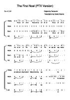
■ ■ ■ ■
■
14
Free body analysis (must know these; practice them): ■ M = F × d (torque/moment) ■ F=M×A Piezoelectric effect: ■ Concave compression side is electronegative. ■ Convex tension side is electropositive. Increase rigidity of external fixators: ■ Bone-to-bone contact (most important). ■ Larger-diameter pins. ■ Additional pins. ■ Decreased bone–rod distance. ■ Increased mass of the rods (or stack the rods). ■ Increased space between pins. ■ Circular fixators. Young’s modulus: ■ Ceramic ■ Cobalt chrome ■ Stainless steel ■ Titanium ■ Cortical bone ■ Polymethylmethacrylate (PMMA) ■ Polyethylene ■ Cancellous bone ■ Tendon ■ Ligament ■ Cartilage Modulus of elasticity: Linear relationship between applied stress and resultant deformation. Toughness: Resistance to fracture. Torsional load in cylindrical bone: Max tensile load if generated at 45 degrees to the long axis. Biomaterials: Stress: Force/area (N/m 2). Strain: (change in length)/(original length) proportion. Young’s modulus E = stress/strain. ■ Ceramic > cobalt chrome > stainless steel > titanium > cortical bone > PMMA > ultra-high-molecular-weight polyethylene (UHMWPE) > cancellous bone > tendon > ligament > skin > cartilage (1) Elastic limit: Point at which stain is no longer recoverable. (2) Yield point: Transition point from elastic to plastic deformation. (3) Yield strength: Amount of stress necessary to produce a specific amount of permanent deformation, usually 0.2%. (4) Ultimate strength: Maximum stress obtained to failure. (5) Elastic (linear) region: Proportional to stress applied. (6) Plastic region: Curve beyond yield point in which stress is not reversible (see Figure 1-2). Ductile: “Tough,” large plastic curve. Brittle: “Hard,” limited ability to deform before failure, small plastic curve. Fatigue failure: Number of cycles to material failure at a specific stress level. Endurance limit: Stress level at which a material can be cyclically loaded an infinite number of times without failing. Torsional bending strength not affected by < 10% of bone diameter. Strength decreases up to 50% if there is a hole that is 20% of the bone’s diameter.
BASIC SCIENCE
Stress–strain graph with references to definitions of elastic limit (1), yield point (2), yield strength (3), ultimate strength (4), elastic region (5), and plastic region (6).
FIGURE 1-2 .
■
■
■
■
Resorbable polymers: ■ Polyglycolide (PGA): Gone by 3 months. ■ Polydioxanone (PDS): Gone by 6 months. ■ Poly-L-lactic acid (PLLA): Gone in years. Wear types: ■ Adhesive: Chemical; fragments from each surface adheres to each other. ■ Abrasive: Mechanical; material mismatch in which the soft surface creates debris. ■ Fatigue: Material; high local stress causes fracture. ■ Third body: Mechanical; wear debris or other particles (cement, metal, bone, etc). Gamma irradiation can increase the number of cross-links in UHMWPE, yet can decrease strength. Ceramics: Best wear resistance, high elastic modulus, high compressive strength, brittle. ■ High conductiveness due to high surface wettability and high surface tension.
15
BASIC SCIENC
NOTES
16
CHAPTER 2
Orthopaedic Anatom y
Shoulder
19
MUSCLE INSERTIONS
19
CORACOACROMIAL (CA) LIGAMENT
19
STERNOCLAVICULAR (SC) JOINT
19
BUFORD COMPLEX
19
SHOULDER STABILIZERS
19
ARCUATE ARTERY
20
AXILLARY ARTERY
20
QUADRANGULAR SPACE
20
TRIANGULAR SPACE
21
TRIANGULAR INTERVAL
21
DISTAL CLAVICLE OSTEOLYSIS
21
Arm/ Forearm
21
Elbow
21
Hand
22
DEEP MOTOR BRANCH OF ULNAR NERVE
23
PARONA’S SPACE
23
CARPAL OSSIFICATION ORDER
23
LUMBRICAL P LUS HAND
23
Spine
23
BRACHIAL P LEXUS
23
VERTEBRAL ARTERY
24
HALO P INS
24
RECURRENT LARYNGEAL NERVE
24
ARTERY OF ADAMKIEWICZ
24
COMMON LUMBAR RADICULOPATHIES
25
INCOMPLETE SPINAL CORD INJURIES
25
COMPLETE SPINAL CORD INJURIES
26
Pelvis
26
HIP APPROACHES
26
ZONES WITH ACETABULAR SCREW P LACEMENT
27
FEMORAL TRIANGLE
27
17
ORTHOPAEDIC ANATOMY
NONTRAUMATIC AVASCULAR NECROSIS (AVN)
27
SUPERFICIAL CIRCUMFLEX ILIAC (SCI) ARTERY
27
Knee ANTERIOR CRUCIATE LIGAMENT (ACL)
28
POSTERIOR CRUCIATE LIGAMENT (PCL)
28
POPLITEUS
28
DISCOID LATERAL MENISCUS
28
Foot and Ankle
18
27
28
HIGH ANKLE SPRAIN
29
P LANTAR FASCIITIS
29
BAXTER’S NERVE
29
SPRING LIGAMENT
29
P ERONEUS LONGUS
29
P ERONEUS BREVIS
29
ANTERIOR TALOFIBULAR LIGAMENT (ATFL) TEARS
30
TALUS BLOOD SUPPLY
30
SHOULDER ■ ■ ■
■
ORTHOPAEDIC ANATOMY
■
Teres major: Innervated by the lower subscapularis nerve. Teres minor: Innervated by the axillary nerve. Pectoralis major: ■ Clavicular head innervated by the lateral pectoral nerve ■ Strenocostal head innervated by the medial pectoral nerve Humeral head is 30 degrees retroverted vs. transepicondylar axis. Glenoid is 5 degrees retroverted.
Mu scle In se rtio n s ■
■
Humerus from lateral to medial: PLT. ■ Pectoralis ■ Latissimus ■ Teres major Coracoid muscle attachments: Short head biceps, pectoralis minor, coracobrachialis.
Co ra co a cro m ia l (CA) Liga m e nt ■ ■
Acromial branch of the thoracoacromial artery runs within. Can be released during subacromial decompression.
Ste rn o cla vicu la r (SC) Jo in t ■ ■
Posterior SC ligaments are the strongest. Posterior SC dislocation is associated with tracheal compression. Be able to arthroscopically
Bufo rd Co m p le x ■ ■ ■
■
Normal variant. Thickened middle glenohumeral ligament (MGHL). Superior labral attachment of the MGHL just anterior to the biceps tendon with concomitant absence of anterosuperior labrum. If mistaken for labral detachment and the MGHL is repaired to the labrum, this inappropriate “repair” will cause loss of external rotation (ER).
Sh ou lde r Sta b ilize rs ■
■ ■
Static stabilizers: Articular anatomy, glenoid labrum, negative pressure, capsule, ligaments. Dynamic stabilizers: Rotator cuff, biceps tendon, scapulothoracic motion. CA ligament: Helps maintains superior joint stability.
SUPERIOR GLENOHUMERAL LIGAMENT (SGHL) ■
Inferior restraint in adduction.
M IDDLE GLENOHUMERAL LIGAMENT (MGHL) ■ ■
Anterior restraint in midrange (45 degrees) abduction. Secondary restraint to inferior translation in the adducted ER humerus. 19
recognize the normal variants (Buford complex and sublabral foramen) and discern them from a pathologic labrum.
INFERIOR GLENOHUMERAL LIGAMENT (IGHL) ■ ■ ■ ■
ORTHOPAEDIC ANATOMY
■ ■ ■ ■
Prevents anterior inferior instability in abduction. Anterior band: Tight at 90 degrees abduction and ER. Posterior band: Tight at 90 degrees abduction and internal rotation (IR). SGHL and MGHL tighten with adduction and ER. Rotator interval prevents flexion and ER of the shoulder. Posterior shoulder arthroscopic portal enters below infraspinatus. If placed too low, axillary nerve is at risk. Posterior approach to the shoulder is between infraspinatus (suprascapular nerve) and teres minor (axillary nerve).
Arcua te Arte ry ■
■
■
Terminal branch of the ascending branch of the anterior humeral circumflex artery. Supplies the humeral head and travels along the lateral border of the bicipital groove. It can be injured when plating a proximal humerus fracture.
Axilla ry Arte ry ■
■
■
Beware when plating proximal humerus fractures that the main blood supply to the humeral head lies in the
■
■ ■
lateral aspect of the bicipital groove.
■ ■
Begins at the lateral border of the first rib as a continuation of the subclavian artery. It changes its name to brachial artery at lower (inferior) border of the teres major. It is broken up into three parts by its relation to the pectoralis minor muscle (see Table 2-1). First part is between the lateral border of the first rib and the medial border of the pectoralis minor. Second part is behind the pectoralis minor. Third part is between the lateral border of the pectoralis minor and the inferior border of the teres major. Thoracoacromial artery runs within the CA ligament. Most tethered at origin of anterior and posterior humeral circumflex vessels.
Qu a dra ngu la r Sp a ce ■ ■
Axillary nerve and posterior humeral circumflex artery pass through this space. Borders: Teres minor (superior), teres major (inferior), long head of triceps (medial), humeral shaft (lateral).
TA B L E 2 - 1 .
Bra n che s of th e Axilla ry Arte ry Divisio n s
AXILLARY ARTERY
20
First division
Suprem e thoracic
Second division
Thoracoacrom ial
Lateral thoracic
Third division
Subscapular
Anterior hum eral
Poste rior hum e ral
circum fle x
circum fle x
Tria n gu la r Sp a ce ■ ■
Circumflex scapular artery passes through this space. Borders: Teres minor (superior), teres major (inferior), long head of triceps (lateral).
■ ■
Radial nerve and profunda brachii artery run through this interval. Borders: Teres major (superior), long head of triceps (medial), lateral head of triceps (lateral).
Dista l Cla vicle Oste o lysis ■ ■ ■ ■ ■ ■
Repetitive trauma (weight lifting). Rheumatoid arthritis. Hyperparathyroidism. Tumor. Cleidocranial dysplasia. Pyknodysostosis.
ARM/ FOREARM ■
■
■ ■ ■
■ ■
Musculocutaneous nerve continues distally between biceps and brachioradialis (BR) as the lateral antebrachial cutaneous nerve. Radial nerve traverses the spiral groove ~13 cm up from elbow joint, pierces septum ~7 cm from epicondyle between brachialis and BR. Posterior approach to humeral shaft splits the medial head of the triceps. Triceps originates from humerus and scapula. Median nerve runs between basilic vein and brachial artery. In median nerve injury, the last muscle to recover is the pronator quadratus. Pacinian corpuscles sense pressure. Ligament of Struthers: ■ Supracondylar process points toward elbow joint. ■ Ligament attaches to medial epicondyle. ■ Can cause median nerve entrapment.
ELBOW ■
■
■ ■ ■
■
■
Medial antebrachial cutaneous nerve runs with basilic vein, can be injured in elbow scopes. Joints with intra-articular metaphysis: Hip, shoulder, elbow (radial head), and ankle (distal fibula). Normal carrying angle of elbow is 7 degrees valgus tilt. Cephalic vein runs with lateral antebrachial cutaneous nerve (laterally). Kocher approach to elbow: Between extensor carpi ulnaris (ECU) (posterior interosseous nerve [PIN]) and anconeus (radial); pronate and flex elbow to protect PIN during approach. Henry approach (volar): Between pronator teres and BR; supinate to protect PIN. Thomspson approach (dorsal): Between extensor carpi radialis brevis (ECRB, radial nerve) and extensor digitorum communis (EDC, PIN). 21
ORTHOPAEDIC ANATOMY
Tria n gu la r Inte rva l
ORTHOPAEDIC ANATOMY
■
■ ■
The volar approach is typically
■
recommended for mid- and distal radius fracture open reduction and internal fixation
■
(ORIF) and the dorsal
■
approach for proximal radius
■
fracture ORIF.
■ ■
■ ■
Anterior bundle of medial collateral ligament (MCL) is primary valgus load stabilizer. Lateral epicondylitis: Primary pathology is in ECRB. Anterior medial portal with elbow scope: Medial antebrachial cutaneous nerve at risk. Radial nerve: ■ Enters forearm between brachialis and BR. ■ At risk with anterior lateral elbow scope portal. Median nerve splits the pronator teres and lies between flexor digitorum superficialis (FDS) and flexor digitorum profundus (FDP). Ulnar nerve lies between flexor carpi ulnaris (FCU) and FDP. Recurrent radial artery: Ligate while exposing proximal radius during anterior approach. Distal biceps rupture: Untreated lose ~40% supination and flexion strength. Superficial radial nerve: Retracting too much on BR risks injury to nerve. Deep radial nerve/PIN: Retracting too much on supinator risks injury to nerve. Posterolateral rotatory instability (PLRI) elbow: Key structure is lateral ulnar collateral ligament.
HAND ■ ■ ■
■
■ ■
■
■
■
■
■ ■ ■ ■
The A2 pulley is the most
■
important pulley to preserve, to prevent bow-stringing,
■
followed by A4.
22
Grayson’s ligaments: Volar to digital artery and nerve (Grayson/Ground). Cleland’s ligaments: Dorsal to digital artery and nerve (Cleland/Ceiling). Sagittal band anchors extensor tendons over the metacarpophalangeal joint (MCP). Triangular ligament connects lateral bands just proximal to terminal tendon insertion at base of distal phalanx. Hook of hamate fracture: X-ray to visualize fracture is carpal tunnel view. Ulnar nerve lesion at or near wrist or base of palm will result in paralysis of hypothenar muscles, all interosseous muscles, part of flexor pollicis brevis (FPB), palmaris brevis, and adductor pollicis. Median nerve innervates lumbricals (index and long), FPB (superficial head), opponens pollicis, abductor pollicis brevis (APB), pronator teres, FDS, flexor carpi radialis (FCR), and palmaris longus. Anterior interosseous nerve (AIN) innervates flexor pollicis longus (FPL), FDP lateral half, and pronator quadratus. Radial nerve innervates BR, ECRB, and extensor carpi radialis longus (ECRL). PIN: ■ Innervates EDC, extensor digiti minimi (EDM), ECU, extensor pollicis longus (EPL), extensor indicis (EI), abductor pollicis longus (APL), and extensor pollicis brevis (EPB). PIN at wrist: Located in floor of fourth compartment. APL has multiple slips. Thumb epiphysis is proximal. Pulleys: A1 at MCP, A3 at proximal interphalangeal joint (PIP), A5 at distal interphalangeal joint (DIP). Scapholunate interosseous ligament: Tear shows scapholunate widening on clenched-fist x-ray view. Froment sign: Weak adductor pollicis (ulnar nerve); thus, to hold paper between thumb and index finger, patient will use the FPL and FPB (median nerve) to compensate.
De e p Mo to r Bra nch o f Uln a r Ne rve ■ ■
Travels with deep palmar arch. Superficial arch blood supply from ulnar artery; deep arch supply from radial artery.
■ ■ ■
Above pronator quadratus and below flexors of forearm. Communication between thumb and small finger. Potential space for communication of pus.
Ca rpa l Ossifica tio n Ord e r ■
■
C-H-Tq-L-S-Tm-Td:
Capitate (6 months), hamate (6 months), triquetrum (3 years), lunate (4 years), scaphoid (5 years), trapezium (5 years), trapezoid (6 years). The ossification order goes counterclockwise looking at the volar surface, starting with the capitate.
Lum brica l Plu s Ha n d
Causes include: ■ ■ ■ ■
Laceration of FDP distal to lumbrical origin. Amputation distal to central slip. Loose tendon graft. Treatment: Lysis of lumbrical to tendon in finger.
SP INE ■ ■ ■ ■ ■ ■ ■
■
T5 has the narrowest pedicles. Aorta is on left, vena cava and azygous on right. 66% of lordosis is from L4–sacrum; most is from the disks. Innervation to spine posterior muscles is segmental. Annulus fibrosis: Mainly type I collagen (hard = bone). Nucleus pulposus: Mainly type II collagen (88% water). Intradiscal pressure: Highest when sitting and flexed forward with weights in hands; lowest is in the supine position. Superior articular facet is lateral and anterior to inferior articular facet.
Bra ch ia l Ple xu s ■
■ ■
■
Four preclavicular branches: Long thoracic, dorsal scapular, suprascapular, and subclavius. Horner syndrome: MAPE (Meiosis, Anhydrosis, Ptosis, Enophthalmos). Erb-Duchenne paralysis: ■ C5/6; arm held in extension IR; paralysis of ERs. ■ Follow biceps (18-month recovery). ■ Best prognosis. Klumpke paralysis: ■ C8–T1; weak wrist flexors, intrinsics. ■ Horner syndrome. ■ Poor prognosis. 23
Come Hug Quigley’s Long Smooth MataDor.
ORTHOPAEDIC ANATOMY
Pa ron a ’s Sp a ce
■ ■ ■ ■ ■
ORTHOPAEDIC ANATOMY
■ ■ ■
■ ■ ■ ■ ■
With nerve injury, the most
■
sensitive test is the threshold
■
(monofilament and vibration)
■
test, and it is the last to come
■
back with nerve recovery.
C2–3: Level of mandible. C3: Level with hyoid. C4/5: Level with thyroid. C6: Cricoid and carotid tubercle. C5–6: Most common disk herniation in cervical spine, most commonly gets C6 root. Neck flexion: 50% at occiput–C1. Neck rotation: 50% at C1–2. Denervation: Electromyographic (EMG) findings; fibrillations and sharp waves. Dermatomes: T4, nipples; T7, xiphoid; T10, umbilicus. Longus colli is anterior to vertebral artery. Cervical ganglion is anterior to longus capitus. Longus capitus is anterior to longus colli. Deepest to anterior: Vertebral artery, longus colli, longus capitis, cervical ganglion. Carotid sheath: Internal carotid artery, common carotid artery, internal jugular vein, vagus nerve (CN X). Neurapraxia: Order of motor and sensory loss. Motor > proprioception > touch > temperature > pain. Regain first to last (APP MMST): ■ Anesthesia > Pressure (proprioception) > Pain (protective) > Moving touch > Moving two-point (Meissner’s) > Static Two-point (Merkel’s).
Ve rte b ra l Arte ry ■ ■ ■
When dissecting near C1, stay
■
within 15 mm of midline to avoid injuring the vertebral artery.
■
■
Branch of subclavian. Runs through C6 to C1 (not C7). Posterior to longus colli. After exiting C1, travels medially on the superior posterior arch of C1 (cephalad) and up through the foramen magnum. Greater occipital nerve: Exits spine at C1–2 (from medial branch of dorsal ramus C2), pierces semispinalis and trapezius. Cruciform ligament: Main stability for occiput–C2.
Ha lo Pin s ■ ■ ■
Anterior pins need to be at least 4.5 cm lateral from midline. Below equator, above supraorbital ridge. Risks injury to supraorbital nerve, supratrochlear nerve, frontal sinus (from lateral to medial).
Re curre n t La ryn ge a l Ne rve When anteriorly exposing the lower lumbar spine, be
■ ■
Unpredictable course on the right side. On left side lies between trachea and esophagus.
cautious of the superior hypogastric plexus that overlies L5. Injury to this plexus can cause retrograde ejaculation.
Arte ry of Ad a m kie wicz ■ ■ ■ ■
24
T8–L1. 80% on left. Arteria radicularis magna. Arterial injury can cause anterior spinal syndrome.
TA B L E 2 - 2 .
LEVEL
Co m m o n Lu m b a r Ra d icu lo p a th ie s
MOTOR WEAKNESS
SENSORY INVOLVEMENT
REFLEX AFFECTED
Tibialis ante rior
Me dial le g and foot
Patellar tendon
L5
EDL and m edial
Middle dorsum of foot
None
Lateral foot
Achilles tendon
ORTHOPAEDIC ANATOMY
L4
ham string (SM and ST) S1
Peroneals, FHL, gastrocs, lateral ham string, and gluteus m axim us
EDL = extensor digitorum longus; FHL = flexor hallucis longus; SM = sem im em branosus; ST = sem ite ndinosis.
Co m m o n Lu m b a r Ra d icu lo p a thie s
The signs and symptoms of lumbar radiculopathies are outlined in Table 2-2. ■ ■
■
■
Isthmic spondylolisthesis L5–S1 gets L5 radiculopathy. Herniated nucleus pulposus (HNP) L5–S1 gets S1 radiculopathy (unless far lateral, then L5). Degenerative spondylolisthesis is most common at L4–5 (get L5 root symptoms). L5 radiculopathy is due to compression between hypertrophic and subluxed inferior facet of L4 and posterosuperior L5 body.
In com ple te Sp in a l Co rd In jurie s CENTRAL CORD SYNDROME ■ ■ ■ ■ ■
Most common. Upper extremity (UE) weakness > lower extremity (LE). Flaccid UE and spastic LE. Intact bladder control. ~75% recovery.
P OSTERIOR CORD SYNDROME ■ ■ ■
Rare. Loss of deep pressure, deep pain, and proprioception. Ambulate with slapping gait (tabes dorsalis).
ANTERIOR CORD SYNDROME ■ ■
■
Worst prognosis. Complete motor and sensory loss except retain trunk and LE pressure/ proprioception. ~10% functional recovery.
BROWN-SÉQUARD SYNDROME ■ ■ ■ ■
Best recovery. Unilateral cord injury. Motor deficit on ipsilateral side. Contralateral pain and temperature loss two levels below injury. 25
■ ■
Most regain bowel/bladder function and can ambulate. > 90% functional recovery.
Co m p le te Sp in a l Cord In ju rie s
ORTHOPAEDIC ANATOMY
■
May have root return of one level in 80% and two levels in 20%. P ELVIS
■ ■
■
■
■ ■
■
For vessels to the femoral
■
head: LAMP (Lateral femoral circumflex is Anterior; Medial femoral circumflex is
■
■
Posterior)
Peroneal division of sciatic nerve is more lateral. Hip pathology can have pain referred to the knee due to continuation of a branch of the obturator nerve (to adductor magnus). Sacroiliac (SI) screws: If placed too anterior, potential for L5 nerve root injury. Femoral nerve: First branch goes to sartorius; last branch goes to articularis genu (see Table 2-3). Internal iliac artery becomes the obturator artery. Vessels to the femoral head: Lateral femoral circumflex is on the anterior neck; medial femoral circumflex is on the posterior neck. Ascending branch of lateral circumflex femoral artery: Encountered in anterior approach to the hip (ligate). Medial femoral circumflex artery is between adductor magnus and brevis medially. Deep external pudendal artery is at risk when doing a percutaneous tenotomy of the adductor longus. Peroneal branch of sciatic nerve runs on the deep surface of long head biceps.
Hip Ap p roa ch e s ■
■
Anterior approach (Smith-Peterson): Interval is between sartorius (femoral nerve) and tensor fascia lata (superior gluteal nerve). True internervous plane. Good for primary total hip arthroplasty (THA) (esp. “mini”), anterior column plating. Lateral femoral cutaneous nerve at risk. Poor visualization of posterior acetabulum. Anterolateral approach (Watson-Jones): Tensor fascia lata (superior gluteal nerve) and gluteus medius (superior gluteal nerve). Primary THA. Risk of damage to tensor fascia lata innervation.
TA B L E 2 - 3 .
Ne rve , Le ve l, a n d Mu scle o f th e Fe m o ra l, Obtu ra to r, a n d Scia tic Ne rve s
NERVE
LEVEL
M USCLE
Fe m oral
L2–4
Iliacus, psoas, sartorius, pe ctineus, quads, articularis ge nu
Obturator
L2–4
Obturator externus, hip adductors, gracilis
Sciatic
L4–S3
Peroneal division: Short head of biceps Tibial division: Sem itendinosus, sem im em branosus, part of adductor m agnus, long head bice ps
26
TA B L E 2 - 4 .
■
■
Birth to age 4
Prim arily m edial and late ral circum flex arte rie s, and ligam entum
Age 4 to adult
Prim arily m edial circum flex, m inim al from lateral and ligam e ntum
ORTHOPAEDIC ANATOMY
■
Fe m o ra l He a d Blo o d Su p p ly
Medial approach (Ludloff): Adductor brevis (obturator nerve) and adductor magnus (obturator and sciatic). Direct lateral approach (Hardinge): Via tensor fascia lata and gluteus medius. Can damage femoral nerve (retractor placement) and superior gluteal nerve (4 cm proximal to greater trochanter). THA. Reduced dislocations in THA compared to posterior approach. Patients can limp. Posterior column plating not possible. Posterior approach: Via gluteus maximus, but innervation is medial to the split so denervation unlikely. THA. Can be extended for posterior column and wall. Higher dislocation risk with THA.
Zo ne s with Ace ta b u la r Scre w Pla ce m e n t ■
■
■
Posterior superior acetabular quadrant (safest zone): Risk injury to sciatic nerve, superior gluteal vessels at risk. Posterior inferior acetabular quadrant: Risk injury to sciatic nerve, inferior gluteal vessels and nerve, internal pudendal vessels and nerve. Anterior inferior acetabular quadrant: Risk injury to obturator nerve, artery, and vein. Anterior superior acetabular quadrant: External iliac vessels.
When placing screws in an acetabular cup, the posterior superior quadrant is the safest zone.
Fe m o ra l Tria n gle ■ ■
Floor (lateral to medial): Iliacus, psoas, pectineus, adductor longus. Femoral canal: Iliacus, femoral nerve, femoral artery, femoral vein, pectineus. Order of Nerves that Pass Below the Piriformis—
No n tra u m a tic Ava scula r Ne cro sis (AVN) ■ ■
Typically affects the anterolateral portion of femoral head. The contribution of the lateral circumflex femoral artery and ligamentum decreases after ~ age 4 (see Table 2-4).
■
■ ■
■
Su pe rficia l Circu m fle x Ilia c (SCI) Arte ry ■
POPS IQ
Landmark for lateral femoral cutaneous nerve (in anterior approach to hip). Used for groin flap.
■
■ ■
KNEE ■
Medial knee: three layers (superficial to deep): ■ I: sartorius, sartorial fascia. ■ II: superficial MCL, posterior oblique ligament (POL), semimembranosus. ■ III: deep MCL, capsule. 27
Pudendal nerve. Nerve to Obturator internus. Posterior femoral cutaneous nerve. Sciatic (2% through piriformis). Inferior gluteal nerve. Nerve to Quadratus femoris.
■
■
ORTHOPAEDIC ANATOMY
■ ■ ■
■ ■
Lateral knee: three layers (less consistent): ■ I: lateral fascia, IT band, biceps tendon. ■ II: patellar retinacullum, patellofemoral ligament. ■ III: capsule, lateral collateral ligament (LCL), arcuate ligament, fabellofibular ligament. Popliteal artery is typically behind the posterior horn lateral meniscus. Lateral inferior geniculate artery runs between LCL and popliteus. Fabella: Located in lateral gastrocnemius (18% of patients). Meniscal repair: ■ Medially, saphenous nerve is at risk. ■ Laterally, peroneal nerve is at risk. Chondrocalcinosis: Calcium-containing crystals in cartilage/meniscus. Menisci: Majority of large collagen fibers are circumferential; hoop stress with compressive loading.
An te rio r Cru cia te Liga m e n t (ACL) ■ ■ ■
■
■
■
Anteromedial bundle: Tight in flexion. Posterolateral bundle: Tight in extension. Classic bone bruise pattern: Mid third of lateral femoral condyle and posterior third of lateral tibial plateau. Lateral meniscus is most common site of acute meniscal tear with ACL injury. Chronic bone scan in an ACL-deficient knee shows increased uptake in medial > lateral > patellofemoral compartment. Segond fracture is associated with an ACL tear; lateral capsular avulsion that can be seen on plain films.
Po ste rio r Cru cia te Liga m e n t (PCL) ■
The key structures in the
■
posterior lateral corner are
■
the popliteus, LCL, and
■ ■
popliteal fibular ligament.
Middle geniculate artery: Main blood supply to PCL and ACL. Anterolateral bundle: Tight in flexion. Posteromedial bundle: Tight in extension. Ligament of Humphrey (anterior to PCL), Wrisberg (posterior to PCL). On magnetic resonance imaging (MRI), “double PCL sign” indicates posterior horn medial meniscus tear displaced into notch.
Po p lite u s ■ ■ ■
Intra-articular. Runs from the posterior tibia to lateral femoral condyle anterior to LCL. Can be visualized during arthroscopy.
Disco id La te ra l Me niscu s ■ ■ ■
Patient may lack full knee extension. ~3% population. Diagnosed by ≥3 sagittal slices with complete bowtie appearance of meniscus. FOOT AND ANKLE
■ ■ ■
28
Anterior talofibular ligament (ATFL) is an intra-articular thickening. Heel spurs originate in the flexor digitorum brevis (FDB). Ankle scope: Anterior lateral portal: Risk to superficial peroneal nerve.
High An kle Sp ra in ■ ■ ■
■
Pla n ta r Fa sciitis ■ ■ ■
First branch of lateral calcaneal nerve. Trapped between abductor hallucis and quadratus plantae. Innervates abductor digiti minimi.
Ba xte r’s Ne rve ■ ■
Lateral plantar nerve. Heel pain.
Sp rin g Liga m e n t ■ ■ ■
Coronoid cavity of calcaneus to inferior (plantar) surface of navicular. Anterior to the sustentaculum tali. Also known as calcaneal-navicular ligament.
Pe ron e u s Lon gu s ■ ■
Paralysis or laceration; can cause a dorsal bunion. Plantar flexes the first ray.
Pe ron e us Bre vis ■
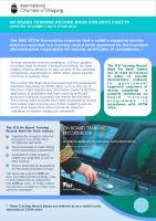
Common to get split longitudinal tears at the fibular groove (see Figure 2-1).
F I G U R E 2 - 1 . Pe ro n e a l te n d o n s. Ope rative photo of the (A) pe roneus brevis and (B) longus
just proxim al to the fibular groove .
29
ORTHOPAEDIC ANATOMY
■
ER mechanism. Injury to the tibiofibular ligament, interosseous ligament. Chronic ankle sprain of anterior inferior tibiofibular ligament (Bassett’s ligament): Get thickening and synovitis. If needed, treat with arthroscopic debridement.
ORTHOPAEDIC ANATOMY
An te rio r Ta lo fib ula r Liga m e nt (ATFL) Te a rs ■ ■
Ankle instability. Treatment: ■ Modified Brostrom: ■ Repair ATFL using local tissue. ■ Reinforce with extensor retinaculum. ■ Chrisman-Snook: Uses half of the peroneus brevis to reconstruct ATFL ■ Watson-Jones and Evans: Uses all of the peroneus brevis to reconstruct ATFL
Ta lu s Blo o d Sup p ly ■
■ ■
30
Main supply is the artery of the tarsal canal (medial), a branch of the posterior tibial artery. Artery of sinus tarsi (lateral), a branch of the dorsalis pedis artery. Deltoid branch, a branch of the artery to the tarsal canal; runs in the deltoid substance.
CHAPTER 3
Orthopaedic Traum a
General Trauma
34
ABCS
34
ADEQUATE RESUSCITATION
34
FLUID RESUSCITATION
34
BASE DEFICIT
35
TRAUMA X-RAY SERIES
35
OPEN FRACTURES
35
FAT EMBOLI SYNDROME
35
TENSION P NEUMOTHORAX
35
COMPARTMENT SYNDROME
36
MANGLED EXTREMITY SEVERITY SCORE (MESS)
36
Spinal Cord/ Plexus Injuries
36
NASOTRACHEAL INTUBATION
36
CRICOTHYROTOMY
36
NEUROLOGIC LEVEL
36
STEROID P ROTOCOL
36
INCOMPLETE CORD SYNDROMES
36
INCOMPLETE SPINAL CORD INJURY
37
CERVICAL TRAUMA
37
C1–2 INSTABILITY
37
C2 (AXIS) HANGMAN’S FRACTURE
37
ODONTOID FRACTURE, TYPE II
37
ODONTOID FRACTURE, TYPE III
38
UNILATERAL C-SPINE FACET FRACTURE
38
BILATERAL C-SPINE FACET SUBLUXATION/ DISLOCATION
39
IPSILATERAL C-SPINE LAMINA AND P EDICLE FRACTURE (FLOATING FACET)
39
ANKYLOSING SPONDYLITIS
39
FLEXION–DISTRACTION INJURY
39
BURST FRACTURE
40
Brachial Plexus Injuries
40
Upper Extremity Trauma
40
STERNOCLAVICULAR (SC) JOINT DISLOCATION
40
CLAVICLE FRACTURES
41
31
ORTHOPAEDIC TRAUMA
ACROMIOCLAVICULAR (AC) INJURIES
41
SCAPULA FRACTURE
41
FLOATING SHOULDER
41
SCAPULOTHORACIC DISSOCIATION
41
GLENOHUMERAL DISLOCATIONS
42
LITTLE LEAGUER’S SHOULDER
43
P ROXIMAL HUMERUS FRACTURE
43
HUMERAL SHAFT FRACTURE
44
DISTAL HUMERUS FRACTURE
45
P EDIATRIC SUPRACONDYLAR HUMERUS FRACTURE
45
P EDIATRIC MEDIAL EPICONDYLE FRACTURE
45
P EDIATRIC LATERAL CONDYLE FRACTURE
45
OLECRANON FRACTURE
46
P EDIATRIC OLECRANON FRACTURE, FLEXION TYPE
46
CORONOID FRACTURE
46
RADIAL HEAD FRACTURE
46
ELBOW DISLOCATION
46
RADIUS/ ULNA FRACTURE
47
RADIUS APPROACHES
47
P EDIATRIC BOTH-BONE FOREARM FRACTURE
47
GALEAZZI FRACTURE
47
MONTEGGIA FRACTURE
48
P IN INJURY
48
DISTAL RADIUS FRACTURE
48
ULNAR STYLOID FRACTURE
49
ULNAR IMPACTION SYNDROME
49
Pelvis and Lower Extremity Trauma
32
49
P ELVIC RING INJURIES
49
SACRAL FRACTURE
50
ACETABULAR FRACTURE
51
HIP DISLOCATION
52
FEMORAL HEAD FRACTURE
53
FEMORAL NECK FRACTURE
53
FEMORAL NECK STRESS FRACTURE
54
INTERTROCHANTERIC HIP FRACTURE
54
SUBTROCHANTERIC HIP FRACTURE
55
FEMORAL SHAFT FRACTURE
56
P EDIATRIC FEMUR FRACTURE
58
DISTAL FEMUR FRACTURES
58
KNEE DISLOCATION
59
PATELLA FRACTURE
60
PATELLA DISLOCATION
61
PATELLA TENDON (LIGAMENT) RUPTURE
61
QUADRICEP TENDON RUPTURE
61
TIBIAL P LATEAU FRACTURE
61
TIBIAL SHAFT FRACTURE
62
TIBIAL P LAFOND (P ILON)
64
65
SYNDESMOTIC DISRUPTION
66
OPEN ANKLE FRACTURE
66
ACHILLES TENDON RUPTURE
66
OS P ERONEUM
67
ACUTE P ERONEAL TENDON DISLOCATION
67
TALAR NECK FRACTURES
67
TALAR P ROCESS FRACTURE
68
SUBTALAR DISLOCATION
68
CALCANEUS FRACTURE
68
NAVICULAR FRACTURE
69
CUBOID FRACTURE
69
LISFRANC FRACTURE–DISLOCATION
70
METATARSAL FRACTURE
70
MTP JOINT DISLOCATION
71
”TURF TOE”
71
P HALANGEAL FRACTURE
71
COMPARTMENT SYNDROME OF THE FOOT
71
ORTHOPAEDIC TRAUMA
ANKLE FRACTURE
33
GENERAL TRAUMA
ABCs ■ ■ ■ ■ ■
Airway Breathing Circulation Disability Evaluation (neurologic status), Exposure, and Environment control
Ad e q u a te Re suscita tio n ■ ■ ■ ■
ORTHOPAEDIC TRAUMA
■
Mean arterial pressure (MAP) > 60 mm Hg. Heart rate (HR) < 100 beats/min. Urine output (UOP) > 0.5–1.0 mg/kg/h. Best predictor of perioperative complications: Lactate levels > 2.5 mmol/L. Interleukin-6 (IL-6) is associated with systemic inflammatory response to trauma. Definitive surgery should be delayed when IL-6 is elevated to avoid multiple organ dysfunction syndrome (MDOS).
Flu id Re su scita tio n ■
■
■ ■ ■
Crystalloid isotonic solutions can be used to correct most extracellular volume deficits. Administered rapidly at 3–4 times the volume of estimated blood loss (EBL). Give blood to those who fail initial fluid boluses. The most common cause of shock is acute blood loss (see Table 3-1). The most common cause of blood transfusion reaction: Clerical error.
T A B L E 3 - 1 . He m o rrh a gic Sh o ck
CLASS I
BLOOD LOSS
HR
PH
RESP
UOP
CNS
Nl
Nl
Nl
Nl
> 30 cm 3
Anxious
Nl
↑
Nl
↑
20–30 cm 3
Irritable,
< 15% (< 750 cm 3 )
II
BP
15%–25% (750–1500 cm 3 )
confuse d, com bative III
25%–40% (1500–2000 cm 3 )
↓
↑
↓
↑
5–15 cm 3
Irritable, lethargic
IV
> 40% (> 2000 cm 3 )
↓
↑
↓
↑
< 5 cm 3
Le thargic, com a
BP, blood pressure; CNS, central nervous system ; HR, heart rate; Nl, norm al; UOP, urine output.
34
Ba se De ficit ■ ■
■
Best measure of adequate resuscitation in first 6 hours after injury. A direct measure of metabolic acidosis and an indirect measure of blood lactate levels. Correlates with organ dysfunction, mortality, and adequacy of resuscitation.
The best test to measure resuscitation in the first 6 hours after injury is
Tra um a X-ra y Se rie s ■ ■ ■
base deficit.
Anteroposterior (AP) chest (mediastinal widening, pneumothorax). Lateral C-spine (must visualize C7–T1 junction). AP pelvis.
Op e n Fra ctu re s ■
Fa t Em b oli Syn dro m e ■
■ ■ ■
Anxiety, confusion, tachycardia, and hypoxemia; usually occurs within 48 hours of injury. Hypoxia (PaO 2 < 60 mm Hg), petechial rash. Treatment: Preventive and supportive. Surgical stabilization of fractures is beneficial.
Te nsion Pn e u m o th ora x ■
■
Tachycardia, hypotension, chest with tympany to percussion, distended neck veins, and deviation of the trachea away from the affected side. Treatment: Urgent needle decompression, typically between second and fourth intercostal space (midclavicular line), followed by definitive chest tube placement.
T A B L E 3 - 2 . Gu stillo Op e n Fra ctu re Cla ssifica tio n
GRADE
SOFT TISSUE WOUND
ANTIBIOTICS
I
< 1 cm
First-generation cephalosporin
II
1–10 cm
First-generation cephalosporin
III
> 10 cm
First-generation cephalosporin, gentam icin; add penicillin if gross contam ination (e .g., farm , bowel)
A
Adequate tissue for closure
B
Needs soft tissue coverage
C
Vascular injury requiring repair
35
ORTHOPAEDIC TRAUMA
■
Need emergent irrigation and debridement (I&D). Appropriate antibiotics based on grade (see Table 3-2) and stabilization.
ORTHOPAEDIC TRAUMA
Co m p a rtm e n t Syn d rom e ■ ■
The most critical clinical sign
■
of compartment syndrome is
■
Have a high index of suspicion. 5 Ps: Pain out of proportion (critical clinical sign), pain with Passive stretch (critical clinical test), Paresthesias, Pallor, and Pulselessness. MAP–compartment pressures < 30 mm Hg critical value to decompress. 4 Cs of viability: Color, Contractility, Capacity to bleed, Consistency.
pain out of proportion.
Ma n gle d Extre m ity Se ve rity Score (MESS) ■ ■
■
Used to predict necessity of amputation after lower extremity trauma. Points are assigned for skeletal/soft tissue injury (1–4), ischemia time (1–6), age of the patient (0–2), and shock defined by hypotension (0–2). MESS score > 7 indicates need for amputation.
SP I NAL CORD/ P LEXUS INJURIES
Na so tra ch e a l In tu b a tio n ■
Treatment of choice in the nonapneic patient with suspected cervical injury with no maxillofacial trauma.
Cricoth yro to m y ■
Preferred approach in adults with facial trauma and possible neck injury.
Ne uro logic Le ve l ■
Most caudal level with normal motor and sensory function; at least 4/5 motor.
Ste ro id Pro toco l ■ ■
■
Methylprednisolone protocol: Initiate within 8 hours. Load 30 mg/kg, then 5.4 mg/kg/h for 23 hours if < 3 hours, continue for 48 hours if 3–8 hours. Steroids are not indicated for nerve root deficits, brachial plexus deficits, or gunshot wounds (GSWs).
In com ple te Co rd Synd ro m e s ■
■
■ ■
36
Central cord: Most common, motor loss upper extremity (UE) > LE, sacral sparing, rare full recovery. Brown-Séquard: Cord hemitransection; good recovery. ■ Ipsilateral loss motor and proprioception. ■ Contralateral loss pain and temperature (two levels lower because these travel up two levels and then cross). Anterior cord: Mechanism flexion–compression; poor prognosis. Posterior cord: Lose proprioception; keep motor, pain, and light touch; rare.
In com ple te Sp in a l Co rd In jury ■
■
■
Best chance for recovery occurs when the canal is cleared and the neural structures are decompressed. Anterior decompression, vertebral body reconstruction, and anterior stabilization have been shown to be highly effective in the treatment of bursttype fractures. Laminectomy alone is contraindicated because of instability.
Akey finding in central cord syndrome is motor weakness that is greater in the upper extremities than the lower.
Ce rvica l Tra u m a ■ ■
■
■
C1–2 In sta b ility ■ ■ ■
■ ■ ■
Secondary to rupture of the transverse ligament. Unstable on flexion–extension views. Atlantodens interval (ADI) > 5 mm (normal: adult, ≤ 3 mm; child, ≤ 5 mm); posterior ADI < 13 mm. Treatment: Posterior spinal fusion. 50% rotation is from C1–2. 50% flexion–extension is occiput–C1.
C2 (Axis) Ha n gm a n ’s Fra ctu re ■ ■ ■ ■ ■
Traumatic spondylolisthesis. Type I (< 3 mm displacement): Cervical collar (4–6 weeks). Type II (> 3 mm displacement): Halo, rare neurologic injury (Figure 3-1). Type IIA: Angulated, extension, no traction, halo. Type III (bilateral facet dislocation, C2 on C3): Reduction and surgical fixation.
Od o n toid Fra ctu re , Type II ■
■
If initial fracture displacement is > 5 mm, angulation > 10 degrees, or > 50 years old, problems with nonunion and malunion are more frequent when treated conservatively. Surgical treatment is with anterior screw or posterior C1–2 fusion.
37
ORTHOPAEDIC TRAUMA
■
Over two-thirds of significant pathology is detected on the lateral view. If the C7–T1 junction is not revealed on radiographs, then a computed tomography (CT) scan with lateral reconstructions should be used to visualize that portion of the spine (10%–15% C-spine injuries at this level). Obtain magnetic resonance imaging (MRI) scans in patients who have a neurologic finding that cannot be explained by bony or ligamentous instability on plain radiographs and dynamic views. While dynamic radiographs are contraindicated in the patient with acute neurologic deficits, they should be obtained in patients who have unexplained neck pain but are neurologically intact. To rule out subacute instabilities that may not have been seen initially, these studies need to be repeated 2–3 weeks after the injury and after cervical spasm has resolved.
ORTHOPAEDIC TRAUMA
Typ e II Ha n gm a n ’s fra ctu re . Tre at with h alo im m o bilizatio n. Also , this film is inad e qu ate , as the C7 – T1 ju n ctio n is n o t visualize d an d add itio n al sp ine in ju rie s co uld b e m isse d. FIGURE 3 -1 .
Od o n toid Fra ctu re , Type III ■
■
Consider operative
■
management of a type II
■
odontoid fracture if initial
This type of fracture has a high union rate following reduction and immobilization (see Figure 3-2). Anterior screw fixation is contraindicated in type III fractures. Treat with halo. Odontoid nonunion: If unstable on flexion–extension x-rays, stabilize and fuse.
displacement is > 5 mm, angulation > 10 degrees, or patient > 50 years old.
Un ila te ra l C-Sp ine Fa ce t Fra ctu re ■ ■ ■
38
Stable bony fracture. Treat with rigid collar for 6 weeks. Flexion–extension x-rays may be obtained at 6 weeks to verify there is no instability.
ORTHOPAEDIC TRAUMA
FIGURE 3 -2 .
Typ e III od o n to id fra ctu re . A high rate of fracture union occurs with halo
im m obilization.
Bila te ra l C-Sp in e Fa ce t Su blu xa tio n / Disloca tion ■
■
■
Skeletal traction and attempt closed reduction if patient is alert and cooperative. This should not be attempted in a patient who is obtunded, comatose, or uncooperative. If any neurological changes are noted during reduction, attempt should be stopped, appropriate radiographic studies obtained (including MRI to evaluate disk), and open reduction and stabilization planned in the operating room.
Ip sila te ra l C-Sp in e La m in a a n d Pe d icle Fra ctu re (Flo a ting Fa ce t) ■ ■ ■
Extremely unstable pattern (involves two motion segments). Treat with lateral mass plates (provides rotational control). Halo does not support the subaxial spine well.
An kylo sin g Sp o n dylitis ■ ■
■
Rigid spines are susceptible to fracture. A high index of suspicion should exist for occult fracture; consider CT scan. Late neurologic deficit likely secondary to hematoma (epidural).
Fle xio n –Distra ctio n In jury ■
■
Closely evaluate for intra-abdominal visceral injury (especially gastrointestinal); initial evaluation should include abdominal CT. Common with lap belt restraints in motor vehicle accidents.
39
ORTHOPAEDIC TRAUMA
■
■
With a flexion–distraction
■
injury (Chance fracture) of the spine, you must rule out intra-
X-rays will most likely reveal an increased interspinous distance and a break in continuity of the pedicle’s cortical margins—empty facet sign. A variety of structural defects may be produced, including pure bone defect (Chance fractures) or bone, ligament, and disk combinations extending over several vertebral segments. Pediatric bony chance fractures are treated with hyperextension casting and TLSO. Bedrest is not necessary. Surgery is required for significant ligamentous injury or progressive kyphosis (> 25 degrees).
abdominal injury.
Burst Fra ctu re
Surgical indications: ■ ■ ■ ■ ■
> 50% loss of height. > 30% kyphosis. > 50% canal compromise. Retained fragments will resorb and do not cause neurologic deterioration. If surgery is needed, anterior decompression with strut graft +/– posterior fusion is required. Laminectomy will further destabilize the spine.
BRACHIAL P LEXUS I NJURI ES ■
■
■
■
■
■
Postganglionic: Preservation of cervical paraspinals and no Horner syndrome, motor and sensory out on electromyography (EMG), nerve conduction velocity (NCV). Preganglionic: Horner syndrome present due to disruption of sympathetics; cell bodies intact, thus motor out and sensory still in on EMG/NCV; triple flare reaction remains intact. Most managed closed; exploration based on clinical and electrical recovery. Primary exploration for penetrating trauma (not GSW), open injuries, progressive neurologic deficit, expanding hematoma, vascular injury. Reconstruction priorities: Elbow flexion > shoulder abduction > hand sensibility > wrist extension > finger flexion > wrist flexion > finger extension > intrinsic function. Reconstructive options: Nerve repair, nerve graft, neurotization, free muscle transfer.
UP P ER EXTREMITY TRAUMA
Ste rn o cla vicu la r (SC) Jo in t Dislo ca tio n ■
■
■
40
X-ray: ■ Serendipity view (40-degree cephalic tilt). ■ CT (study of choice). Anterior dislocation: ■ Reduce under general anesthesia with abduction, extension, and direct pressure. ■ Unstable reduction: Accept deformity (preferable) vs. resect medial clavicle. Posterior dislocation: ■ May present with stridor, tachypnea, dysphagia.
Reduce under general anesthesia with abduction, extension, towel clip on medial clavicle; have thoracic surgery available. ■ Unstable reduction: Excision of medial clavicle (preferable) vs. surgical stabilization. Medial clavicle resection: Do not resect more than 1.5 cm, avoid costoclavicular ligaments. Medial clavicle physeal fractures: < 25 years old, Salter-Harris I or II, nonoperative unless symptomatic; will remodel. ■
■
■
Cla vicle Fra ctu re s ■ ■
■ ■
Acrom io cla vicu la r (AC) In ju rie s ■ ■
■
Primary treatment: Sling for ~3 weeks. Nonoperative treatment for types I, II, and III (some advocate operating on type III). Indications for surgical intervention: ■ Superior displacement of clavicle > 100% (type V). ■ Posterior displacement through trapezius (type IV). ■ Inferior lateral displacement under coracoid (type VI).
Sca p u la Fra cture ■
■ ■
High-energy trauma with 80%–90% associated injury; thoracic rib fractures most common, pulmonary contusions. Treat most with sling for 3–6 weeks. Indications for surgical intervention: ■ Involvement of > 25% glenoid with humeral subluxation (Figure 3-3). ■ Intra-articular stepoff > 5 mm or major gap. ■ > 40 degrees angulation or 1-cm translation of neck fracture, excessive medialization of glenoid.
Flo a tin g Sh o uld e r ■ ■
If unstable, will need ORIF (usually of just the clavicle). Nonsurgical management of the majority of combined injuries in patients with < 10 mm of displacement and stable has resulted in excellent shoulder function.
Sca p u lo th o ra cic Disso cia tio n ■ ■
■
Closed traumatic forequarter amputation. Chest x-ray: Nonrotated shows scapula edge laterally displaced > 1 cm from contralateral side (measure from spinous processes). Vascular injury common: Subclavian most common. 41
ORTHOPAEDIC TRAUMA
Primary treatment: Sling for 6 weeks. Indications for surgical intervention (indications expanding): Open fractures, significantly displaced fractures (tenting skin), vascular injury, lateral third with unstable medial fragment. For atrophic nonunion: Open reduction and internal fixation (ORIF). Options include: 3.5-mm AO plate, modified Hagie intramedullary (IM) fixation.
ORTHOPAEDIC TRAUMA
Fra ctu re th ro u gh th e gle n o id a n d in to th e sca pu la . The significant gle noid involvem ent and articular stepoff re quire ORIF. FIGURE 3 -3 .
■ ■
■
Arteriogram and, if needed, immediate repair. Brachial plexus injury common; complete deficit has poor outcome, partial has guarded outlook. Must stabilize the dissociation: ORIF clavicle and AC joint.
Gle n oh u m e ra l Dislo ca tio n s ■ ■ ■ ■
■
■
42
Radiographic evaluation: AP/scapular/axillary. Westpoint view: Help evaluate anterior glenoid margin (bony Bankart). Stryker notch view: Help evaluate Hill-Sachs defect of humeral head. Anterior dislocation: ■ If > 40 years old, high incidence of rotator cuff tears. ■ If < 20 years old, high recurrence rate (instability). ■ Surgical treatment: Labral repair, > 95% success. ■ TUBS: Traumatic Unilateral Bankart Surgery. Inferior dislocation—“luxatio erecta”: ■ Greater incidence of neurovascular injury (axillary nerve and artery). ■ May resolve after reduction. ■ Be aware of late thrombosis. ■ Reduce and immobilize for a short period of time. Posterior dislocation: ■ Rare compromising < 5%. ■ Often associated with lesser tuberosity fracture. ■ Clinically unable to externally rotate the shoulder. ■ Must be ruled out by complete radiographic series (including axillary and scapular Y).
■
■
■ ■
■
Common following seizures and electrical shocks (stronger internal rotation [IR] vs. the weaker external rotation [ER]). 50% have associated impaction fracture between articular margin and lesser tuberosity. If impaction < 20%: Closed reduction in extension and ER. If 20%–40% defect: Transpose lesser tuberosity with subscapularis into defect (Neer modification). If > 40%–50% defect: Consider hemiarthroplasty.
Must obtain an axillary lateral to evaluate location of dislocation or rule out posterior dislocation.
Little Le a gu e r’s Sh ou ld e r ■ ■ ■
Rotational stress fracture of proximal humeral physis. Widening of physis (compare with opposite side). Treat with cessation of throwing activities with rehab of shoulder girdle muscles and evaluate pitching technique. If proximal physeal stress
■
■ ■
■ ■
Anterolateral branch of the anterior humeral circumflex artery is the main blood supply to humeral head; runs in intertubercular groove. 45% incidence of nerve injury (axillary most common). Two-part: ■ Surgical neck: Sling unless > 45-degree angulation; anterior and varus poorly tolerated. ■ Greater tuberosity: Sling if < 5-mm displacement, ORIF if greater. ■ Lesser tuberosity: Usually with posterior dislocation, ORIF if large fragment, excision with cuff repair if small. ■ Anatomic neck: Rare; ORIF in young, ORIF in elderly vs. acute hemiarthroplasty. Three-part: ORIF for young patient, hemiarthroplasty for elderly. Four-part: ORIF for young patient, hemiarthroplasty for elderly.
A FIGURE 3 -4 .
shoulder) is suspected, obtain x-rays of contralateral proximal humerus to compare
B A thre e -p a rt p roxim a l h um e rus fra ctu re in a yo u ng p a tie n t. The treatm e nt is ORIF.
43
width of growth plates.
ORTHOPAEDIC TRAUMA
injury (Little Leaguer’s
Proxim a l Hu m e ru s Fra ctu re (Figu re 3-4)
■
■
Four-part (valgus impacted): ■ Good results with ORIF. ■ Posteromedial component intact, preserving intraosseous blood supply. ■ Low rate of avascular necrosis (AVN). ■ Minimally displaced fractures have a better outcome when physical therapy is initiated within 2 weeks, regardless of the age of the patient or greater tuberosity involvement. ■ Prolonged immobilization results in stiffness. Pediatric proximal humerus fracture: ■ Sling and swathe for 3 weeks, followed by gradual motion and strengthening. ■ Bayonet apposition can produce good results.
Hu m e ra l Sh a ft Fra ctu re
ORTHOPAEDIC TRAUMA
■
■
■
■ ■ ■
■
Shoulder pain is the most
■
common complication after
■
IM nailing of a humerus fracture.
■
■
Nonsurgical is the usual treatment of choice. See Table 3-3 for surgical indications. Initial: Coaptation splint for 7–10 days, then functional brace; low nonunion rate. Functional brace contraindications: ■ Severe soft tissue injury or bone loss. ■ Unreliable patient. ■ Inability to obtain or maintain reduction (segmental fractures). Plating is current standard. IM nail: Increased shoulder pain. Neurovascular examination critical for preoperative or prereduction assessment. Radial nerve highest risk is in middle or distal one-third fractures. Neurapraxia most common with > 70% recovery over 3 months. Transverse fractures are most commonly associated with neurapraxia. Spiral fractures of distal one-third most commonly associated with laceration or entrapment (Holstein-Lewis). Radial nerve palsy with closed fracture on presentation. ■ Observe initially. ■ EMG/NCV at 6 weeks: Fasciculations, continued observation, fibrillations (denervation), then exploration.
T A B L E 3 - 3 . Hu m e ru s Fra ctu re Su rgica l In d ica tio n s a
a
■
Ope n fracture
■
Bilateral fractures
■
Flo ating e lbow
■
Polytraum a
■
Vas cular injury
■
Brachial plexopathy
■
Intra-articular fracture
■
Ne rve injury post re duction
■
Failure of closed treatm e nt
■
Pathologic fracture
■
Large body habitus
■
Ne urom uscular conditions
■
Segm ental fractures
Boldface indicate s absolute indications.
44
Closed fracture with anatomic reduction: Observe. ■ Irreducible fracture or evidence of soft tissue interposition: Explore. If radial nerve is in on presentation and then out after reduction, observe (unless new pain; then explore acutely). Humerus nonunion: Plate and screw fixation (consider dual plating) with autogenous bone grafting. ■
■
■
Dista l Hu m e ru s Fra ctu re ■
■
■ ■ ■
Distal metaphyseal fractures: Disrupt medial and lateral columns, ORIF (dual plating). Dual plating: 90-degree offset posterolateral and medial has the greatest rigidity. Intra-articular: ORIF; most common nerve injured is ulnar nerve. Must visualize joint; transolecranon osteotomy is best for visualization. Most common complication: Stiffness. Medial displacement: Radial
Pe dia tric Su pra co n d yla r Hu m e rus Fra cture ■ ■ ■
■
■ ■
■ ■ ■
■
■
Extension type is the most common (~96%). Treatment: Closed reduction and percutaneous pinning. Anterior interosseous nerve (AIN) is the most common palsy with extension type. Medial and lateral pins are the most stable construct (be aware of ulnar nerve with medial pin). Peak incidence in children who are ~7 years old. Distal supracondylar fractures can disrupt the lateral ossification center of the trochlea, leading to local osteonecrosis and disruption of the articular surface. Fracture malalignment is the most common cause of cubitus varus. Physeal damage is rare after supracondylar fractures. Angular malalignment corrects slowly and incompletely in the distal humerus, especially in the coronal plane. If pulses are absent but the hand is warm and pink: Closed reduction and pinning followed by observation. If hand is dysvascular: Surgical exploration to free entrapped vessels is warranted.
Posterolateral displacement: Brachial artery and median nerve injuries.
AIN is the most common nerve injured with supracondylar humerus fractures (extension type).
Pe dia tric Me d ia l Ep ico nd yle Fra cture ■
■
Unless the fragment is entrapped or significant valgus loading is anticipated, nonoperative treatment is indicated. Range of motion (ROM) should be started within 1 week.
Pe dia tric La te ra l Co n d yle Fra ctu re ■
■ ■
Closed reduction and pin fixation should be attempted if they are displaced more than 2 mm. If this is not successful, open reduction should be performed. Nonunion may develop if there is displacement > 2 mm because the fracture is almost completely intra-articular.
45
ORTHOPAEDIC TRAUMA
nerve injury.
Ole cra n o n Fra ctu re ■
■ ■ ■ ■
Nondisplaced: Rare, nonoperative; splint with elbow between 45 and 90 degrees; frequent radiographic follow-up. Stable and not comminuted: ORIF (tension band vs. screw). Comminuted: Contoured 3.5 dynamic compression plate (DCP). Olecranon fracture can excise up to 50% before instability. The most common complication is painful hardware.
Pe dia tric Ole cra n o n Fra ctu re , Fle xio n Typ e ■ ■
■
ORTHOPAEDIC TRAUMA
■
Extensor mechanism disrupted. Treatment: Oblique screw across the fracture, with a tension band. Can use heavy absorbable suture as the tension band. No significant growth disturbance noted across apophysis with fixation. AO technique with parallel pins and wire tension band can be used but requires more extensive dissection for removal.
Co ro n oid Fra ctu re ■ ■ ■ ■
Usually with elbow dislocations. Associated with recurrent instability after dislocations. ORIF when associated with instability. Nothing inserts on tip of coronoid, so type I fractures are shear injuries, not avulsions.
Ra d ia l He a d Fra ctu re ■
■
■
If patient has a minimally displaced radial head fracture, check pronation/supination. If
■ ■
no block to motion, treat with
■
sling and early ROM.
■
Mechanism of injury is usually axial load on to pronated forearm. Evaluate the distal radioulnar joint (DRUJ) and wrist. Most common is anterolateral due to no articular cartilage or subchondral bone. If minimally displaced, treat with early ROM if no mechanical block (may need to aspirate and inject local anesthetic). If displaced or mechanical block, treat with ORIF. If comminuted, treat with ORIF when possible; resect only if no wrist or DRUJ injury, otherwise replace with titanium head. Safe zone for plate is lateral with forearm in neutral (110 degrees). Essex-Lopresti: Radial head fracture with intraosseous membrane disruption.
Elb o w Disloca tion ■ ■ ■
■
The elbow joint should never
■ ■
be immobilized for more than 2–3 weeks.
46
Posterolateral elbow dislocation is most common (80%). “Terrible triad”: Elbow dislocation, coronoid fracture, radial head fracture. Primary stabilizers: Ulnar-trochlear articulation, anterior band of medial collateral ligament (MCL), ulnar portion of lateral collateral ligament (LCL). Secondary stabilizers: Radial head, capsule, muscles (flexors—biceps, brachialis, brachioradialis (BR); extensors—triceps). Never immobilize adult elbow more than 2–3 weeks (will get stiff). Simple dislocation’s most common complication is elbow stiffness, and the patient should be aware of this prior to reduction. Begin early active motion exercises.
Ra d iu s/ Uln a Fra ctu re ■
■
■
■
■
Nightstick fracture (ulna): Nonoperative treatment if nondisplaced isolated ulna fracture. Distal two-thirds ulna fracture with < 50% displacement and < 10 degrees angulation: Functional fracture brace with good interosseous mold. Proximal one-third ulna fracture or more displaced distal ulna fracture: ORIF. Radial shaft or both bone fractures require plating both: Immediate motion can be initiated, with function rapidly restored. Separate approach to ulna and radius decreases synostosis rate (see Table 3-4).
Ra d iu s Ap p roa che s ■
■
■ ■ ■
Pe dia tric Both -Bo n e Fo re a rm Fra ctu re ■ ■
Bayonet apposition is acceptable, as long as the angulation is < 10 degrees. Rotation must be acceptable as well.
Ga le a zzi Fra ctu re ■ ■ ■
Distal one-third radius fracture with DRUJ injury. If DRUJ is irreducible, ECU is probably entrapped. Treat with ORIF of radius and pinning of DRUJ vs. splinting in supination.
T A B L E 3 - 4 . Syn ostosis Risk Fa cto rs with ORIF o f Both -Bo n e Fo re a rm Fra ctu re s ■
Operate after 2 weeks
■
One incision
■
Fractures at the sam e le ve l
■
Long screws and on interosseous m e m brane
■
High e ne rgy
■
Bone graft on inte rosseous m e m brane
■
Closed head injury
■
Infection
47
ORTHOPAEDIC TRAUMA
■
Volar (Henry): Interval is BR and pronator teres (PT)/flexor carpi radialis (FCR); extensile, fasciotomies if needed, when exposing the proximal radius, full supination of the forearm is necessary to prevent damage to the posterior interosseous nerve (PIN). Dorsal (Thompson): Interval is extensor digitorum communis (EDC) and extensor carpi radialis brevis (ECRB); PIN at risk (from retraction) in proximal third (pronate to protect PIN). Ulna: Surgical interval is extensor carpi ulnaris (ECU), PIN, and flexor carpi ulnaris (FCU) (ulnar nerve). DCP 3.5 mm—standard AO technique. Restoring radial bow is important to maintain supination and pronation. To evaluate reduction, radial styloid should be 180 degrees to the bicipital tuberosity.
ORTHOPAEDIC TRAUMA
Mo nte ggia Fra cture ■ ■ ■
Radial head dislocation with ulna fracture. Most common nerve injury is PIN (~20%). Most common reason for recurrent subluxation of the radial head: Malreduction of the ulna.
Pin In jury ■
■ ■
■
■ ■
Unable to extend fingers at metacarpophalangeal joint (MCP) and lag in extension of thumb. It emerges from the supinator ~8 cm distal to the elbow joint. The nerve supplies branches to the ECRB and supinator before turning dorsally. After passing through the supinator, branches are supplied to the extensor digitorum, extensor digiti minimi (EDM), ECU, and interossei. Radial nerve innervates mobile wad. With PIN injury, wrist will actively dorsiflex and radially deviate from the mobile wad (branches to ECRB, extensor carpi radialis longus [ECRL], BR).
Dista l Ra dius Fra ctu re ■ ■
■
■
■
■
■
■
■ ■ ■
48
Lateral x-ray: Normal palmar tilt ~11 degrees. Posteroanterior (PA) view: Normal radial height ~12 mm, inclination ~23 degrees. Ulnar styloid fracture denotes higher-energy fracture and greater radial displacement. Distal radius fractures (and spinal fractures) in the elderly are predictive of future hip fractures. Acceptable reduction (adult): ■ Change in palmar tilt < 10 degrees from normal. ■ Radial shortening < 2 mm. ■ Change in radial inclination < 5 degrees. ■ < 1–2 mm articular stepoff. Treatment options: ■ Cast: Follow closely for displacement and skin problems; repeat reductions with < 50% satisfactory results. ■ Percutaneous pinning: Good results when used in appropriate situations. ■ Maintain sagittal alignment in unstable extra-articular fractures when stable volarly. ■ Does not maintain length when volar or bicortical comminution is present. ■ External fixation has a high complication rate. ■ ORIF for articular marginal impaction fractures and complex comminuted intra-articular fractures. Neuropathy in 1%–12%, 30% in high-energy fractures; most common is median nerve. Decompress nerve if paresthesias are progressive or do not respond to reduction and last > 24–48 hours. Beware of compartment syndrome. Carpal tunnel pressure is lowest with wrist in neutral position. Reflex sympathetic dystrophy (RSD) (chronic regional pain syndrome [CRPS]) association is not understood and has wide array of symptoms.
■
■
Tendon problems: ■ DRUJ injuries may entrap ECU or EDM. ■ Extensor pollicis longus (EPL) is the most common tendon to rupture due to attrition; EIP transfer for EPL ruptures. ■ Tenosynovitis (first and third dorsal compartments): EPL requires early release. ■ Other peritendinous adhesions rarely require release unless refractory. Malunion and nonunion: ■ Takedown for intra-articular malunion > 6 weeks. ■ Extra-articular malunion requires opening wedge osteotomy, ORIF, and bone grafting. ■ Ulnar shortening should be added if radial length is not restored. ■ The primary factor in preventing disability related to the DRUJ is anatomic reduction of the distal radius.
Distal radius fracture with a volar shear component (volar Barton’s) will need to be treated with a volar plate.
Uln a r Stylo id Fra ctu re ■
■ ■
Reflects high degree of initial fracture displacement. Fractures through the base are often associated with rupture of the triangular fibrocartilage complex (TFCC) and instability. Can have painful hypertrophic nonunion in the absence of instability. ORIF if there is associated instability.
Uln a r Im p a ctio n Syn d rom e ■
■
Causes: ■ Radial shortening: Shorten by 2 mm and increase forces across ulna by 42%. ■ Positive ulnar variance. ■ Altered mechanics. ■ Lunate chondromalacia. ■ TFCC degenerative tears. Treatment: ■ Radial osteotomy. ■ Ulnar shortening. ■ TFCC debridement. ■ Distal ulna resection. P ELVIS AND LOWER EXTREMITY TRAUMA
Pe lvic Rin g In ju rie s ■ ■
■
■
■
Hemorrhage is the leading cause of death. Initial team must always examine the perineum, vagina, and rectum to rule out occult open injuries. If diagnostic peritoneal lavage (DPL) is being performed with a pelvic ring injury, it should be done through a supraumbilical incision. Pelvic crush injury: ■ If gross hematuria, a bladder rupture must be ruled out. ■ An appropriate workup would include a cystogram with scout and postevacuation radiographs. X-rays ■ AP view. ■ Inlet view shows AP translation. 49
ORTHOPAEDIC TRAUMA
■
Outlet shows vertical translation. ■ Iliac oblique: Posterior column, anterior wall. ■ Obturator oblique: Anterior column, posterior wall. Urogenital injuries: ■ Retrograde urethrogram (RUG) should be performed with severely displaced anterior ring injuries, high-riding prostate, or blood at meatus. ■ Negative RUG with persistent hematuria should be followed by cystogram. Open pelvis fracture may require a diverting colostomy, in addition to the usual orthopaedic treatment. Anteroposterior compression (APC) injury: Be aware of visceral (pubic symphisis) injury, retroperitoneal hemorrhage. Lateral compression (LC): High incidence in closed head injury and intra-abdominal injury. Vertical shear is associated with intrapelvic hemorrhage. Arterial injury from fracture: ■ Most common is superior gluteal > internal pudendal > obturator > lateral sacral. ■ Internal pudendal artery tends to give most symptomatic bleeds. ■ If hemodynamically unstable after adequate resuscitation, pelvic binder, and thoracic abdominal sources ruled out, then needs angiography and embolization. ■ Early aggressive pelvic clamp, pelvic binder, or external fixation (if displaced anterior ring with stable posterior ring) to help control retroperitoneal hemorrhage. Sacroiliac (SI) joint dislocations require anatomic reduction and stabilization. SI fracture–dislocation: ■ When placing percutaneous SI screws, potential exists for L5 nerve root injury. ■ Pain is the most common reason for poor outcome. ■ Poor outcome is associated with: ■ SI incongruity. ■ High degree of initial displacement. ■ Malunion or residual displacement > 1 cm. ■ Leg length discrepancy > 2 cm. ■ Nonunion. ■ Neurologic injury. ■ Urethral injury. Deep venous thrombosis (DVT) 60% incidence with pelvic fracture patients. 2% pulmonary embolism (PE) rate vs. 0.2% in polytrauma without pelvis fracture. ■
■
Patients with anterior pelvic ring injuries need to have a complete exam of perineum,
■
■
vagina, and rectum to rule out occult open fractures.
■
■
ORTHOPAEDIC TRAUMA
■
■
■
The L5 nerve root is at risk for injury when placing percutaneous SI screws.
■
■
Sa cra l Fra ctu re ■ ■
■ ■
50
This is a highly missed injury. Lower sacral root function: ■ Anal sphincter tone/voluntary contracture. ■ Bulbocavernosus reflex. ■ Perianal sensation. CT scan is the study of choice. Denis classification, three zones:
Zone I (50%): ■ Location: Alar fracture. ■ Neurologic injury ~6%. ■ L5 nerve root most commonly injured. ■ Nondisplaced: Nonoperative with protected weight bearing. ■ Displaced: Consider anterior ring stabilization. ■ Vertically unstable: Consider anterior and posterior stabilization. ■ Zone II (34%): ■ Location: Transforaminal. ■ ~28% neurologic deficits. ■ Nerve roots most commonly injured: L5, S1, S2. ■ If foramen is patent: Nonoperative with protected weight bearing, frequent follow-up films. ■ If unstable and/or foraminal compromise: Indirect or direct decompression with anterior and posterior stabilization. ■ Zone III (16%): ■ Location: Spinal canal. ■ ~57% neurologic deficit. ■ Injury is usually caudal and associated with bowel, bladder, and sexual dysfunction. ■ If stable and neurologically intact: Nonoperative (dictated by pelvic ring). ■ If neurologic deficit: Decompression with anterior and posterior stabilization. ■ Unilateral sacral root preservation is adequate for bowel/bladder control. Transverse fractures: ■ Upper S1–3: Higher incidence of bladder dysfunction. ■ Lower S4–5. ■ Treatment if displaced: Lateral mass plates. ■
Ace ta b u la r Fra ctu re
Table 3-5 outlines the Letournel classification of acetabular fractures. ■
AP pelvis x-ray: ■ Iliopectineal line (anterior column) ■ Ilioischial line (posterior column)
T A B L E 3 - 5 . Le tou rne l Cla ssifica tion
ELEMENTARY FRACTURES
COMBINATION FRACTURES
■
Posterior wall
■
Posterior colum n + posterior wall
■
Posterior colum n
■
Transverse + posterior wall
■
Ante rior wall
■
T-type
■
Ante rior colum n
■
Ante rior with posterior he m itransve rse
■
Transverse
■
Both colum ns (“floating acetabulum ”)
51
ORTHOPAEDIC TRAUMA
■
“Teardrop” ■ Weight-bearing dome ■ Anterior wall ■ Posterior wall Iliac oblique (45 degrees): ■ Posterior column ■ Anterior wall Obturator oblique (45 degrees): ■ Anterior column ■ Posterior wall CT scan to evaluate: ■ Articular surface involvement ■ Posterior wall size ■ Marginal impaction ■ Loose bodies ■ Preoperative planning Nonoperative treatment: ■ Non- or minimally displaced fracture (< 2 mm). ■ Displaced fracture with roof arcs > 45 degrees on AP and both Judet views. ■ Posterior wall fracture < 20%. ■ Secondary congruence in both column fractures. Surgical indications: ■ Displacement of dome > 2 mm. ■ Posterior wall fracture > 40%. ■ Marginal impaction. ■ Loose bodies in joint. ■ Irreducible fracture–dislocation. Posterior approach (Kocher-Langenbach): ■ Posterior wall, posterior column, transverse, transverse + posterior wall, T-type. ■ Risks: Sciatic nerve iatrogenic injury (2%–10%), damage to femoral head blood supply. Anterior approach (ilioinguinal): ■ Anterior wall, anterior column, anterior column with posterior hemitransverse, both columns, transverse. ■ Risks: Femoral nerve injury, lateral femoral cutaneous nerve (LFCN) injury, femoral vessel thrombosis, laceration of corona mortis. ■ Lowest incidence of heterotopic ossification (HO). ■ Corona mortis connects external iliac and obturator systems. Extensile approaches (triradiate, extended iliofemoral): HO (common), possible posterior gluteal muscle necrosis. Posttraumatic degenerative joint disease (DJD): ■ Anatomic reduction essential to prevent DJD. ■ Treatment: Hip fusion vs. total hip arthroplasty (THA). ■ Results of THA after acetabular fracture not as good as for osteoarthritis (OA). ■
■
■
■
ORTHOPAEDIC TRAUMA
■
■
The appropriate surgical approach is critical to achieve
■
adequate reduction and fixation of acetabular fractures. ■
Outcome after acetabular fracture correlates with
■
accuracy of reduction. ■
Hip Dislo ca tio n ■
52
Physical exam: ■ Posterior dislocation (much more common): Slight flexion, adduction, and IR. ■ Anterior dislocation: Flexion, abduction, and ER.
■ ■
■
■
■
Postreduction CT scan is indicated for all traumatic hip dislocations. Rule out femoral head fracture, loose body, nonconcentric reduction, acetabular fracture. Treatment: ■ Emergent closed reduction. ■ Reduce within 6 hours. ■ Patient supine; traction in line with deformity. ■ Assess hip stability post reduction. Treatment post reduction: ■ Remove incarcerated fragments if needed. ■ ORIF of femoral head and/or acetabular fractures if needed. ■ Stable hip joint without associated injuries: Protected weight bearing for 2–4 weeks. Complications: ■ Osteonecrosis of the femoral head (15%). ■ Posttraumatic arthritis (most common). ■ Recurrent dislocation rare.
ORTHOPAEDIC TRAUMA
Fe m o ra l He a d Fra ctu re ■
■ ■ ■
■ ■
Indications for ORIF: ■ Inadequate fracture reduction (> 1 mm stepoff) if above fovea. ■ Loose bodies within joint. ■ Associated neck or acetabular fracture. ■ Polytrauma patient. Surgical approach. Anterior approach (Smith-Peterson). Internervous plane: Superior gluteal and femoral (tensor fascia lata [TFL] and sartorius). Better visualization of head fragment. No increased risk of AVN.
Fe m o ra l Ne ck Fra ctu re ■ ■ ■
■ ■
■
■
Main blood supply to femoral head: Medial femoral circumflex. MRI (study of choice) for occult fractures. Displaced: ORIF if young (< 50), hemiarthroplasty vs. THA if old (consider activity level) (see Figure 3-5). Nondisplaced or valgus impacted: Percutaneous pinning. Hemiarthroplasty: ■ Cemented results > uncemented. ■ Posterior approach (increased risk dislocation) vs. anterolateral (abductor weakness). Osteonecrosis: ■ Incidence: 10%–45%. ■ Increased risk with increased initial displacement, increased time to reduction, and nonanatomic reduction. Nonunion: ■ No healing at 12 months. ■ Incidence 10%–30%. ■ MRI to evaluate for osteonecrosis. ■ Valgus intertrochanteric osteotomy if head is viable. ■ Older patient: Prosthetic replacement.
53
MRI is the study of choice to rule out an occult femoral neck fracture.
ORTHOPAEDIC TRAUMA
A FIGURE 3 -5 .
B Displaced fem oral neck fracture (A). Treated with a cem ented hem iarthroplasty (B).
Fe m o ra l Ne ck Stre ss Fra ctu re ■
■
Compression type: ■ Inferior femoral neck. ■ Usually stable. ■ Treat with protected weight bearing. Tension type: ■ Disruption of the superior neck. ■ Treat acutely with cannulated screws.
In te rtro ch a nte ric Hip Fra ctu re ■
■
54
Classification: ■ Stable: Will resist medial compressive loads once reduced. ■ Unstable: Will collapse into varus, or shaft will displace medially despite axial reduction of fragments. Treatment: ■ Surgical stabilization is indicated for previously ambulatory patients. ■ Early surgery within 48 hours if medically stable; associated with a decreased 1-year mortality. ■ Goal is to obtain neck–shaft axial alignment and correct translation. ■ Anatomic reduction of intermediate fragments is not necessary. ■ Medial displacement osteotomy is of no benefit. ■ Sliding hip compression screw with side plate: ■ Dynamic interfragmentary compression. ■ Can get medial displacement of shaft in unstable fracture patterns. ■ “Center/center” position of screw is best, with lowest rate of cutout. ■ Within 1 cm of subchondral bone, tip-to-apex distance < 25 mm. ■ Consider mild valgus overreduction in unstable fracture patterns. ■ Cephalomedullary hip screw: Possibly better for unstable fractures. ■ IM fixation (less torque). ■ Resists excessive fracture collapse and medialization. ■ “Percutaneous” insertion.
Possible faster rehabilitation. ■ Risk of shaft fracture at tip. ■ Reverse obliquity fracture: ■ Do not use sliding hip screw. ■ 95-degree fixed-angle device indicated (blade plate, dynamic condylar screw [DCS]), or cephalomedullary nail. Postoperative treatment: ■ Weight bearing as tolerated. ■ Cognitively intact will “auto-protect” unstable fractures. ■ The most common complication is implant failure. ■
■
Acenter/center position with a tip-apex distance (TAD) of < 25 mm will help prevent compression screw cutout.
Su btro cha n te ric Hip Fra cture ■ ■ ■
■ ■ ■ ■
■
T A B L E 3 - 6 . Su b tro ch a n te ric Fra ctu re Tre a tm e n t Op tio n s
SUBTROCHANTERIC FRACTURE
TREATMENT OPTIONS
Below lesser trochanter
First-generation IM nail
Extends above lesser trochanter, without
Second-generation IM nail or plate device
piriform is e xte nsion Below lesser trochanter, with piriform is
Cephalom edullary nail or plate device
extension Extends above lesser trochanter, with
Plate device
piriform is e xte nsion
55
ORTHOPAEDIC TRAUMA
■
Usually high-energy trauma, younger patients. Lesser trochanter to 5 cm distally. Strong muscular forces with long lever arms and increased bending moments. High compressive forces medially and tensile forces laterally. Proximal fragment goes into abduction, flexion, and ER. Transition from cancellous to cortical bone. High rate of implant failure. Russell-Taylor classification: ■ Type I: No extension into piriformis fossa. ■ Type II: Extension into greater trochanter with involvement of the piriformis fossa. ■ Look on lateral x-ray for piriformis fossa extension. Treatment (Table 3-6): ■ IM fixation: ■ Preserves vascularity. ■ Load-sharing implant. ■ Bone graft effect of reaming. ■ Significantly stronger construct in unstable fracture patterns. ■ Insertion of nail cannot reduce fracture. ■ May be contraindicated in type II fractures. ■ IM nail has lower reoperation at 1 year, compared to fixed-angle device and side plate.
ORTHOPAEDIC TRAUMA
Extramedullary fixation: ■ Fixed-angle device with long side plate. ■ Compromises vascularity of fragments. ■ Less strong implant in unstable fractures. ■ Consider bone graft if medial comminution. ■ Indicated for type II fractures. Complications: ■ Implant failure/loss of fixation. ■ Nonunion (defined at ~6 months). ■ Malunion (length and rotational differences). ■
With subtrochanteric fractures, be aware there is a
■
high rate of implant failure before union occurs.
Fe m o ra l Sh a ft Fra cture ■ ■ ■ ■
■
■ ■
56
High-energy mechanism of injury. Associated injuries are common. Bilateral femur fractures are at increased risk of complications. Early stabilization of femoral shaft fractures (within 24 hours) associated with: IM. ■ Decreased pulmonary complications. ■ Decreased thromboembolic complications. ■ Increased rehabilitation of patients. ■ Decreased costs of hospitalization. If severe closed head injury, may benefit from longer delay before surgery (to prevent hypotension and a possible second hit to the brain). HO in abductors after antegrade femoral nail is related to reaming. Treatment options: ■ External fixation: ■ Indications: Unstable polytrauma patient, severe open fracture, associated vascular injury. ■ Complications: Pin tract infections, knee stiffness. ■ May be safely converted to IM fixation within 2–3 weeks. ■ Plate fixation: ■ Usually reserved for special cases (e.g., neck–shaft fracture). ■ Higher incidence of infection, nonunion, and implant failure. ■ IM nailing: ■ Treatment of choice for vast majority of femoral shaft fractures. ■ Union rate ~98%–99%. ■ Complications are infrequent. ■ Be aware of possible pudendal nerve injury from compression of perineal post on fracture table. ■ If significant head injury, consider treating with delayed IM nailing. ■ Unreamed nails: Decreased union rate and increased time to union. ■ Reamed insertion is the preferred technique. ■ Reamer design and technique are important: Sharp reamers, deep flutes, small reamer shafts; advance slowly. ■ Consider unreamed technique in a patient with bilateral chest injury. ■ Retrograde IM nail (Figure 3-6): ■ Current accepted indications: ■ Obesity. ■ Ipsilateral tibial shaft fractures (floating knee). ■ Ipsilateral neck–shaft fractures.
ORTHOPAEDIC TRAUMA
FIGURE 3 -6 .
Dista l third com m inute d fe m ur fra cture . Treat with a locked ream ed retrograde
fem oral nail.
Ipsilateral acetabular fractures. ■ Traumatic knee arthrotomy (clean). ■ Bilateral femur fractures. ■ Open femur fractures: ■ Emergent surgical debridement. ■ Reamed IM nailing. ■ Delayed wound closure. ■ Results comparable to closed fractures. ■ Gunshot femur fractures: ■ Low velocity: Immediate reamed nailing. ■ High velocity: Aggressive soft tissue management; external fixation vs. unreamed nail. ■ Ipsilateral femoral neck fracture: ■ Overall incidence: ~2%–6%. ■ Missed ~30% of time. ■ Usually seen with comminuted midshaft fractures. ■ Neck fractures are given priority in treatment and should have separate fixation. ■ If missed, should be treated with percutaneous lag screws anterior to the nail. Complications: ■ Infection: Rare (< 1%); treat with nail removal after fracture healing, reaming of canal. ■ Delayed union: Treat with dynamization +/− bone grafting. ■ Nonunion: Treat with reamed exchange nailing. ■ Nerve injury: Pudendal (from perineal post pressure). ■ HO is the most frequent complication (25%); rarely clinically important. ■
■
57
The treatment of choice for almost all femoral shaft fractures is a reamed IM nail.
ORTHOPAEDIC TRAUMA
■
Malunion: ■ Nail supine: Increased chance of IR. ■ Nail lateral: Increased chance of ER. ■ Nail with traction: Increased chance of being too long. ■ Nail without traction: Increased chance of being too short.
Pe dia tric Fe m u r Fra cture ■ ■ ■
Piriformis entry nails risk injuring the lateral ascending
■
vessels of the femoral neck and can lead to osteonecrosis of the femoral head.
■ ■
■
■
0–6 months of age: Treat with Pavlik harness. 6 months to 6 years: Closed reduction and spica cast. Surgical fixation (flexible nails) indications: ■ > 6 years old ■ Severe comminution ■ Polytrauma patient ■ Floating knee ■ Vascular injury Rigid IM nail indications: ■ Mature patient (closed physes) ■ Obese patient (aids in rotational stability) Closed reduction and flexible IM nailing for transverse fracture. Interlocking nails with piriformis entry runs the risk of greater trochanteric growth disturbance and/or osteonecrosis of the femoral head. Plate fixation, while effective, requires considerable tissue dissection for insertion and removal. Distal femoral physis has the highest rate of growth arrest following injury (50%), followed by the distal tibia (32%) and distal radius (4%); almost never occurs at distal and proximal humerus.
Dista l Fe m u r Fra ctu re s ■
■
■ ■ ■
58
High-energy trauma in young patients, with typically significant displacement. Low-energy trauma in older patients, with typically less displacement and osteoporotic bone. Potential for injury to popliteal artery with significant displacement. Angiography indicated if no pulses after gross alignment is restored. Treatment: ■ Nondisplaced fractures: ■ Hinged knee brace. ■ Non–weight bearing for 6 weeks. ■ Start immediate motion of knee. ■ Displaced fractures: ■ Anatomic reduction of joint surfaces (lag screws). ■ Stable fixation of articular segment to diaphysis. ■ Preserve vascularity of fragments. ■ Start early motion of knee joint. ■ Surgical options: ■ Fixed-angle plate devices: ■ Most stable construct; union rates 92%–96%. ■ Need ~2 cm from joint for blade and ~4 cm for DCS. ■ May be contraindicated with coronal (Hoffa’s) fracture. ■ Direct reduction via lateral approach. ■ Indirect reduction with percutaneous plating for complex fractures.
■
■
Non-fixed-angle plating: ■ Condylar buttress plate. ■ Useful in severely comminuted fractures (coronal plane fractures). ■ Tendency for varus malalignment. Retrograde IM nails: ■ Useful for supracondylar fractures without significant comminution (see Figure 3-6). ■ Preferred implant in osteoporotic bone and periprosthetic fractures. ■ Consider retrograde in multitrauma (especially ipsilateral ankle or leg). ■ Less axial and rotational stability. ■ Increased knee pain postoperatively.
Kne e Disloca tion ■
FIGURE 3 - 7.
Po ste rio r kn e e d islo ca tio n . Treat with em ergent reduction and vascular exam .
59
ORTHOPAEDIC TRAUMA
■
High incidence of associated injuries: ■ Vascular injury: ~30%. ■ “Hard” signs: Absent pulses, bleeding, expanding hematoma, bruit, thrill. ■ “Soft” signs: Diminished pulses, decreased capillary refill, hypesthesia, decreased leg temperature. ■ Neurologic injury (peroneal nerve): ~23%. Treatment: ■ Orthopaedic emergency. ■ Reduce knee and reexamine neurovascular status (see Figure 3-7). ■ Early vascular consultation; consider angiogram.
Ligament repair. ■ Important to repair posterior cruciate ligament (PCL), posterolateral corner. ■ Augmentation/substitution is often necessary. ■ Treat MCL nonoperatively. Complications: ■ Rarely have “normal” knee. ■ Stiffness most common. ■ Neurologic/vascular injury. Treatment of vascular injury: ■ If hard signs: No preoperative arteriogram needed, straight to OR. ■ If soft signs post reduction: Arteriogram indicated. ■ Revascularize within 6 hours. ■ Reverse saphenous vein graft. ■ Fasciotomy is typically indicated after vascular repair. ■
If hard signs of a vascular
■
injury exist after reduction of knee dislocation, patient should go straight to the OR
■
with a vascular surgeon, not to the angiography suite first.
ORTHOPAEDIC TRAUMA
Pa te lla Fra cture (Figu re 3-8) ■
■
■
Inability to actively extend the knee is indicative of a clinically significant extensor mechanism injury. Classification: ■ Transverse ■ Vertical ■ Comminuted (stellate) Treatment: ■ Nonsurgical: ■ If nondisplaced fracture or minimally displaced with intact extensor mechanism. ■ Hinged knee brace for 4–6 weeks, weight bearing as tolerated with brace locked in extension. ■ Surgical: ■ ORIF (tension band) vs. partial patellectomy for displacement of 3mm or 2-mm articular surface displacement.
A FIGURE 3 -8 .
B Transverse patella fracture fixed with screws .
60
Preserve patella whenever possible. ■ No role for complete patellectomy. ■ Partial patellectomy: ■ Extra-articular distal pole fractures (distal 30%). ■ Severely comminuted fractures. ■ Preserve largest pieces and reattach patella tendon anteriorly. ■ Open fractures: Treat as closed fracture (ORIF) following debridement. Complications: ■ Symptomatic hardware (common). ■ Loss of reduction (up to 20%): Technical errors, patient noncompliance. ■ Nonunion: < 5%. Patella sleeve fracture: ■ Patella tendon avulsion from the inferior patella, along with the retinaculum and articular cartilage. ■ Common in children between ages 8 and 10 years. ■ Treatment: Anatomic reduction and repair of the extensor mechanism. ■
■
■
■ ■ ■ ■
Typically adolescents and young adults. Reduce with knee full extension. High redislocation rate. Injury to medial patellofemoral ligament.
The key structure injured with a patella dislocation is the medial patellofemoral ligament.
Pa te lla Te n d o n (Liga m e n t) Ru p tu re ■ ■ ■ ■ ■
Typically active adult patients (< 40 years old). Patellar tendinitis is a risk factor. X-ray: Patella alta. Ligament typically avulses off distal pole of patella. Treatment: ■ Direct primary repair indicated with nonabsorbable suture. ■ Patellar drill holes or suture anchors (consider using ACL guide to direct your drill holes). ■ Consider supplementation with cerclage wire or tape.
Qu a drice p Te n d on Rup tu re ■ ■ ■ ■
Older adults (> 40 years old) with predisposing medical risk factors. Diagnosis is more difficult (consider MRI). Most common location of rupture is ~2 cm above proximal pole. Treatment: ■ Surgical repair is indicated for loss of active knee extension. ■ End-to-end primary repair. ■ Worse results with late repair.
Apatient < 40 years old is more likely to have a patella tendon rupture; quadricep rupture is more typical in a patient > 40 years old.
Tib ia l Pla te a u Fra cture ■ ■ ■
Mechanism of injury: Varus–valgus loading with axial compression. Lateral plateau > bicondylar > medial plateau. Associated injuries: ■ 50% incidence of soft tissue injuries. ■ Meniscal tears: 47%. 61
ORTHOPAEDIC TRAUMA
Pa te lla Dislo ca tio n
MCL > ACL. ■ Compartment syndrome. Classification: Schatzker ■ I: Split fracture lateral. ■ II: Split-depressed fracture lateral. ■ III: Pure depression fracture lateral. ■ IV: Medial plateau fracture. ■ V: Bicondylar fracture. ■ VI: Metaphyseal–diaphyseal disassociation. CT scan is useful to identify articular depression, fracture comminution, and in preoperative planning. Treatment: ■ Nonoperative: ■ Indications: < 3-mm stepoff, stable knee full extension (< 10-degree varus-valgus instability). ■ Hinged brace with early ROM and delayed weight bearing. ■ Operative indications: ■ Articular stepoff 3 mm. ■ Condylar widening > 5 mm. ■ All medial plateau fractures. ■ All bicondylar fractures. ■ The posteromedial approach is between the semimembranosus and medial gastrocnemius. ■ Avoid varus malalignment (poor outcome). ■ Plate fixation: ■ Direct anatomic reduction. ■ Achieve rigid fixation. ■ High incidence of complications. ■ Must respect soft tissues. ■ Avoid “dead bone sandwich.” ■ External fixation: ■ Best for bicondylar fractures. ■ Minimal soft tissue insult. ■ Used with limited open or percutaneous fixation of articular segments. ■ Keep thin wires >14 mm from joint; avoid septic knee. ■ Bridging external fixation: ■ Temporary stabilization. ■ Can restore length, angular, and rotational alignment. ■ Can affect indirect joint reduction. ■ Indications: Significant soft tissue injury, polytrauma. ■ Outcomes: ■ Increased risk of posttraumatic arthritis (5–7 years). ■ Worse results with ligamentous instability. ■ Worse results with menisectomy. ■ There are fewer wound complications if surgery is delayed 10–20 days. Other outcome parameters are the same. ■
■
■
ORTHOPAEDIC TRAUMA
■
Correct overall alignment is most important for outcome after fixation of a tibial plateau.
Tib ia l Sha ft Fra ctu re ■ ■
62
Soft tissue injury is critically important. Treatment of closed fractures: ■ Cast immobilization: ■ Best for low-energy fractures, transverse fractures in children. ■ Closed reduction/long leg cast.
Early transition to functional brace. ■ High success rate if acceptable alignment: ■ Varus-valgus angulation ≤ 5 degrees. ■ Sagittal plane angulation ≤ 10 degrees. ■ Cortical apposition ≥ 50%. Shortening ≤ 1 cm. Rotational alignment within 10 degrees. In children, metaphyseal fractures can result in late genu valgum. Treatment is observation. ■ IM nailing: ■ Unacceptable alignment with reduction and casting. ■ Significant soft tissue injury. ■ Segmental fracture. ■ Ipsilateral limb injury. ■ Polytrauma. ■ Bilateral tibia fractures. ■ Morbid obesity. ■ Decreased time to union and increased union rate vs. closed treatment for displaced fractures. ■ Statically interlock for rotational stability. ■ Reaming causes damage to endosteal vascularity, but increases periosteal flow. ■ Reamed technique superior (decreased time to union, increased union rate, less hardware failure). ■ Reamed IM nails are contraindicated in children with open physes. ■ Compartment pressures are greatest when constant traction is applied. ■ Complications: ■ Anterior knee pain, incidence ~50%. ■ Higher with patellar tendon splitting. ■ Pain relief after nail removal is unpredictable. ■ External fixation: ■ May be useful in proximal and distal metaphyseal fractures. ■ Pin tract infections are common. ■ Higher incidence of malalignment compared to IM nailing. Treatment of open fractures: ■ Emergent surgical debridement. ■ Remove all devitalized tissue, including cortical bone if necessary. External fixation vs. IM nailing: ■ No difference in infection rates or time to union. ■ IM nailing is associated with decreased malalignment, decreased secondary surgeries, and decreased time to weight bearing. Relative indications for amputation: ■ Warm ischemia time > 6 hours, no plantar sensation, severe ipsilateral foot trauma. ■ Lower Extremity Amputation Prevention (LEAP) study: No significant difference in functional outcomes with salvage vs. amputation. Complications: ■ Most common complication is knee pain and ankle stiffness. ■ Nonunion or delayed union: ~6–9 months. ■ Treatment options: ■ Nail dynamization (axially stable). ■ Exchange nailing (not axially stable). ■ Posterolateral bone grafting (bone loss). ■ Noninvasive (electrical stimulation, ultrasound). ■
■ ■ ■
■
■
■
63
associated with higher reoperation rates in tibial shaft fractures.
ORTHOPAEDIC TRAUMA
■
Cortical apposition < 50% is
IM nailing is the treatment of choice for high-energy and/or unstable tibial shaft fractures.
■
Compartment syndrome: ■ High index of clinical suspicion with both open and closed fractures. ■ On exam, key finding is pain with passive stretch and pain out of proportion. ■ Most sensitive indicator: Compartment pressure within 30 mm Hg diastolic blood pressure (BP). ■ Treatment is an emergent fasciotomy.
Tib ia l Pla fon d (Pilo n ) ■ ■ ■ ■ ■ ■
ORTHOPAEDIC TRAUMA
■
A
High-energy trauma (axial loading). Characteristically have articular impaction and comminution. Significant associated soft tissue injury. Associated musculoskeletal injuries. Increasing incidence. Almost always displacement. Treatment: ■ Surgery is indicated for displaced fractures. ■ Temporary bridging external fixator followed by ORIF at 10–14 days (see Figure 3-9).
B
(A) Pilon fracture treated with acute spanning external fixation and fibular plating. (B) De laye d de finitive treatm ent with anatom ic articular reduction and internal fixation. FIGURE 3 -9 .
64
■ ■ ■ ■ ■
■
■
An kle Fra ctu re
Figure 3-10 shows radiographic images of ankle fracture. ■ Classification: ■ Lauge-Hansen: ■ Supination–adduction ■ Supination–ER ■ Pronation–abduction ■ Pronation–ER ■ Danis-Weber (based on level of fibular fracture): ■ A: Infrasyndesmotic ■ B: Transsyndesmotic ■ C: Suprasyndesmotic ■ Most important when assessing for surgical treatment is the position of the talus in the mortise. ■ Regarding swelling, the best determinant for surgical timing is the wrinkle sign at the site of incision. ■ Any abrasion should be epithelialized before operating through this skin. ■ Treatment: ■ Goal of treatment is anatomic reduction of talus in mortise. ■ 1-mm lateral talar shift: 42% increased tibiotalar contact stress. ■ Any talar displacement is indication for ORIF. ■ Fix posterior malleolus if > ~25% of articular surface or stepoff > 2 mm. ■ Keep diabetics non–weight bearing for 3–6 months. ■ Complications: ■ Wound problems: ~5%. ■ Deep infection: ~2%. ■ Posttraumatic arthrosis, rare following surgical reduction and fixation. ■ Diabetes: High complication rate with deep infection rate up to 20%. ■ Posttraumatic ankle arthrosis: ■ Treat with ankle arthrodesis. ■ Long-term sequelae of arthrodesis is ipsilateral hindfoot and midfoot arthrosis. 65
There is a high incidence of wound problems with immediate ORIF of pilon fractures; spanning external fixation with limited initial fixation is preferred.
ORTHOPAEDIC TRAUMA
■
High incidence of soft tissue complications with ORIF. Full-thickness flaps are essential; must be gentle to soft tissue. Need at least a 7-cm skin bridge. No benefits to acute fixation. For postoperative wound breakdown, the treatment of choice is free flap because rotation flaps in poor tissue are not as good. Complications: ■ Wound slough (~10%). ■ Deep infection. ■ Malunion, most commonly varus. ■ Nonunion. ■ Posttraumatic arthrosis. External fixation: ■ Decreased incidence of wound complications and deep infection compared to ORIF. ■ No significantly increased ankle ROM demonstrated with nonspanning fixators. Complications: ■ Pin and/or wire tract infections. ■ Neurovascular injury. ■ Loss of ankle motion.
ORTHOPAEDIC TRAUMA
A
B
(A) AP of ankle fracture. (B) Patient with significant osteoporosis treated with anatom ic reduction of m ortise and internal fixation. FIGURE 3 -10 .
Syn de sm otic Disru p tio n ■ ■
Intraoperative stress testing is
■
the most reliable means of assessing syndesmotic injury.
Assess stability intraoperatively. Fixation typically not required when fibula fracture is within 4.5 cm of joint. Treatment: ■ One or two cortical screws (not lag) 2 cm above joint, 3 or 4 cortices. ■ Dorsiflexion of ankle not necessary. ■ 30% risk of screw breakage with weight bearing. ■ Do not remove screws before 3 months.
Op e n An kle Fra cture ■ ■ ■
Emergent operative debridement. Immediate ORIF. Results for lower-grade open fractures similar to closed fractures.
Achille s Te n d on Ru p tu re ■ ■
66
2–4 cm above calcaneal insertion (most common location). Diagnosis: Thompson test.
■
■
Missed diagnosis up to 25%, often called an “ankle sprain”; must do Thompson test. Treatment: ■ Surgical repair vs. casting. ■ With surgery there is a decreased rerupture rate and increased plantar flexion strength. ■ Percutaneous repair is weaker, yet fewer wound complications. For chronic Achilles tear
Os Pe ro ne um ■
■
(> 1 month), consider
Accessory ossicle located within the peroneus longus, typically located at the level of the cuboid groove in the lateral hindfoot and midfoot region. Proximal migration indicates disruption of the tendon.
supplementing with flexor hallucis longus (FHL) transfer or a turn-down flap.
Acute Pe ro n e a l Te n d o n Dislo ca tio n
Consider acute repair of periosteal attachment of the peroneal sheath and the superior peroneal retinaculum.
Ta la r Ne ck Fra ctu re s
See Table 3-7 for talar neck fracture classification. ■ Most common of talus fractures. ■ Mechanism: Forced dorsiflexion with axial load. ■ High incidence of osteonecrosis. ■ Treatment: ■ Displaced fractures require urgent treatment (ORIF). ■ Anatomic reduction is necessary. ■ Dorsomedial comminution is common. ■ Two 4.0-mm screws is the common accepted construct. ■ Posterior-to-anterior screws provide better construct. ■ Non–weight bearing for 10–12 weeks. ■ Complications: ■ Subtalar arthritis (most common). ■ Tibiotalar arthritis. ■ Varus malunion: Treatment is triple arthrodesis. ■ Osteonecrosis: Treatment is tibiocalcaneal fusion.
T A B L E 3 - 7 . Ta la r Ne ck Fra ctu re Cla ssifica tio n
TYPE
FRACTURE PATTERN
% CHANCE AVN
Hawkins I
Nondisplaced
0%–13%
Hawkins II
Displaced with subtalar dislocation
20%–50%
Hawkins III
Displaced with talar body dislocation
20%–100%
Hawkins IV
Displaced with talar body and head dislocation
100%
AVN, avascular necrosis.
67
ORTHOPAEDIC TRAUMA
■
ORTHOPAEDIC TRAUMA
Ta la r Pro ce ss Fra ctu re
Lateral > medial. Often misdiagnosed as an ankle sprain. Nondisplaced: Short leg cast for 6 weeks; non–weight bearing. Displaced: ORIF of large fragments or excision of small fragments. Snowboarder’s fracture: Lateral process of talus, due to dorsiflexion, axial load, inversion, and ER.
■ ■ ■
Hawkins sign is seen as a
■
subchondral lucency of AP
■
x-ray at 6–8 weeks after talus fracture; it is indicative of intact vascularity (no AVN).
Su bta la r Disloca tio n
High energy; often open. Medial > lateral. Treatment: Closed reduction; short leg cast for 4–6 weeks. If irreducible lateral dislocation, the most common structure preventing reduction is the posterior tibial tendon (flexor digitorum longus also can block reduction). If irreducible medial dislocation, it is the extensor digitorum brevis or extensor retinaculum, or navicular fracture. Most subtalar dislocations result in hindfoot stiffness, usually asymptomatic. Painful degenerative arthrosis is the most common serious complication.
■ ■ ■ ■
■ ■
Ca lca n e u s Fra ctu re
See Table 3-8 for Sanders classification of calcaneus fractures. Primary fracture line in an axial loading fracture of the calcaneus occurs from superior lateral to inferior medial. Calcaneus separates into the sustentacular and tuberosity fragments and typically enters the subtalar joint through the posterior facet. CT scan performed in the axial and semicoronal planes. Sustentaculum tali remains in its anatomic position during calcaneus fractures. The FHL runs within the sustentaculum. Subchondral lag screws placed across the posterior facet exit the medial side of the calcaneus in the FHL groove, which can cause triggering of the FHL. When reducing, the primary fracture line is key, and the fracture is typically reduced to the sustentacular fragment, which is typically maintained in the appropriate location.
■
■
■ ■
■ ■
■
T A B L E 3 - 8 . Sa n d e rs Cla ssifica tio n, Ba se d o n Co ro n a l CT Im a ge s o f Po ste rior Fa ce t
TYPE
68
FRACTURE PATTERN
I
Nondisplaced posterior facet
II
Single fracture line posterior facet
III
2 fracture lines with 3 posterior facet fragm ents
IV
3 fracture lines with 4+ posterior facet fragm ents
■
■
■
■
Na vicu la r Fra ctu re ■
■
■ ■
Dorsal lip avulsions are most common; symptomatic treatment for most (short leg cast). Tuberosity fractures: Posterior tibial tendon insertion; ORIF if displaced or articular surface is involved. Body fractures: Intra-articular; ORIF indicated. Stress fractures: Central one-third; short leg cast, non–weight bearing for 6 weeks.
Cu b o id Fra ctu re ■ ■
Avulsions: Symptomatic treatment. “Nutcracker”: Compression fracture between calcaneus and metatarsal; shortens lateral column; treat with ORIF vs. external fixation. 69
The sustentaculum tali is the constant fragment; it remains in its anatomic position during calcaneus fractures.
ORTHOPAEDIC TRAUMA
■
Posttraumatic subtalar arthrosis following calcaneus fracture: ■ If no hindfoot deformity, in situ subtalar joint arthrodesis is the treatment of choice. ■ Calcaneal osteotomy or distraction bone block arthrodesis is beneficial in patients with severe dorsiflexion or malunion of the calcaneal body. Treatment: ■ Nonoperative: ■ Nondisplaced or extra-articular fractures. ■ Cast vs. cast-boot; early ROM. ■ Closed reduction cannot restore articular congruity. ■ Operative: ■ Better outcomes: ■ Age < 40 ■ Female ■ Unilateral injury ■ Not job related ■ Worse outcomes: ■ Worker’s compensation patients ■ Age > 40 ■ Smoking ■ Vasculopathies ■ Increased trend toward surgical intervention. ■ Goal is anatomic reduction. ■ Low-profile implants. ■ Wound complications ~10%–20% (increased risk with tobacco and diabetes). Complications: ■ Compartment syndrome: 10%. ■ Heel deformities/malunion (varus hindfoot/shortening). ■ Subtalar arthrosis. ■ Factors associated with poor outcome: Age > 50, obesity, manual labor, worker’s compensation. Tongue-type fracture (Essex-Lopresti) can be treated with closed reduction and pinning. Anterior process fracture: ■ If < 25% of the joint is involved with minimal displacement, treat with a walking cast or removable boot. ■ If > 25% joint involvement with joint incongruity, treat with ORIF.
ORTHOPAEDIC TRAUMA
Lisfra n c Fra ctu re –Disloca tion ■ ■ ■ ■ ■
■ ■ ■
■
■
■
■ ■ ■ ■
Injury to tarsometatarsal joint(s). Secondary high-energy trauma. Usually associated with fractures. Subtle injuries may be missed. Ligament runs from medial cuneiform to the base of the second metatarsal, plantar side. The ligament tightens with pronation and abduction of the forefoot. This maneuver causes pain in acute ruptures. Diagnosis with plain films: ■ AP: Alignment of second metatarsal with middle cuneiform. ■ Oblique: Alignment of fourth metatarsal with cuboid. ■ Lateral: Dorsal displacement of metatarsophalangeal (MTP) joints. Classification: ■ Homolateral ■ Isolated ■ Divergent Treatment: ■ ORIF is most commonly indicated. ■ Goal: Anatomic reduction and stable fixation. Postoperative treatment: ■ Early ROM midfoot. ■ Protected weight bearing. ■ Hardware removal: ■ K-wires: 6–8 weeks. ■ Screws: 3–6 months. Improved outcomes with anatomic reduction. Common to have altered gait, posttraumatic arthrosis. May be a source of significant long-term disability and pain. Midfoot arthrodesis for posttraumatic arthrosis that fails conservative therapy.
Me ta ta rsa l Fra ctu re ■ ■ ■
■
■
70
Common, must rule out Lisfranc injury. Commonly treated nonoperatively. Nonoperative treatment: ■ Cast/healing shoe. ■ Weight bearing to comfort. Operative indications: ■ Open fractures. ■ First, fifth metatarsal fractures (get internal splint with central metatarsal fractures). ■ Multiple metatarsal fractures. Jones fracture ■ Proximal metadiaphyseal junction fifth metatarsal (fracture into the proximal fourth and fifth metatarsal joint). ■ High incidence of nonunion. ■ Treatment: ■ Conservative: Short leg cast, non–weight bearing for 6–8 weeks; 12% require late surgery. ■ Athlete: Percutaneous screw fixations; faster healing and return of function.
MTP Jo in t Dislo ca tio n ■ ■ ■ ■
Relatively uncommon injury pattern. Dorsal is most common. First MTP > lesser toes. Treatment: Closed reduction and short leg walking cast or hard sole postop shoe.
”Tu rf To e ” ■ ■ ■ ■
Hyperextension injury of first MTP. Plantar plate injury. May result in loss of motion. Treatment: Conservative, taping.
Ph a la n ge a l Fra ctu re ■ ■ ■
Almost all phalangeal fractures are best treated nonoperatively. Treatment: Buddy taping, weight bearing as tolerated, ROM exercises. Surgery is occasionally indicated for intra-articular fractures of the hallux. Conservative treatment for intra-articular fractures of lesser toes.
Co m p a rtm e n t Syn d ro m e o f th e Foo t ■ ■
■
■ ■ ■
Crushing, high-energy trauma. Associated with: ■ Multiple metatarsal fractures. ■ Lisfranc injuries. ■ Calcaneal fractures. Diagnosis ■ High clinical suspicion. ■ Compartment pressure measurements. Compartments of foot (9). Medial, superficial central, deep central, lateral, interossei (4), adductor. Treatment: ■ Surgical fasciotomy. ■ Two dorsal incisions +/− medial incision. ■ Late sequela of missed compartment syndrome: Claw toes.
71
ORTHOPAEDIC TRAUMA
■
NOTES
72
CHAPTER 4
Joint Arthroplasty
Total Hip Replacement
75
BIOLOGIC FIXATION
75
CEMENT FIXATION
75
TECHNIQUES
76
STRESS SHIELDING
76
DEVELOPMENTAL DYSPLASIA OF THE HIP (DDH)
76
REVISION THA
77
HIP/ P ELVIS OSTEOTOMY
79
ARTHRODESIS
79
DISLOCATION IN THA
81
FEMORAL P ERIPROSTHETIC FRACTURE
82
ACETABULAR P ERIPROSTHETIC FRACTURE
82
OSTEOLYSIS
82
THA WEAR
83
SEARCH FOR IDEAL BEARING SURFACE
84
HIP LOOSENING
85
AVASCULAR NECROSIS (AVN)
85
HIP ARTHROSCOPY
86
MISCELLANEOUS
86
Total Knee Arthroplasty (TKA)
88
SURGICAL TECHNIQUE
88
P ROSTHETIC DESIGN AND P OSITIONING
88
COMPONENT POSITIONING
89
WEAR
90
PF ARTICULATION
91
VALGUS KNEE
91
VARUS KNEE
91
BILATERAL TKA
91
UNICOMPARTMENTAL KNEE ARTHROPLASTY (UKA)
91
REVISION TKA
91
P ERIPROSTHETIC FRACTURE
92
KNEE OSTEOTOMY
93
ARTHRODESIS
94
73
MISCELLANEOUS TKA
95
LIGAMENT BALANCING
95
Shoulder Arthroplasty TECHNIQUE
96
P ROSTHETIC DESIGN
97
HEMIARTHROPLASTY
97
TSA
99
ARTHRODESIS
99
Miscellaneous Reconstruction Topics
JOINT ARTHROPLASTY
INFECTION
74
96
99 99
HETEROTOPIC OSSIFICATION (HO)
100
WEAR
101
COMPONENT FIXATION
101
BIOMECHANICS
101
HEMATOLOGY
102
CARTILAGE
102
DEFINITIONS
102
ASSOCIATIONS
102
ANATOMY
102
FAT EMBOLI SYNDROME
103
MISCELLANEOUS
103
TOTAL HI P REP LACEMENT
Bio lo gic Fixa tio n ■
■
■
In-growth: ■ Must have rigid fixation and cortical contact. ■ Pore size and pore depth: 50–300 microns. ■ Surface area (porosity): If too porous will shear off, optimum is 40%–50%. ■ Gaps < 50 microns between prosthesis and bone. ■ Rigid fixation is required. ■ Motion > 150 microns gets fibrous in-growth. ■ Prefer micromotion < 50 microns. On-growth: Grit blast: Bone on-growth gets divets in metal, and bone grows on to the divets. Press-fit total hip arthroplasty (THA) femur: Circumferential proximal porous coating and rigid initial fixation are key elements.
In-growth and on-growth technology depends heavily on exact manufacturing specifications (thickness) as well as surgical technique.
Ce m e n t Fixa tio n ■ ■ ■
■
■
■
■
■
■
TA B L E 4 - 1 .
Ce m e n tin g Te ch n iq u e
First generation
Finger packing, no plugs, no canal preparation.
Second generation
Medullary plug, cem ent gun, retrograde filling, canal lavage.
Third ge ne ration
Porosity re duction, ce ntrifugation (vacuum m ixing), ce ntralize r, pre ssurize ce m e nt, surface m odifications of fem oral com pone nts (no titanium ste m s).
75
Cement fixation is a grout with limited remodeling potential. Irradiated areas of the body will have limited ability for ingrowth, and cement is preferred (e.g., irradiated pelvis and need for cemented acetabular components).
JOINT ARTHROPLASTY
■
The cementing technique is outlined in Table 4-1. Cement is a grout with limited remodeling potential. Cement resists compression much better than tension. ■ Monomer strength variable: Can vary due to temperature, heat, humidity, pressure, etc. THA cement mantle: Keep > 2 mm (more cement mantle fractures if < 2-mm mantle). Cemented cups fail at a higher rate than cemented stems because of higher shear and tension forces. Prior pelvic irradiation: Use a cemented cup or risk early loosening (Figure 4-1). Cemented femoral stems: Poor results if varus stem position, poor cement mantle, nonrounded stems (sharp corners), stovepipe bone (Dorr C). Mantle defects are associated with increased loosening (stem touching cortical bone is bad). Stem breakage: Cantilever bending; more with cemented varieties because smaller stems (r4). Fatigue failure of prosthetic femoral stem is associated with stable stem fixation distally.
F I G U R E 4 - 1 . Bila te ra l THA with ce m e n te d cu p s a n d ste m s. Use was due to the patient’s
prior pelvic irradiation.
JOINT ARTHROPLASTY
Te chn iq u e s ■
Press-fit: ■ Can cause fracture due to underreaming. ■ Compression hoop stresses are responsible for initial stability. ■ Long-term stability is biologic fixation. ■ Surface coating of hydroxyapatite: Osteoconductive only; optimum thickness is 50 microns.
Stre ss Sh ie ld in g DDH (or congenital dysplasia
■
of the hip [CDH]) hips have lengthening concerns for nerve injury as well as
■
component placement
■
concerns. Cup position should return to anatomic location, and stem version will be
■
Proximal location with large diameter, extensively porous, coated, uncemented stems: ■ Amount is due to implant rigidity (stiffness). ■ Greater stiffness will have greater stress shielding. Collared stems can decrease proximal stress shielding by ~10%. More preoperative osteopenia will worsen stress shielding. Usually progresses for 1–2 years and then stabilizes without treatment.
De ve lo p m e n ta l Dysp la sia o f th e Hip (DDH) ■
significantly different than the disease state. ■ ■
76
DDH THA planning: Goal is anatomic cup location (medialize, can leave 20% uncovered); desire maximum host bone coverage but place cup anatomically. Lengthen no more than 5 cm (usually no more than 3.5 cm). If lengthening is necessary, then no block and keep knee flexed with hip extended to relax sciatic nerve and slowly straighten leg while patient is alert and cooperative (over days).
A
B
(A) DDH hip with previous oste otom y followe d by (B) THA with the m odular system . Treatm ent of choice was due to the patient’s altered socket and fem oral anatom y. FIGURE 4 -2 .
■ ■ ■
Alternative is femoral shortening osteotomy. 13% nerve injury (none in those lengthened < 4 cm). Femurs can have excessive anteversion (a modular stem may be necessary). Dislocated vs. subluxed DDH: More revision chance and nerve palsy with dislocation (Figure 4-2).
Re visio n THA ■
■
■
Risk factors: ■ Obesity. ■ Steroids, systemic or local pulmonary. ■ Smokers >> nonsmokers. ■ EtOH elevates dislocation risk. Bone graft (allograft): ■ In osteoarticular allograft failure, the most common complication is graft collapse during revascularization. ■ Femoral head allograft is not preferred for the proximal femur due to inability to match femoral head to cup size, yet better abduction is possible. Bone defects: ■ Segmental ■ Cavitary ■ Combined deficiency ■ Malalignment 77
JOINT ARTHROPLASTY
■
Stenosis ■ Femoral discontinuity Acetabulum: ■ Host bone needs to have > 50% coverage for cementless cup in revision THA; otherwise, the failure rate is 70% at 5 years. ■ Acetabular bone graft: Problem with graft resorption and/or collapse. ■ Use a ring or cage. ■ Can cement in cup. ■ Can even use jumbo cup (tantalum if needed) and cage and cement in liner. ■ Pelvic discontinuity: ■ Use a cage with cemented polyethylene (PE) cup or cemented shell, then place PE. ■ Large cup +/− cemented liner (Figure 4-3). Femur: ■ Extended trochanteric osteotomy: ■ Efficient for removing well-fixed femoral implants and distal cement plugs. ■ Keeps vastus lateralis blood supply attached. ■ Reduces the torque that the femur can tolerate compared to an intact cortex (need partial weight-bearing during early rehab). ■ Loose cemented stem: Revise long stem and include cables and struts if needed. Bypass cortical defects in femur by 2 cortical diameters. ■
■
JOINT ARTHROPLASTY
■
Re visio n THA. Note utilization of acetabular augm e nts and calcar replacing fully porous-coated stem . FIGURE 4 -3 .
78
■
Impaction grafting: ■ Impaction grafting with cement; the most common complication is subsidence (50%), yet loose only 5%. ■ Banks on early subsidence; the most common short-term complication is fracture (proximal > distal). ■ No difference in results vs. extensively coated stems, yet it is a difficult technique to master. ■ Postoperatively, get viable trabecular bone from incorporation and remodeling of allograft. ■ Greater fracture risk and subsidence. ■ Major benefit is the possibility of reconstituting bone stock. ■ Negatives: Fracture, perforation, cost (possibly).
Impaction grafting is a technical skill relying on early subsidence, yet risks early fracture.
Hip/ Pe lvis Oste o tom y ■
■ ■ ■
■
■
JOINT ARTHROPLASTY
■
Periacetabular osteotomy: ■ Problem of anterior displacement of the joint (therefore limited flexion). ■ Treatment option for symptomatic acetabular dysplasia in young adults. ■ Indications: ■ Closed triradiate. ■ Minimal subluxation. ■ Joint congruity is essential. ■ C ontraindications: ■ Open triradiate cartilage. ■ Advanced osteoarthritis (OA). ■ High subluxation or complete dislocation. ■ Different acetabular cup and femoral head sizes. Dial or spherical osteotomy: Leaves teardrop in original position and redirects acetabulum. Chiari: Salvage for noncongruent subluxed joint. Pemberton: Open triradiate cartilage required. Femoral osteotomy for hip dysplasia will help pain [not range of motion (ROM) or abductor strength]. Varus intertrochanteric osteotomy: Increased abductor arm, decreased hip joint reactive forces; same center-edge angle. Developmental dysplasia: Periacetabular osteotomy if hip reduced, congruent, and no degenerative joint disease (DJD).
Arth rod e sis ■
■
■ ■
■
■
Hip fusion: 25–30 degrees flexion, neutral abduction/adduction (or slight, 5–10 degrees, adduction, no abduction ), slight (5–10 degrees) external rotation (ER). Low back pain associated risk of hip fusion (also, ipsilateral knee laxity, degeneration of contralateral hip). Hip fusion takedown can alleviate low back pain. Hip fusion converted to THA: Walking ability depends on abductor function. Hip fusion and contralateral THA: Total hip side has 40% more mechanical failure and loosening. Consider anterior approach to THA if contralateral arthrodesis, as this can help minimize dislocation risk when sitting. Use caution in patient with osteonecrosis since this is usually a bilateral problem. 79
Hip fusion takedown can alleviate preoperative low back pain, but overall improvement will depend on abductor function.
COMPLICATIONS ■
■
■
■
■
■
JOINT ARTHROPLASTY
■
■ ■
■
■
■
■
■
Acetublar screw anterior superior quadrant is the “Zone of Death,” and one should avoid screw placement here. The posterior superior quadrant is the ideal location
Deep venous thrombosis (DVT) is most common. ■ Occurs in 58% of unprotected patients and 20% of protected patients (with some sort of prophylaxis). ■ Fatal pulmonary embolus ~0.6% no matter what preventative measures are taken. ■ More bleeding risks with enoxaparin (Lovenox) and warfarin (Coumadin) than with aspirin, yet less DVT chance. Infection ~1%: ■ Suspect infection with THA if loose in short amount of time. ■ Infection risk elevated in diabetes mellitus (DM), systemic lupus erythematosus (SLE), sickle cell disease (SCD), and psoriatic arthritis. ■ THA aspirate > 25,000 equals infection (some now say > 2500, not thousand). Nerve injury: ■ THA postoperative peroneal nerve palsy is highest with hip dysplasia in women. ■ Femoral nerve palsy after THA—errant retractor placement or prolonged hip extension. ■ Sciatic nerve injury: Do not lengthen more than 6%; average length is 75 cm, thus keep lengthening < 4.0 cm. Nickel hypersensitivity exists; beware of this with implant selection. Use titanium or ceramic implants. THA cemented femoral component failure: Most common is varus stem position ; less proximal medial and distal lateral. Anterior approach negatives include abductor lurch for 6 months and leg shortening. Hardinge approach (direct lateral): Postoperative limp is the most common complication. Cement pressurization of femoral component increases emboli. Ceramic femoral heads and skirted femoral necks have high catastrophic failure rates. Cementing of the femoral component has more often acute hypotension, hypoxemia, cardiac arrest, and sudden death. Acetabular fracture in THA is associated with an underreamed cup; refrain from underreaming > 2 mm. If posterior column is fractured and displaced, a recon plate or acetabular cage is required to reestablish pelvic continuity. THA cementless after radiotherapy (XRT)—loosening is a major problem. Calcar fracture during stem implant: Treat with calcar cable; patient bears full weight if stem is stable.
ZONE/ QUADRANT RISKS ■
■
■ ■
if screws are necessary.
80
Posterior superior: Superior gluteal nerve, artery, and vein; sciatic nerve. This is the safest zone for cup screws. Posterior inferior: Inferior gluteal nerve, artery, and vein; internal pudendal nerve, artery, and vein; sciatic nerve. Anterior inferior: Obturator nerve, artery, and vein. Anterior superior: External iliac artery and vein; femoral nerve. External iliac vessels are in anterior superior quadrant (“Zone of Death”).
Dislo ca tio n in THA ■ ■
■
■
■
■ ■ ■ ■
■
Dislocation < 5% accepted rate, female > male 2:1 (Figure 4-4). A revision THA in which only the poly liner was exchanged puts patients at higher risk for dislocation. An elderly patient undergoing THA for failed dynamic hip screw (DHS) has a very high dislocation rate. Most occur within the first month; recurrent dislocation is more likely if it occurs after 3 months. C omponent design: Head size, neck thickness, cup/neck modifications. Larger head in THA means greater excursion distance (ED = head diameter) and thus lower dislocation. Increasing primary arc range (thus improving stability): ■ Greater head size ■ Narrow neck Head–neck ratio: Large ball, narrow neck gives best ROM, less levering out. Abductor complex: Key to stability (gluteus medius and minimus). Low neck cut can lead to trochanteric impingement. THA posterior approach: Repair external rotators and capsule to improve dislocation rate. Though some say this repair fails by the time the patient reaches the recovery room, it remains the standard of care. Anterior hip dislocations have more femoral head impaction than posterior hip dislocations. Anterior approaches have lower dislocation rates and greater postoperative limp.
JOINT ARTHROPLASTY
FIGURE 4 -4 .
Dislo ca te d THA.
81
■
■ ■
Larger femoral heads will improve the functional range of motion and hopefully dislocation rates (i.e., the metal-on-metal 38-mm heads).
THA: Parkinson vs. OA, equivalent aseptic loosening rates; higher dislocation and mortality with Parkinson. EtOH is associated with higher dislocation rates in THA patients. Management: ■ Trochanteric advancement for recurrent dislocations with well-positioned components. ■ Recurrent dislocations in THA via anterolateral approach could be due to too much cup anteversion. ■ Constrained liner option only if soft tissue cannot be tightened. ■ Spinal stenosis: Use larger head. ■ Last resorts: Constrained liners, convert to hemiarthroplasty, or girdlestone. ■ Risk of recurrent dislocation: ■ 40% if first dislocation < 5 weeks post op. ■ 60% if first dislocation > 5 weeks post op. ■ 75%–80% if more than one dislocation. ■ Revision for dislocation has highest failure rate of all revisions.
Fe m o ra l Pe ripro sthe tic Fra ctu re VANCOUVER CLASSIFICATION ■ ■
■
JOINT ARTHROPLASTY
■
Type A: Trochanteric region; osteolysis is the most frequent reason. Type B1: Located around the stem or at the tip of a well-fixed stem. ■ Type B2: Located around or at the tip of a loose stem. ■ Type B3: Fracture with loose stem and proximal femur is comminuted or of poor quality. Type C : Fracture distal to the stem. Treatment: ■ Revise stem if loose or unstable due to fracture. ■ Use a cementless stem to achieve bone ingrowth (cement cannot be contained). ■ Otherwise, keep stem and use plates and/or struts as needed (Figure 4-5). ■ Bypass defect by at least 2 cortical diameters. ■ Strut grafts. ■ Cable plates.
Ace ta b u la r Pe rip ro sth e tic Fra cture ■ ■ ■ ■
Mainly due to underreaming. Ream to within 2 mm of cup size (0–1 mm if good bone). Allows for more impaction (press-fit). Consider screws if ream line-to-line.
Oste o lysis ■
■ ■ ■ ■
82
Loose THA = start-up pain (groin = cup, thigh = stem), especially internal rotation (IR) is painful. Loose femoral component has macrophages around it. THA effective joint space and osteolysis (the border of everything metal). Check the psoas sheath for osteolytic debris carefully. Well-fixed acetabular cup with osteolysis around a screw: Treat with liner exchange and bone graft lesion (leave cup) through holes in cup and/or around the cup.
A
B
(A) Revision fe m oral ste m utilizing allografts and long-ste m prosthe sis. (B) Fracture through the allograft and cortical bone due to weight bearing too early. FIGURE 4 -5 .
JOINT ARTHROPLASTY
Consider preoperative computed tomography (CT) to evaluate extent of the bone loss. ■ Order custom cages or have available revision devices if significant bone loss is possible. Threaded uncemented cups = higher loosening rates—no longer recommended. Noncircumferential porous coating increases distal femoral osteolysis via greater effective joint space. Diaphyseal osteolysis is common with noncircumferentially coated femoral components. THA extensive coating, cementless: Osteolytic fracture occurs most commonly at the greater trochanter. ■
■ ■
■
■
Revision for osteolysis with a well-fixed cup requires screw removal and bone grafting the osteolytic areas after removing the entire osteolytic substance (check the psoas sheath carefully).
THA We a r ■
■
■ ■
Osteolysis depends on volume of particles; number of wear cycles more important than implant time (more active = more wear). Abrasive and adhesive wear: ■ Volumetric wear after open reduction (OR) that lowers with time. ■ Volumetric wear: Radius squared. ■ Screws with cups increase backside wear. THA 0.1 mm/yr wear rate is the current industry standard. High risk when wear > 0.2 mm/yr. 83
■
■
JOINT ARTHROPLASTY
■
Titanium on poly in THA (vs. cobalt chrome): Surface hardness is less. ■ More easily scratched and thus more wear debris and loosening. ■ Avoid titanium heads—too soft and scratch too easily. ■ No cemented titanium stems either—generate third body particles. ■ Press-fit titanium stems are actually very good with a porous-coated surface. C eramics: ■ Ceramic–ceramic THA offers fewer neck length options. ■ Ceramics are the best coefficient of friction in hip data. ■ Risk of ceramic component fracture due to material’s brittleness. ■ Case reports of “squeaky” ceramic hips. ■ Most likely due to impingement of neck on cup during swing phase of gait. ■ Seen on fluoroscopic studies, and the “squeak” may occur during relocation of the head into the cup. Ultra-high-molecular-weight polyethylene (UHMWPE): ■ Ram bar extrusion with machining. Machining has potential to worsen wear rates via surface delamination. ■ Hot isostatic pressing. ■ Compression molding: ■ Direct compression molding, no machining (best wear rates; see Table 4-2 and Figure 4-6). ■ See Biomet’s AGC TKA system and Arcom PE. ■ Highly cross-linked: Better wear rates yet lower mechanical properties—reduced Young’s modulus, yield strength, fracture toughness, fatigue crack resistance. ■ Acetabular cups: Every 1-mm decrease in thickness of metal-backed PE cups, gets 25% increase in wear. ■ Zone 1, Gruen: At greater trochanter is place of first site of osteolysis. ■ Zone 3 in the DeLee acetabulum is most common.
Se a rch fo r Id e a l Be a rin g Surfa ce ■ ■
■ ■ ■
There is a continuous give-
■
and-take in the biomaterials forum as the perfect bearing surface is yet to be employed.
■
■
Continuous give-and-take in search for perfect bearing surface. Metal on metal has less osteolysis, larger heads for better ROM and less dislocation, yet risk of metal ions is unknown. Metal ions are smaller, more numerous, and excreted by the kidneys. Metal ion concentration highest at 12–24 months. Cancer has not been linked to metal ions from arthroplasty. Ceramics have the better wear rates yet limited modularity and catastrophic fracture risk and squeaking. Cobalt-chrome on PE has excellent modularity but risk of osteolysis and dislocation. Titanium on PE has worse wear.
TA B L E 4 - 2 .
84
THA: We a r Ra te s
Ce ram ic on ceram ic
0.5–2.5 µm / yr (m ade of alum ina)
Me tal on m etal
2.5–5.0 µm / yr
Poly on m etal
75–250 µm / yr
THA PE wear of the socket as visualized on the radiograph by the superior m igration of the fem oral he ad. Also note the vertical orientation of the ace tabular prosthesis, which will heighten the stress on the PE and increase wear potential. FIGURE 4 -6 .
JOINT ARTHROPLASTY
Hip Lo ose ning ■
■ ■
THA cemented with midstem fracture: Due to loosening (infection 16% simultaneous). Hip loosening: Can diagnose via serial x-ray; subsidence, pedestal. C alcar hypertrophy below an extensively porous-coated femoral stem means loosening.
Ava scu la r Ne cro sis (AVN) ■ ■
■
■ ■
■
■
MRI has the highest sensitivity and specificity for AVN of the femoral head. Anterolateral region of head (in nontraumatic cases) is the most common site of disease. Once collapsed, THA, not core decompression, should be done (some may attempt a free-fibula transfer). AVN and degenerative changes to acetabulum: Do THA. AVN rate is low (< 2%) after an elective hip dislocation for relief of femoral–acetabular impingement. Free vascularized fibula: The most common complication is donor site morbidity—sensory deficit (12%), motor weakness (2.7%), flexor hallucis longus (FHL) contracture (2%), DVT (1%). AVN is associated with human immunodeficiency virus (HIV) and protease inhibitors. 85
Free fibula transfer is an excellent alternative technique for treating AVN in the young patient, but has mixed results outside of the developing center’s realm.
Hip Arth ro sco p y ■
■
Hip scope, anterior portal: Risk injury to lateral femoral cutaneous nerve medial branch. Torn labrum: Pain and click with hip flexion, adduction, and IR.
Misce lla n e o u s ■
■
JOINT ARTHROPLASTY
■
A
Templating (Figure 4-7): ■ Preoperative templating enables the surgeon to have necessary components available. ■ Also enables surgeon to have intraoperative steps thought out before the surgery. Trauma: ■ THA after OR and internal fixation (ORIF) of acetabular fracture: Failure most often due to acetabular loosening. ■ Previous acetabular fracture treated with ORIF = less need for bone graft with THA, yet more bleeding, instability, longer procedure. ■ Elderly with preexisting DJD hip and intertrochanteric fracture: Do a THA. ■ Femoral neck fracture in a young patient: ORIF, can accept 10 degrees of posterior angulation and 15 degrees of valgus. ■ Hip fracture: Increased risk if increased trunk muscle activity (relaxed fall is safer); fall to side. ■ Spontaneous hip fracture occurs in 25% due to increased hip muscle activity. ■ If a femoral neck fracture heals in 90 degrees of varus, do Pauwel’s intertrochanteric valgus osteotomy. ■ Traumatic hip dislocations: 5%–10% cannot be reduced closed. Rheumatoid arthritis (RA) acetabular protrusion: Place cup in normal position (move center to anterior and inferior) as disease moves center posteromedial. ■ RA patients with THA have lower functional results than those with OA, yet better patient satisfaction.
B
(A) Preoperative tem plating for THA. Stem and cup orientation and size are m arked in addition to neck cut. Ideal cup position is 45 degrees abduction, and stem s need to have equal leg lengths postoperatively. (B) Metal-on-m e tal postope rative radiographs revealing acceptable com ponent position and leg lengths. FIGURE 4 - 7.
86
RA with acetabular protrusion: Use cancellous autograft with cementless acetabular component. Idiopathic transient osteoporosis of the hip (ITOH): ■ Transient osteoporosis: Women, third trimester; men fifth decade. ■ MRI: Diffuse signal changes throughout the femoral head and neck (osteonecrosis has segmental changes in the head). ■ Treat with activity modification, weight-bearing restrictions, and time (some say calcitonin helps with pain). Sickle cell patients have more loosening (and infection) after THA. Recommend porous-coated components with SCD in THA. Thrombosis (as indicated by elevated markers of thrombotic generation and fibrin formation) initiated at time of femoral canal preparation. Hip OA loses IR first (groin pain with hip IR is pathopneumonic). Faux profile view: Assesses anterior coverage. Periacetabular bone mineral density loss in first 2 years after noncemented cup is from altered stress patterns. Abductor lurch (limp) after THA: Treat with abductor strengthening. Highest risk of HO: Anterolateral and direct lateral approaches. Treat with Indocin or 700 Gy radiation (single dose). THA and normal baseline hemoglobin: No need for blood donation. E-poetin only for patients with preop hemoglobin (Hgb) below 10–12. More offset: Fewer joint reactive forces, yet more torsion on the stem. If < 60 years old, use a cementless, porous-coated cup (current recommendation). Hip resurfacing: Major complication is notching of the femoral neck leading to femoral neck fracture (Figure 4-8). Paget: Wait until disease is quiescent, then do THA. Do not cement components. ■
■
■
■
■ ■ ■
■ ■
■ ■ ■ ■
■
■
Hip approaches show anterolateral with less dislocation yet more limp, and posterior with less limp but greater dislocation risk.
JOINT ARTHROPLASTY
It is critical to avoid notching the fem oral neck during hip resurfacing. Notching is associated with fem oral neck fracture. FIGURE 4 -8 .
87
TOTAL KNEE ARTHROP LASTY (TKA)
Su rgica l Te ch n iq u e ■ ■
■
■
■ ■
■
Goal is to restore mechanical alignment. Femoral cut: Valgus cut angle: Anatomic axis of the femur (AAF)–mechanical axis of the femur (MAF) (distal femoral cut is perpendicular to MAF). Normal 6-degree valgus cut (short patient needs more, tall patient needs less). Tibial cut: Bisect tibial canal. Cut perpendicular to mechanical axis of the tibia (MAT) (MAT and anatomic axis of the tibia [AAT] are usually coincident in tibia). Anterior tibial slope diminishes knee flexion: ■ Knee flexion requirements: 67 degrees to walk, 83 degrees to ascend stairs, 90 degrees to descend stairs. ■ 5 degrees of anterior tilt to tibial component in TKA that has 0–60 degrees ROM. There is often a need to revise the tibia. Maximum joint line should be raised or lowered is 8 mm. Lateral release puts patellar blood supply at risk, gets the superior lateral geniculate artery. Varus positioning increases rate of failure (Figure 4-9).
Pro sthe tic De sign a n d Po sitio n in g ■
JOINT ARTHROPLASTY
■
Avoid a hinged prosthesis if at all possible (for test response). Constrained component: Use when medial collateral ligament (MCL) is stretched out, lateral collateral ligament (LCL) attenuation, and/or flexion gap laxity (i.e., use when knee is unstable).
FIGURE 4 -9 .
88
TKA with varus positioning setting up for early failure .
■
■
■
■
■
■
Co m p o ne n t Position in g ■
■
■ ■
■ ■
Avoid IR of tibia or femoral components—this causes lateral subluxation of the patella. IR tibial component leads to relative ER of the tibial tubercle and greater Q-angle and thus poor patellar-femoral tracking. Do not place femoral component medially. Consider lateralizing femoral component in PS knee (slight decrease in Q-angle with this position). Elevation of joint line may limit knee flexion in TKA. A PS knee should not be loose in flexion; this can allow the cam to jump the post, dislocating the knee.
89
JOINT ARTHROPLASTY
The difference in radius of curvature of the native knee (LFC > MFC) causes the distal femur to pivot around the MFC as the knee goes from extension to flexion. C ruciate-retaining (C R) systems: ■ Paradoxical anterior femoral translation and lateral liftoff (less rollback than with posterior stabilized [PS] TKA). ■ Does not improve proprioception. ■ Posterior cruciate ligament (PCL) sparing is often preferred if hyperflexion is needed. C ruciate-sacrificing (PS) systems: ■ PS TKA for patellectomy patient. Prior patellectomy necessitates a PS TKA. ■ Better formal rollback with PS TKA. ■ For true rollback, anterior cruciate ligament (ACL) and PCL must be linked. CR knees actually have sliding instead of rolling. ■ PS more closely reproduces normal joint kinematics (some say). ■ Better overall ROM. ■ Equivalent stair testing with PS and CR TKA. Press-fit TKA vs. cemented TKA: ■ Cemented designs have less osteolysis overall. ■ Cementless tibial fixation has been more difficult than on the femoral side. ■ Future seems to be press-fit, but data is still early. Poly design: ■ Increased conformity TKA: Lower peak contact stresses and lower wear rates. ■ Conformed mediolateral articular surface in TKA has a favorable impact on wear. ■ More constraint, limits motion, and more stress at knee–cement interface. ■ Metal-backed tibial component in TKA decreases compressive stress on underlying cancellous bone. ■ Monolithic metal-backed tibial components lessen backside wear, thus less osteolysis. Compression-molded systems seem to have best wear characteristics. ■ Minimal poly thickness in TKA 6–8 mm (UHMWPE). Some advocate 4-mm poly if compression-molded, anatomic-graduated, nonmodular components are used. ■ A mobile-bearing TKA has no improved survivorship over staticbearing TKA.
■ ■ ■
IR of the femoral and tibial
■
components in TKAmust be avoided to prevent patellar
■
maltracking.
Consider medializing the patella component (keeps Q-angle low). Smaller button has less lateral release need. Too thick (patella height) tightens lateral patellofemoral (PF) ligament and thus pulls patella laterally. Small patella dome placed superiorly can be helpful (effectively elevates joint line). Patellar clunk in PS knees only: At 30 degrees of flexion, as standing, patient feels a snap. Treat with debridement. Lateral release increases the occurrence of patella osteonecrosis and patella component failure.
We a r ■ ■ ■ ■ ■ ■
■ ■ ■
JOINT ARTHROPLASTY
■
Large-particle wear in TKA is a macroscopic problem. THA has smaller particles and increased macrophage response. Direct-compression molding gives best wear in TKA. Wear in TKA: Subsurface delamination and fatigue cracking with pitting. Minimal wear with monoblock compression-molded systems. PE sterilized with gamma irradiation in air causes oxidation and decreases wear performance. Higher conforming polyethylent inserts reduce contact stress on the poly. PS TKA systems can have rotational impingement wear to the PE post. Press-fit TKA osteolysis: 30% chance of osteolysis if press-fit both components, 10% if cement tibia and press-fit femur, 0% if both are cement. Always think of infection if there are radiolucent areas on TKA radiographs (see Figure 4-10).
FIGURE 4 -10 .
tibial tray.
90
TKA osteolysis as seen on radiograph by the radioluce nt are as be low the
PF Articu la tio n ■
■
■
Deep, curved anatomic femoral trochlear groove has lowest contact stresses. PF problems are the most common cause for reoperation. Patella thickness < 12 mm increases risk of stress fracture. Patellar instability: Valgus malalignment, IR of femur or tibia, medial femoral component, no lateral release.
PF complications are the number one reason for poor TKAoutcomes.
Va lgu s Kn e e ■ ■
■
■ ■
Peroneal nerve palsy occurs in ~3%. Valgus TKA with postoperative neurologic changes necessitates dressing removal and knee flexion. Epidural is a risk factor for postoperative peroneal nerve palsy in valgus TKA patients. MCL advancement is a surgical option. Cause often to lateral femoral condyle hypoplasia.
Va ru s Kn e e ■
■
Tight medial structures: Be prepared for incompetent MCL by having a CCK device around. Avoid varus—failure rate of the TKA is 168 times higher if placed in as little as 3 degrees of varus.
Bila te ra l TKA ■
■
Bilateral TKA patients have a higher 30-day mortality rate. Congestive heart failure (CHF) is more common in 80-year-olds who have bilateral TKA vs. single TKA. Acute delirium is more common in the bilateral group.
Un ico m p a rtm e nta l Kn e e Arth rop la sty (UKA) ■
■ ■
■
Indications (these indications are constantly being pushed and altered): ■ > 60 years old ■ Sedentary ■ Unicompartmental OA ■ > 90-degree preoperative flexion ■ < 15-degree flexion contracture ■ < 5-degree fixed varus deformity ■ Competent ACL The main concern of revision of a UKA to a TKA is bone loss. Oxford mobile-bearing medial compartment UKAs have 98% success at 10 years with proper positioning. Complications can include medial tibial plateau failure and other compartment decompensation (Figure 4-11).
Re visio n TKA ■ ■
The most common reason to reoperate is patella problems (8%–35%). Infection, 1%–2%; joint instability, 6%. 91
Varus in TKAhas 168 times failure rate (as little as 3 degrees).
JOINT ARTHROPLASTY
■
JOINT ARTHROPLASTY
A
B
Medial com partm ent unicom partm ental knee arthroplasty. (A) Good im plant position of Oxford m obilebearing system . (B) Another film showing faile d ORIF of m e dial tibial plate au fracture be low UKA. The subseque nt TKA was a m uch m ore difficult procedure requiring augm e nts and ste m s. FIGURE 4 -11 .
■
■
■
■ ■
Includes patellar instability, fracture, loosening, surface erosion, and pain. Factors with lower failure rates: Primary TKA, RA, > 60 years old, and metal-backed tibia with condylar prosthesis. C ementless TKA (see Figure 4-12): The most frequent late complication is osteolysis (~30%). If patella ≤ 10-mm bone thickness, then do not place a component. Abnormal joint line problems: ■ Elevated joint line: Patella baja, PF tracking problems, decreased knee scores. ■ Lowered joint line: Lack of full extension, flexion instability.
Pe rip rosth e tic Fra ctu re ■
■
■ ■
92
Nondisplaced periprosthetic fracture of the patella: Treat with knee extension and immobilization. Femur fracture: Consider cast vs. flexible rods vs. ORIF (Less Invasive Stabilization System [LISS] plate vs. dynamic condylar screw [DCS], etc.) vs. intramedullary nails vs. revision prosthesis (tumor prosthesis if needed, hinged if needed) (Figure 4-13). Complex issue, but component stability and bone stock must be assessed. If prosthesis is loose, then revision system.
FIGURE 4 -12 .
Revision TKA syste m ne ce ssitating ste m s and scre w augm e ntation.
Kne e Oste o to m y ■
JOINT ARTHROPLASTY
Indications for high tibial osteotomy (HTO): ■ Young age. ■ Pain and disability in medial compartment only. ■ ≤ 15-degree varus. ■ ROM at least 90 degrees. ■ Flexion contracture < 15 degrees. ■ Motivated and compliant patient with good vascular status.
Supracondylar fem ur fracture above a TKA. This fracture was successfully treated with percutaneous screws and retrograde nail.
FIGURE 4 -13 .
93
JOINT ARTHROPLASTY
■
HTO often does very well for
■
5–7 years and then the result
■
declines precipitously.
■
■ ■ ■ ■
■
C ontraindications for HTO: ■ Narrow lateral compartment. ■ Lateral tibial subluxation > 1 cm. ■ Medial compartment bone loss > 3 mm. ■ Flexion contracture > 15 degrees. ■ Knee flexion < 90 degrees. ■ Inflammatory arthritis. ■ > 20-degree correction needed. ■ Ligament instability. ■ Advanced PF disease. ■ Obese patient (variable definitions, usually body mass index [BMI] > 35). ■ Varus thrust on visual gait inspection. HTO failure is common if there is varus (lateral tibial) thrust preoperatively. HTO is associated with patella infera. HTOs do well for 5 years (80%–90%), then good results taper off dramatically. Best results with overcorrection to valgus of 8–10 degrees and nonoverweight patients. If overweight and undercorrected, then 60% failure after 3 years. TKA after HTO: Patella baja, difficult exposure, and poor ROM. If tibial deficiency is large, then correct just below the tubercle (minimizes baja). Distal femoral varus osteotomy (lateral opening wedge) for young, large person with wicked valgus. HTO for valgus not good if valgus > 12 degrees.
Arth rod e sis ■ ■
■
■
Ideal position 0–7 degrees valgus and 10–15 degrees of flexion. Convert fusion to TKA: < 50% good results (infection, 14%; total complication rate, 57%). Only 29% satisfactory outcome, must have extensor mechanism. Knee fusion is warranted if there is loss of extensor mechanism and instability or gram-negative infection. Knee external fixation pins need to be at least 14 mm from joint to minimize joint contamination and infection.
COMPLICATIONS ■
■
■
94
Infection: ■ If intraoperative cultures show coagulase-negative Staphylococcus 2 days after surgery of TKA revision, keep components in and treat with antibiotics alone. ■ If C-reactive protein (CRP) and erythrocyte sedimentation rate (ESR) are negative, infection is unlikely. ■ An aspirate with an absolute leukocyte count of < 1700 with < 50% neutrophils in an uncompromised patient who has not taken antibiotics rules out infection. ■ Two-stage exchange is the gold standard but a single-stage exchange or liner exchange can be done depending on circumstances. Acute atherosclerotic plaque dislodgment: Consider this diagnosis in a pulseless limb after TKA. Well-fixed and aligned TKA with pain: ■ If no infection, then leave alone because pain relief success rate is only 40% with revision surgery for this reason.
Tibial base plates with honeycomb back seem to have poor cement–plate interface and do well with revision. ■ Medial overhang of the tibial plate has been associated with postoperative pain. Patellar tendon rupture in TKA is most devastating in that it is extremely difficult to regain knee extension. Patella fracture after TKA: If transverse, then cylindrical cast; if vertical, early motion. Reflex sympathetic dystrophy (RSD) following TKA: ■ Nonanatomic distribution of pain. ■ Trophic changes. ■ Treat with sympathetic block, oral medications, physical therapy (active-assist ROM). Obese patients have more wound complications (definition is variable but BMI > 35 is often seen). Stiffness after TKA: ■ Rule out infection. ■ Rehab early. ■ Manipulate by 6 weeks if needed. ■ Arthroscopic arthrolysis, open arthrolysis with poly exchange, and revision TKA gain little arc of motion and have poor outcomes. ■ Patient satisfaction is higher if knee extension is improved. ■
■
■
■
■
■
Painful TKAwith no discernible cause has a 40% success rate at revision.
REHABILITATION ■
■
■ ■
Preoperative ROM is the best indicator for postoperative ROM.
Misce lla n e o u s TKA ■
■
■
■
■ ■
■ ■
AVN: Elderly female with knee pain and relatively benign-appearing radiograph: MRI will show the true extent of the osteonecrotic lesion. Beware of medial tibial stress syndrome, which mimics AVN versus meniscal tear in clinical appearance, except for pain below the joint line on the proximal medial tibia. Treat with conservative measures first. High failure rates with heat-pressured UHMWPE used with porous-coated anatomic tibial inserts. UHMWPE TKA: This type of PE has decreased ductility and fatigue strength, yet better wear. Contraindication to TKA: No extensor mechanism. Paget disease and TKA: Obtain a 50% reduction in alkaline phosphatase before considering surgery. TKA and tourniquet use does not increase risk of pulmonary embolism. Screw augmentation with cement for bone defects has proven just as effective as more expensive prosthetic augments (Figure 4-14).
95
POLO: Pull Out (loose), Lift Off (tight).
JOINT ARTHROPLASTY
■
Extensor lag (more passive than active knee extension) after TKA: Treat with physical therapy and quadriceps strengthening. Postoperative ROM = preoperative ROM +/− 10 degrees. Use of a CPM postoperatively may increase early ROM but it does not affect final ROM after TKA. Manipulation should be done around 6 weeks if needed. Manipulation under anesthesia for stiff TKA: Still get good results at 4 months postoperatively; can gain 30 degrees.
Standard 30–40 m m screws can be used as Rebar in cem ent to help fill bone defects both on the fem oral and tibial side in prim ary and revision cases.
JOINT ARTHROPLASTY
FIGURE 4 -14 .
Liga m e n t Ba la ncing ■ ■ ■
Symmetric issue, work on tibia; otherwise, work on
■
■
femur.
Main Principle 1: Symmetric flexion and extension gaps, then work on tibia. Main Principle 2: Asymmetric flexion and extension gaps, then work on femur. Tight laterally in extension—iliotibial band (ITB) alter +/− posterior lateral capsule and adjust thickness of PE as needed. Tight laterally in flexion—popliteus alter +/− LCL and adjust thickness of poly as needed. Ligament balancing is the number one key for a long-term good result. A gap guide is shown in Table 4-3. SHOULDER ARTHROP LASTY
Te ch n iq u e ■ ■
Subscapularis avulsion is a major complication of
■
■
shoulder arthroplasty.
96
Humeral head cut should approximate the native cartilaginous surface. Place stem in retroversion, ideally 30 degrees. If placing glenoid component, assess preoperative version (CT if needed) (Figure 4-15). Repair subscapularis tendon: Bone to bone, tendon to tendon, or tendon to bone.
TA B L E 4 - 3 .
Liga m e n t Ba la n cin g Ga p Gu id e
Flexion good Extension good
Keep it a go!
Flexion tight Extension tight
Cut m ore tibia
Flexion loose Extension loose
Thicker poly
Flexion good
Cut m ore distal fe m ur
Extension tight
Release posterior capsule
Flexion good
Add distal fem oral augm ents
Extension loose
Sm aller com ponent (AP less), now sym m etric problem and tibia thicker Thicker PE, now deal with tight flexion
Flexion loose
Large r fe m oral com pone nt (large r AP diam eter)
Extension good
Posterior fem oral augm ents or cem ent Thicker PE and now flexion good but extension tight, then cut m ore distal fem ur
Flexion tight
Re ce ss and/ or re le ase PCL.
Extension good
Add m ore posterior slope to tibia Cut m ore posterior fe m ur (go to sm alle r com pone nt)
Flexion tight
Multistep
JOINT ARTHROPLASTY
Extension loose Flexion loose
Multistep
Extension tight AP, anteroposterior; PCL, posterior cruciate ligam ent; PE, polye thyle ne .
Pro sth e tic De sign ■
■
Cement vs. press-fit: Cemented total shoulder arthroplasty (TSA) has promising results. ■ Difficult revision as humeral bone stock is often attenuated. ■ Some prefer press-fit and claim excellent results. Hemi vs. total: The debate rages on, yet there are testable topics.
He m ia rth ro p la sty INDICATIONS (VS. TSA) ■
■
Concentric glenoid to contain humeral head: ■ Insufficient bone stock to support a glenoid component. ■ Obtain preoperative axillary lateral radiograph to assess glenoid stock (or CT scan). High-riding humerus due to cuff tear arthropathy (CTA): ■ Cuff integrity is most important predictor of outcome for shoulder hemi. 97
FIGURE 4 -15 .
■ ■
JOINT ARTHROPLASTY
■
A
Pre operative CT scan asse ssing gle noid version.
Possible CTA implant or reverse prosthesis (see Figure 4-16). Preserve the coracoacromial arch (CA ligament) or else the CTA hemi has no support. Rotator cuff tear can lead to glenoid failure if a TSA is done—“rocking horse” phenomenon.
B
(A) Pre ope rative radiograph re ve aling rotator cuff deficie ncy due to high-riding hum e ral head. (B) Postoperative radiograph of a CTA prosthesis showing the hum eral CTA head articulating with the acrom ion. Save the CA ligam ent if at all possible. FIGURE 4 -16 .
98
■ ■ ■
■
History of joint infection. Heavy demands. Three-, four-part proximal humerus fracture: Hemiarthroplasy in the elderly and ORIF in the young if possible. The most common complication is glenoid arthritis.
shoulder with arthritis merits
TSA ■
■
Rotator cuff–deficient a hemiarthroplasty; consider
Rheumatoid patients do better with TSA (in some series); must still assess bone stock and glenoid anatomy. The most common complication is glenoid loosening.
CTA.
REHABILITATION ■ ■
Passive ROM initially and make sure elbow and hand do not get stiff. Delay active IR or extreme passive ER (protect subscapularis repair).
The most common complication in
REVISION TSA ■ ■ ■ ■
hemiarthroplasty is glenoid
Options depend on bone stock and soft tissue envelope. Press-fit the humeral stem if possible. A glenoid component often cannot be used. Options: CTA, reverse prosthesis (constrained).
arthritis.
P ERIPROSTHETIC FRACTURE ■ ■
Similar protocol as with femoral stems. Plates, cables, struts, casting.
Shoulder fusion at 30-30-30 (30 degrees flexion, IR,
COMPLICATIONS ■ ■
Subscapularis rupture Recurrent dislocations
Arth rod e sis ■
■
Ideal position is 30 degrees abduction, 20 degrees forward flexion, 40 degrees IR. Some advocate the 30-30-30 rule (30 degrees for all positions).
MISCELLANEOUS RECONSTRUCTION TOP ICS
In fe ctio n ■
■
Risk factors for infection in total joints: ■ Previous surgery (but not arthroscopy). ■ Immunocompromised patients (RA, DM, psoriasis, HIV). ■ Posttraumatic. ■ Use of highly constrained components. Early pain after total joint replacement: Infection must be ruled out. ■ Check white blood cell (WBC) count, ESR, CRP, then aspirate. ■ If CRP and ESR are negative, infection is unlikely.
99
JOINT ARTHROPLASTY
and abduction).
CRP is the best indicator of infection. ■ Aspiration is most reliable to diagnose infection. Hip aspiration falsepositive rate is 15%. Biofilm or glycocalyx: All bacteria make a biofilm. “C” wound: Do two-stage exchange. Periosteal new bone raises concern for infection. > 10 polymorphonuclear neutrophils (PMNs) is level for infection via intraoperative section. Transplant patients undergoing a joint replacement have an infection rate ~20% (due to immunosuppression). Phemister triad (tuberculosis arthritis): Juxta-articular osteoporosis, peripheral erosions, joint space narrowing. PROSTALAC for TKA and THA infections is a local antibiotic delivery system that maintains soft tissue tension while providing some functional ROM (see Figure 4-17). ■
■
CRP returns to normal value much quicker than ESR in the
■ ■ ■
postoperative period. ■
■
■
Previous surgery at the site of arthroplasty is a significant risk factor for infection at the time of joint replacement.
He te ro to p ic Ossifica tion (HO) ■
JOINT ARTHROPLASTY
■
Risk factors: Male, bilateral hypertrophic OA, ankylosing spondylitis, traumatic OA, prior hip fusion, diffuse idiopathic skeletal hyperostosis (DISH), Paget disease. HO prophylaxis: ■ Radiation: 700 rads, one dose, either pre- or postop (must be within 72 hours of surgery); shield noncemented components.
A FIGURE 4 - 17.
B (A) Prefabricated antibiotic and soft tissue tem porary spacer clinical im age at 6-week rem oval and (B) in situ
radiograph.
100
■
■ ■
■
Indocin: 50 mg bid for 2 weeks or 75 mg qd for 2–6 weeks. ■ Variable duration recommendations—at least a week. ■ Take with gastrointestinal prophylaxis. Excise when mature (trend is to earlier excision). Ankylosing spondylitis: ■ HO is most common complication after THA. ■ 20%–60% without and 10% with treatment. Higher infection and HO rate than found for OA; also fewer good results long-term.
We a r ■ ■ ■
■
■
■ ■
■
■
■ ■
■
■ ■
Gamma irradiation in air is a poor PE sterilization method manifested in subsurface delamination and early failure.
Co m p o ne n t Fixa tio n ■
■
Precoated stems with polymethylmethacrylate (PMMA): Better bonding between stem and cement mantle; no difference in survival; more revisions possible. Bone in-growth: Pore size 100–400 µm, micromotion < 50 µm, need intimate bone-implant contact, circumferential porous coating (noncircumferential has less stability).
Bio m e ch a n ics ■
■
Elastic modulus: “Cir C oby Stands Trial while Courting Pending Punishment for C anceling Team Luncheons Continuously” (Ceramic, C O Balt, Stainless steel, Titanium, Corticalbone, PMMA, PE, C ANC ELlous bone, Tendon, Ligament, Cartilage) (greatest to least). Radius to the fourth power—bending rigidity. 101
Bending rigidity is calculated by radius to the fourth power.
JOINT ARTHROPLASTY
■
Mode 1: Two intended surfaces (head and liner). Mode 2: Second surface, nonintended wear (head on shell). Mode 3: Primary surface and third body (bone chip, cement, metal fragment) → abrasion. Mode 4: Two secondary surfaces (cup–liner, shell–screw, stem–cement). Backside wear requires local osteolysis. Average wear rate for non–cross-linked UHMWPE is 0.1–0.2 mm/yr. Particle size for UHMWPE is < 1 µm; easily phagocytized by macrophages. PE wear is the same for constrained and unconstrained PEs. Sterilizing UHMWPE with radiation in an inert environment prevents immediate and shelf-life oxidative degradation. Gamma irradiation in air worsens PE oxidation degradation and produces fusion defects and a subsurface white band in UHMWPE. Metal on metal: Nonlinear wear rate, smaller particle size, < 0.6 mm 3 metallic debris per year; needs polar contact more than equatorial contact (or risk seizing). Alumina (ceramic) is hardest, most resistant to scratching. Highly cross-linked UHMWPE has decreased tensile strength and fatigue strength vs. UHMWPE. Osteolysis is affected by head size, implant design, third-body wear. Knee poly wear: Fatigue cracking and delamination stress are maximum just below the poly surface. Hip poly wear: Volumetric wear due to adhesion and abrasion; wear is at the surface of the poly. Volumetric wear increases with femoral head size. “Bedding-in” phenomenon: Wear rates decrease the longer they are in.
JOINT ARTHROPLASTY
He m a to lo gy ■
■ ■
Preoperative Hgb is the most important factor in
■ ■
determining transfusion risk postoperatively; some
■
advocate allowing Hgb levels of 7 in asymptomatic patients.
■
■
Preoperative Hgb level is the most important factor regarding postoperative transfusion. Lower blood transfusion if preoperative erythropoietin is used. If transfusion risk is < 10%, then no preoperative autologous donation is required. Transfusions may increase infection risks due to currently undefined reasons. Anemia can cause elevated heart rate and cardiac output, as well as coronary blood flow requirement; lowered peripheral vascular resistance and blood viscosity. Low-molecular-weight heparin (LMWH): Same risk for DVT as warfarin (3.6% vs. 3.7%). ■ Principal action inhibits factor Xa (bind AT-III to factor Xa). ■ Thrombocytopenia is less common with LMWH than with unfractionated heparin. ■ Better bioavailability than heparin (thus improved efficacy). Heparin: Anticoagulation effect via interaction with AT-III. Conformation change accelerates ability to inactivate thrombin factor (II), factor IXa, and factor Xa. Warfarin inhibits posttranslation carboxylation of vitamin K–dependent factors (II, VII, IX, and X).
Ca rtila ge ■ ■
Tidemark resists shear stress,
■ ■
and the superficial cartilaginous zone resists
Articular cartilage, 65%–80% water; type II collagen, 95% water. Hyaluronate natural compound is the backbone of the central core of the proteoglycan aggregate. Tidemark resists shear stress. Superficial zone (parallel collagen fibers) resists tensile forces.
De finitio ns
tensile forces. ■ ■ ■ ■
Fatigue: Repetitive loading. C revice corrosion: Occurs at a taper interface. Galvanic corrosion: Junction of two different metals. Torsional moments longitudinal axis THA—100% more with stairs than walking; similar to jogging.
Asso cia tio ns ■ ■
■ ■ ■ ■ ■
HO: Paget disease, ankylosing spondylitis. Infection: Psoriatic arthritis, SLE, organ transplant, renal failure, diabetes, SCD. Bleeding: SCD, Paget disease. Joint contractures: Parkinson disease (TKA). Dislocations: Parkinson disease (THA and hemiarthroplasty). Loosening: SCD, hemophilic arthropathy, renal failure, organ transplant. Osteonecrosis: Antiphospholipid syndrome, alcohol use, steroid use, pregnancy, sickle cell (hemaglobinopathy).
An a to m y ■
The saphenous nerve is on the posterior aspect of the sartorius at the level of the knee.
102
■ ■
■ ■
The gracilis is above the semitendinosus (“Grace before Tea”). Superior gluteal nerve: Gluteus medius, gluteus minimus, tensor fascia lata (L4–S1). Transverse acetabular ligament: Obturator nerve, artery, and vein. Popliteal tendon inserts on to lateral epicondyle just in front of LCL (distal and anterior).
Fa t Em b oli Syn dro m e ■
■
Greatest risk at time of cemented femoral component position. Also occurs during acetabular reaming and hip dislocation. Major criteria: ■ Axillary conjunctival petechia. ■ Transient 4–6 hours hypoxia. ■ Central nervous system (CNS) depression. ■ Pulmonary edema.
Misce lla n e o u s ■ ■
■
■
■
■ ■ ■
■ ■
■
103
Beware of pseudo-obstruction of the colon in postoperative joint patients; daily abdominal palpation is warranted to detect pseudo-obstruction earlier.
JOINT ARTHROPLASTY
■
Osteoporosis is best diagnosed by T score, bone mineral density scores. Postoperative ileus (3%): Hypokalemia, prolonged bed rest; Ogilvie syndrome: Acute pseudo-obstruction of colon. Use cane on opposite side. If carrying a weight, use on same side (actually fewer hip forces this way). Onlay strut grafts more easily incorporate into host bone than bulk segments femur allografts; never completely, but do provide structural support. Pediatric patient with open physis with osteochondral defect of the medial femoral condyle and attached: Cast in flexion. RA: Symmetric joint space narrowing, periacetabular and femoral erosions, diffuse periarticular osteopenia; superior femoral head migration (4.5 mm/yr) and medial migration (2.5 mm/yr). OA has asymmetric joint space narrowing. Psoriatic arthritis: Infection risk is higher. For hemophilia patients, recommend porous-coated THA components, but cement all TKA parts regardless. Monomer from PMMA: A vasodilator and a myocardial depressant. Spot welds are a sign of stable (in-growth) extensively porous-coated femoral components. Preoperative cardiac risk factors: Myocardial infarction or pulmonary edema within 6 months, unstable angina, aortic stenosis, angina with minimal exertion.
JOINT ARTHROPLASTY
NOTES
104
CHAPTER 5
Sports Medicine
Upper Extremity
107
SHOULDER
107
ANATOMY
107
ROTATOR CUFF TEARS
107
SUPERIOR LABRAL ANTERIOR–POSTERIOR (SLAP) LESIONS
108
ADHESIVE CAPSULITIS (FROZEN SHOULDER)
108
CALCIFIC TENDONITIS
109
ANTERIOR INSTABILITY
109
CHRONIC POSTERIOR GLENOHUMERAL DISLOCATION
110
SCAPULAR WINGING
110
OVERHEAD ATHLETE
111
ACROMIOCLAVICULAR INJURIES
111
STERNOCLAVICULAR DISLOCATION
111
SHOULDER ARTHROPLASTY
112
ADDITIONAL KEY SHOULDER FACTS
112
ELBOW
113
ANATOMY
113
POSTEROLATERAL ROTATORY INSTABILITY
113
DISTAL BICEPS RUPTURE
113
ELBOW DISLOCATION
113
LATERAL EPICONDYLITIS (TENNIS ELBOW)
113
ADDITIONAL KEY ELBOW FACTS
113
HAND
114
MALLET FINGER
114
KIENBÖCK DISEASE (AVN OF THE LUNATE)
115
GAMEKEEPER’S THUMB
115
ADDITIONAL KEY HAND FACTS
115
Lower Extremity
115
HIP
115
FEMORAL NECK STRESS FRACTURE
116
KNEE
116
ANTERIOR CRUCIATE LIGAMENT (ACL)
116
POSTERIOR CRUCIATE LIGAMENT (PCL)
118
105
119
LATERAL COLLATERAL LIGAMENT (LCL)
119
LIGAMENT HEALING
119
MENISCUS
119
PATELLA
121
ILIOTIBIAL (IT) BAND FRICTION SYNDROME
121
ADDITIONAL KEY KNEE FACTS
121
CARTILAGE LESIONS
121
FOOT AND ANKLE
121
WINDLASS MECHANISM
121
SYNDESMOSIS INJURY
122
TURF TOE
122
ACUTE P ERONEAL TENDON DISLOCATION
122
TALAR OSTEOCHONDRAL LESION
122
NAVICULAR STRESS FRACTURE
122
LATERAL P ROCESS OF THE TALUS FRACTURE
122
ADDITIONAL KEY ANKLE FACTS
122
SPINE AND HEAD INJURIES
123
CONCUSSION
123
MISCELLANEOUS SPORTS TOPICS
123
COLLAGEN TYPES
124
SPORTS MEDICINE
POSTEROLATERAL CORNER
106
UP P ER EXTREMITY
SHOULDER An a to m y ■ ■ ■ ■
■
■ ■ ■ ■ ■ ■
■
■ ■ ■
■
■
Inferior glenohumeral ligament (IGHL). Superior glenohumeral ligament (SGHL). Middle glenohumeral ligament (MGHL). Posterior band IGHL: Most important at preventing posterior subluxation with the shoulder at 90 degrees abduction and internal rotation (IR). Anterior band IGHL: Resists anterior/inferior subluxation with 90 degrees abduction and maximum external rotation (ER). SGHL: Resists inferior translation with arm adducted at side. MGHL: Resists anterior translation at midabduction with ER. Subscapularis prevents posterior subluxation in ER. Coracohumeral (CH) ligament at neutral prevents posterior subluxation. Supraspinatus contributes little to anterior–posterior shoulder stability. SGHL and CH ligament are primary restraints to inferior glenohumeral translation with 0 degrees abduction. Internal rotators of the shoulder: ■ Subscapularis ■ Pectoralis major ■ Teres major ■ Latissimus dorsi Upper subscap: Belly press; innervation from C5 (upper subscap nerve). Lower subscap: Lift off test; innervation from C5–6 (lower subscap nerve). Quadrilateral space syndrome: Axillary nerve, posterior humeral circumflex artery. Sublabral foramen or Buford complex with cordlike MGHL. ■ Anatomic variants ■ No treatment needed Rotator interval: ■ Superior glenohumoral and coracohumeral ligaments between supraspinatus and subscapularis. ■ Injury leads to posterior-inferior shoulder instability. ■ Overtightening can limit ER.
Ro ta to r Cu ff Te a rs ■
■
■
■
■
107
SPORTS MEDICINE
■
During throwing, the rotator cuff helps prevent superior migration and controls anterior–posterior shoulder translation by depressing the humeral head. Cuff tear is associated with long head of biceps avulsion in those > 45 years old. Preserve coracoacromial arch (CA ligament) if irreparable cuff; to prevent anterior superior escape. Revision cuff surgery is good for pain relief but not for improving strength. Chronic massive cuff tears can lead to superior migration of the humeral head and degenerative changes (Figure 5-1). Margin convergence repair decreases strain at the free margin of the rotator cuff. Internal impingement: The most consistent scope finding is articular side rotator cuff tears; supraspinatus abrades posterior superior glenoid in cocking positions.
SPORTS MEDICINE
F I G U R E 5 - 1 . Ro ta to r cu ff a rth ro p a th y. Massive chronic rotator cuff tear can le ad to superior
m igration of the hum eral head and degenerative changes.
■
■
Do not resect CAligament in patients with irreparable cuffs;
External impingement: Scope findings are biceps fraying, CA ligament fraying, and acromial spurs. Cuff repair rehab: ■ Postop: Pendulum and passive forward flexion and ER. ■ Intermediate: Closed kinetic chain. ■ 12 weeks: Open kinetic chain resistance. ■ For return to play: Plyometrics.
this ligament will help prevent anterior superior escape.
Su pe rio r La b ra l An te rior–Poste rio r (SLAP) Le sion s ■ ■ ■
■
Debride types I and III. Repair types II and IV. SIAD lesion increases strain in anterior band IGHL by 100% (to ER forces). SLAP associated with spinoglenoid cysts (Figure 5-2).
Ad he sive Ca psu litis (Froze n Sh o u ld e r) ■ ■ ■ ■
More common and more severe in diabetics. Three phases: Freezing, frozen, thawing. Initial treatment: Physical therapy. Surgical intervention (manipulation under anesthesia [MUA], possible arthroscopic lysis of adhesions). ■ Considered when there is no improvement in pain or shoulder motion after an appropriate course of physical therapy and anti-inflammatory medication. ■ Caution manipulation of frozen shoulder if bone is osteoporotic due to fracture risk.
108
F I G U R E 5 - 2 . Spin o gle n o id cyst. The se cysts occur in conjunction with a SLAP te ar and can
cause suprascapular nerve com pression and isolated infraspinatus atrophy.
Ca lcific Te n do n itis (Figu re 5-3) ■ ■ ■ ■ ■
An te rio r In sta bility ■
■ ■
■
■
Age is the most significant factor for recurrence after shoulder dislocation. The younger the patient, the higher the chance of recurrence. If > 45 years old, rotator cuff tear is often associated with shoulder dislocation. TUBS: Traumatic, Unilateral, Bankart = Surgery (Bankart repair, possible capsular plication). AMBRI: Atraumatic, Multidirectional, Bilateral = Rehab; if extensive rehab fails, consider Inferior capsular shift. Relocation test: Accurate when there is apprehension with abduction 90 degrees and ER; this is relieved with posterior pressure on the humeral head.
109
SPORTS MEDICINE
■
Calcified deposits within rotator cuff—H[2]O, CO[3], and PO[4]. Typically seen in fourth decade. More common in diabetics. Acute and chronic phases. Acute symptoms normally resolve spontaneously in 7–14 days. Treatment: For prolonged symptoms, cortisone injection, percutaneous needling, or arthroscopic removal of bursa and debulking.
SPORTS MEDICINE
F I G U R E 5 - 3 . Pla in x-ra y o f ca lcific te nd on itis o f th e ro ta to r cu ff. Acute sym ptom s
typically resolve within 2 weeks.
■
■
Anterior shoulder dislocations in young patients (especially < 20 years old) have an
■
■ ■
extremely high rate of
Bankart lesion: Anterior capsulolabral avulsion from anterior glenoid. Relative contraindication to arthroscopic Bankart repair: > 25% anterior inferior glenoid rim defect or engaging Hill-Sachs lesion (Figure 5-4). Humeral avulsion glenohumeral ligament (HAGL): Failure to recognize may lead to recurrent anterior instability. Bristow procedure (mostly historical): Concern for musculocutaneous nerve injury. Thermal capsular shrinkage: Complications → capsule ablation. Anterior shoulder stabilization: Overtightening can cause long-term IR contracture and subsequent posterior glenoid wear.
redislocation.
Ch ro n ic Po ste rio r Gle n o h um e ra l Dislo ca tio n ■ ■ ■
Posterior dislocation is more common following a seizure
< 20% humeral head defect: Reduce. 20%–40% defect: Transfer subscapularis and lesser tuberosity to articular defect. > 40%–50% defect: Humeral arthroplasty.
Sca p u la r Win ging ■
or an electrocution. ■ ■
■
Serratus anterior winging: ■ Injury to long thoracic nerve. ■ Causes elevation and inferior medial rotation of inferior scapular pole. Tendon transfer: Pectoralis to inferior boarder of scapula. Trapezius winging: ■ Injury to spinal accessory nerve. ■ Causes depression and inferior lateral scapular rotation. Tendon transfer: Levator scapulae and rhomboid major and minor.
110
F I G U R E 5 - 4 . CT sh o ws a Ba n ka rt le sio n th a t in vo lve s p a rt o f th e a n te rio r gle n o id (“b o n y
Ba n ka rt”). The bony fragm ent is < 25% of the ante rior glenoid rim and can be re paired arthroscopically.
Ove rh e a d Ath le te ■ ■ ■ ■
■
Commonly posterior-superior pain and labral pathology. Limited ER. IR contracture treated with posterior capsule stretching. Pain over coracoid: Pec minor contracture secondary to scapular dyskinesis. Treat with pec minor stretching. Pitching’s highest shoulder torque occurs during deceleration. ■ Eccentric muscle contractions. ■ May require retraining of pitching motion.
Acrom io cla vicu la r In ju rie s ■
Primary treatment (types I, II,+/− III): Sling –3 weeks. Indications for surgical intervention: ■ Superior displacement of clavicle > 100% (type V) (Figure 5-5). ■ Posterior displacement through trapezius (type IV). ■ Inferior lateral displacement under coracoid (type VI).
SPORTS MEDICINE
■
Ste rn o cla vicu la r Dislo ca tio n ■ ■ ■ ■
X-ray: 40-degree cephalic tilt. Best evaluated with computed tomography (CT). Posterior dislocation: Closed reduction with thoracic surgeon available. Anterior dislocation: Symptomatic treatment.
111
SPORTS MEDICINE
A
B
F I G U R E 5 - 5 . Typ e V a crom io cla vicula r jo in t se p a ra tio n . (A) Clinical photo showing the significant depression of the right
shoulder. (B) X-ray shows > 100% displacem ent of the acrom ioclavicular joint.
Sh ou lde r Arth ro p la sty ■
■
When plating the proximal humerus, do not disrupt the ascending branch of the
■
■
anterior humeral circumflex artery running in the lateral
■
aspect of the bicipital groove, as it could lead to AVN.
■
Total shoulder arthroplasty (TSA) has improved pain relief vs. hemiarthroplasty. Do not resurface glenoid if there is an irreparable cuff tear; concern for early glenoid loosening (rocking horse effect). Hemiarthroplasty for avascular necrosis (AVN) unless there is stage V (i.e., glenoid involved), then TSA. Proximal humerus fracture: ■ Three- and four-part proximal humerus fracture: Hemiarthroplasty in elderly. Avoid early active range of motion (ROM) after three- and four-part proximal humerus fractures treated with arthroplasty. Rheumatoid shoulder: ■ Hemiarthroplasty when end stage and cuff deficiency and/or glenoid deficiency. ■ TSA with intact cuff and sufficient glenoid bone stock.
Ad ditio n a l Ke y Sh o uld e r Fa cts ■
■ ■
■
■
■
Humeral head main blood supply: Ascending branch of anterior humeral circumflex artery (arcuate artery, or artery of Liang). Milwaukee shoulder: Inflammatory cause, hydroxyapatite (phosphate). Tumoral calcinosis: Associated with hyperphosphatemia; chronic renal failure (CRF). Excise if interfering with function or causing discomfort. Mumford failure (distal clavicle) with scope: Due to leaving anterior– lateral bone. Shoulder AVN: Stage of disease (nontraumatic variety) is the most significant prognostic factor. Pectoralis major rupture: Inferior fibers of sternal head maximum stretch final 30 degrees of humeral extension; likely to rupture with forced humeral flexion (e.g., at initiation of bench press).
112
ELBOW An a to m y ■ ■
■ ■
Axis of rotation: Center of trochlea and capitellum. Primary elbow restraints: Ulnar lateral collateral ligament (LCL), anterior band of medial collateral ligament (MCL), and the coronoid. Secondary stabilizer to elbow valgus is the radial head. Functional ROM: ■ Flexion/extension: 30–130 degrees. ■ 50 degrees of supination and 50 degrees of pronation. ■ Pronation tightens medial elbow structures; supination tightens lateral elbow structures.
The primary restraint to valgus load on the elbow is the anterior band of the MCL.
Po ste ro la te ra l Ro ta tory In sta b ility ■
■
Apprehension test is performed by flexing elbow with valgus load from an extended supinated position. Ulnar LCL is the main stabilizer to varus stress.
Dista l Bice p s Ru ptu re (Figu re 5-6) ■
■
The posterior interosseous nerve (PIN) travels within the supinator and can be injured when repairing biceps (greater risk with one-incision technique). Anterior approach to biceps repair at elbow: The most common nerve injury is lateral antebrachial cutaneous.
Elb o w Disloca tion ■ ■ ■
Reduce in supination, then splint in pronation. The most common postreduction complication is flexion contracture. Severity of contracture is associated with length of immobilization.
La te ra l Ep icon d ylitis (Te n n is Elb o w) ■ ■
■
Involved muscle: Extensor carpi radialis brevis (ECRB). Pathology: Angiofibroblastic hyperplasia or hyaline degeneration, not inflammation. Due to eccentric overload (commonly with tennis backhand).
■
■
■
■
■
Cubital tunnel syndrome: Night splint with upper extremity in 45 degrees elbow flexion and neutral forearm rotation helps protect the ulnar nerve. Supracondylar process: Associated with median nerve compression, ligament of Struthers. Heterotopic ossification (HO) after open reduction and internal fixation (ORIF): Some now advocate earlier release with heterotopic bone resection (–8 weeks). Floating elbow: ORIF of both fractures to promote earlier ROM and prevent elbow stiffness. Bicolumn distal humerus fracture: Medial and posterolateral plates; either dynamic compression plate (DCP) or 3.5-mm reconstruction plates placed at 90 degrees to each other. 113
SPORTS MEDICINE
Ad ditio n a l Ke y Elb o w Fa cts
SPORTS MEDICINE
A
B
C F I G U R E 5 - 6 . Dista l b ice p s ru p tu re . (A a n d B) MRI dem onstrating distal biceps rupture . Note re traction of the tendon and
significant edem a. (C) Single-incision repair of a distal biceps rupture dem onstrating the end of the tendon.
■
■
■ ■
■ ■
Congenital radial head dislocation: ■ Dome-shaped radial head on radiographs. ■ Flexion loss, while extension is typically maintained. ■ Posterior subluxation on lateral film. ■ No treatment needed unless symptomatic. Pitcher’s elbow: Posteromedial osteophytes; due to ulnar collateral ligament insufficiency and valgus extension overload. MCL avulsion in throwing athlete: Surgery. Adolescent throwers: OCD of capitellum is treated with rest until symptoms resolve. Elbow arthroscopy: Anterolateral portal places the radial nerve at risk. Monteggia fracture: The most common nerve injured is the PIN.
HAND Ma lle t Fin ge r ■ ■
Disruption of extensor tendon insertion into distal phalanx. Typically from forced flexion on fingertip or laceration on extensor surface near distal interphalangeal joint (DIP).
114
■
Treatment: ■ Splint in hyperextension for 6 weeks. ■ If DIP is volarly subluxated, may need early operative fixation.
Kie n b ö ck Dise a se (AVN o f the Lu n a te ) ■ ■ ■ ■
■
Often causes little disability. Boggy synovitis around lunate. Decreased wrist ROM. The radiologic findings and symptoms do not correlate well. Ulnar negative wrist is a frequent finding. Treatment: ■ Conservative first; no surgical procedure has been conclusively shown to prevent progression. ■ Surgery is indicated only when pain and disability cannot be managed with conservative measures.
Ga m e ke e p e r’s Th u m b ■
■
Ulnar collateral ligament tear at the thumb metacarpophalangeal (MCP). Stenner lesion is typical if > 30 degrees opening with stress or > 15 degrees difference from the other side. Adductor aponeurosis is entrapped.
Ad ditio na l Ke y Ha nd Fa cts ■ ■ ■ ■ ■
Hook of hamate fracture: Usually occurs in a golfer or baseball player. Diagnose with carpal tunnel view. Cast if diagnosed early. Excise if late finding (symptomatic nonunion). Pisiform fracture: Attempt nonoperative treatment first.
Hook of hamate fractures can be evaluated on the carpal tunnel view.
LOWER EXTREMITY
HIP ■ ■
■ ■ ■
■
■
■
115
SPORTS MEDICINE
■
Traumatic hip dislocation (90% posterior). AVN rate following traumatic dislocation is 10%–20%. Piriformis syndrome: Localized posterior hip pain and radicular symptoms in sciatic distribution. Athletic pubalgia: Lower abdominal and adductor pain. Osteitis pubis: Symphysis pubis inflammation and pain. Iliac apophysis: Three to five times weaker than tendon and therefore risk of tendon avulsion. Beware of pelvis radiograph with bone fragment near the anterior-superior iliac spine (ASIS). Hamstring proximal avulsion in athlete: Early operative treatment, prolonged rehab, and then return to sports. Femoral-acetabular impingement: ■ Femoral neck impinges on anterior-superior acetabulum. ■ Associated with minor trauma in young active adults. ■ Associated with dysplasia of femoral neck and acetabulum.
SPORTS MEDICINE
Associated with early osteoarthritis (OA). ■ Provocative maneuver: Hip flexion, IR, adduction. ■ Treatment: Hip arthroscopy or surgical dislocation ■ Surgical dislocation of the hip does not increase risk of AVN. Snapping hip (coax saltans): ■ Internal: Psoas snapping over iliopectineal line or femoral head. ■ Produced by hip flexion/extension with ER and abduction. ■ Treatment: Conservative then psoas tendon lengthening if necessary. ■ External: Snapping of IT band or gluteus maximus over trochanter. ■ Most common. ■ Produced by hip flexion/extension with IR and adduction. ■ Treatment: Conservative then iliotibial (IT) band lengthening if necessary. ■
■
Fe m o ra l Ne ck Stre ss Fra cture (Figu re 5-7) ■ ■
Compression side: Treatment is protected weight bearing. Tension side: Percutaneous pinning.
KNEE An te rio r Cru cia te Liga m e n t (ACL) ■
■ ■ ■ ■
■
■
■ ■
■
■
With an acute ACL tear the posterior horn of the lateral meniscus is most commonly torn. In an ACL-deficient knee, the medial meniscus is most likely to be torn.
■
■
■
Anteromedial bundle tight in flexion; posterolateral bundle tight in extension. Highest tension in full extension. Injury with extension, deceleration, cutting/pivoting (Figure 5-8). Main cruciate blood supply is from the middle geniculate artery. Segond sign: Lateral capsule avulsion with fleck of bone—consistent with ACL tear. Decreased tears in females with implementation of plyometric and neuromuscular training. Rehab after ACL reconstruction: No active isometric quadriceps exercises for 6 weeks. Strongest graft is quadruple looped semitendinosus and gracilis. If more anterior drawer with ER: Worrisome for posterior-medial capsule injury. Valgus load primary stabilizer is MCL (secondary is ACL and posterior cruciate ligament [PCL]). ACL tibial graft placement: Most reliable landmark is PCL (footprint is 10–11 mm anterior to PCL). ■ Another landmark is the posterior margin of the anterior-horn lateral meniscus, but this has twice the variability. ■ ACL reconstruction: ■ For exam, two-bundle ACL reconstruction will not be the answer. ■ If too anterior on femoral side, ACL will be tight in flexion and lax in extension. ■ Cyclops lesion: Due to a remnant of ACL and/or drilling remnants. Treatment/prevention: Remove ACL remnant and debris, +/− more notch-plasty. Pull-out strength of interference screws decreases as they diverge from the bone plug. ACL-deficient knee: ■ The most significant factor for developing OA is meniscal integrity. ■ “Quad avoidance” from 45 degrees to full extension during stance phase; an adaptation to prevent anterior tibial translation (an ACL function).
116
A
B
C
D F I G U R E 5 - 7 . Fe m o ra l n e ck stre ss fra ctu re . (A) X-ray showing com pression side stress fracture. (B) Noncom pliant patient
■
■
■
ACL reconstruction with allograft: Possible development of pretibial cysts and/or tunnel enlargement. Rehab following AC L reconstruction: Avoid early open-chain knee extension (extension from 60 degrees to 0 degree increases anterior tibial translation). Acute ACL injury alters the strain patterns in the lateral meniscus; chronic ACL insufficiency increases the strain in the medial meniscus and often results in tears of the posterior horn.
117
SPORTS MEDICINE
continued to run and com pleted the fem oral neck fracture. (C) X-ray illustrates a failed pinning and nonunion, which was treated with a Pauwel’s valgus intertrochanteric osteotom y (D).
A
B
F I G U R E 5 - 8 . Ante rio r cru cia te liga m e n t (ACL) in ju ry. (A a n d B) CT scans showing ACL avulsion from tibial spine.
Po ste rio r Cru cia te Liga m e n t (PCL) (Figu re 5-9) ■
■
SPORTS MEDICINE
■
Anterolateral bundle tight in flexion, posteromedial bundle tight in extension. Ligament of Humphry is anterior to PCL. Ligament of Wrisberg is posterior to PCL.
F I G U R E 5 - 9 . PCL te a r. Sagittal MRI showing a disruption near the fem oral insertion.
118
■
■
■
Main cruciate blood supply is from the middle geniculate artery, same as ACL. On examination, a patient with posterior lateral corner injury will have more tibial ER at 30 degrees than 90 degrees (Dial test). ■ More rotation at 90 degrees indicates isolated PCL injury. ■ If increased during both maneuvers, then both are injured. ■ More valgus in knee extension is concerning for possible PCL injury. Treatment: ■ Grade I/II PCL tear: Protected weight bearing and quad rehab; return to play in 2–4 weeks. ■ Grade III PCL tear: Extension immobilization for 2–4 weeks, then quad rehab. ■ Approach for PCL reconstruction inlay technique is a posteromedial incision between the semimembranosus and the medial head of the gastrocnemius. ■ Popliteal artery is 2 cm from bone block on the tibia. ■ PCL double-bundle technique: Anterolateral bundle tension at 90 degrees flexion and the posteromedial bundle tension in extension.
Po ste ro la te ra l Co rn e r ■ ■ ■
Popliteofibular ligament: Resists posterolateral rotation of tibia on femur. Early repair preferred; if > 3 weeks since injury, consider reconstruction. ER test is best performed with a prone patient.
La te ra l Co lla te ra l Liga m e n t (LCL) ■ ■ ■ ■
Primarily resists varus at 30 degrees knee flexion. Secondarily resists posterolateral rotation with flexion 200%. In the ACL-deficient knee, menisci are the primary stabilizers to anterior translation. Meniscal repair risks: ■ Medial: Saphenous nerve and vein, popliteal vessels. ■ Lateral: Peroneal nerve, popliteal vessels. Fibrochondrocyte is the cell responsible for meniscal healing.
120
Pa te lla ■
■
■
■
Symptomatic lateral tilt failing conservative management: Consider arthroscopic lateral release. Symptomatic lateral tilt and subluxation failing conservative management: Consider lateral release and anteromedial transfer of tubercle. Lateral patellar dislocation: Disruption of the medial patellofemoral ligament, which is the major static soft tissue restraint (often while batting). Patellar tendinitis (jumper’s knee): ■ Pain at inferior patellar pole; common in basketball players. ■ Initial treatment is quadriceps and hamstring strengthening. ■ If conservative treatment fails and mucoid degeneration is present within the tendon, consider operative debridement. ■ Be leery of placing steroids in tendons.
Ilio tib ia l (IT) Ba n d Frictio n Syn d rom e ■ ■ ■ ■ ■
Pain over distal IT band and lateral femoral epicondyle. Pain is greatest at –30 degrees flexion. Common in runners, especially those running on sloped surfaces. Physical therapy is successful in the majority of patients. Rarely, debridement of an ellipse of the IT band is required to provide relief.
Ad ditio n a l Ke y Kn e e Fa cts ■ ■
■
■ ■
■
The knee capsular reflection is most distal posterior to the fibula. Reflex sympathetic dystrophy (RSD) is treated with: ■ Alpha-blocking agents ■ Sympathetic blockade ■ Active-assist ROM Prepatellar abscess: Attempt treatment with aspiration, compression, and antibiotics. Osteochondral defect (OCD) prognosis is dependent on physeal age. Tibial tubercle avulsion: Beware of concomitant anterior compartment syndrome. A knee brace during sports can reduce MCL injuries.
Ca rtila ge Le sio n s ■
■ ■ ■ ■ ■
FOOT AND ANKLE Win d la ss Me ch a n ism ■
■
Dorsiflexing the great toe while standing accentuates the rigidity of the transverse tarsal joint. Dorsiflexing the great toe will tighten the plantar fascia and stabilize the longitudinal arch. 121
SPORTS MEDICINE
■
Proteoglycans retain fluid in cartilage matrix. Chondrocyte modulation is via mechanotransduction. IL-1 is part of the pathogenesis of OA. Tidemark = calcified cartilage (rests on subchondral bone). A lesion that does not penetrate subchondral bone will not heal (avascular). Deep lesions that cross the tidemark heel with Type I cartilage. Most lesions heal with fibrocartilage.
SPORTS MEDICINE
■
■
Supination makes the talonavicular and calcaneocuboid joints nonparallel, which accentuates the rigidity of the transverse tarsal articulation. Heel goes into varus, resulting in obligatory tibial ER.
Syn de sm o sis In jury ■ ■ ■
■ ■
Dorsiflexion ER ankle sprain can cause syndesmosis injury. Sideline evaluation: Diagnose with inability to do single-leg hop. The best determinant for return to play is amount of tenderness along the syndesmosis, as measured from the distal tip of the fibula up the syndesmosis. Klieger ER test indicates syndesmosis injury. Can result in tibiofibular synostosis 6–12 months after injury. Treat conservatively until it is “cold” on bone scan. Operate only if it continues to be painful.
Tu rf To e ■ ■ ■
Rupture of plantar plate. Caused by forceful hyperextension of the first MTP joint. Treatment: Initially conservative; consider stiff-sole shoe, rocker bottom.
Acute Pe ro n e a l Te n d o n Dislo ca tio n ■ ■ ■
Treat with cast immobilization and partial weight bearing. Consider acute superior retinacular repair in a high-level athlete. If chronic and symptomatic, consider fibular groove–deepening procedures.
Ta la r Oste och o nd ra l Le sio n ■ ■
Anterolateral talar dome is a common location from trauma. Posteromedial talar dome is a common location from chronic injury.
Na vicu la r Stre ss Fra ctu re ■ ■
Cast and non–weight-bearing immediately. Consider ORIF in an athlete.
La te ra l Pro ce ss o f th e Ta lus Fra ctu re ■ ■ ■
Snowboarder’s fracture. Obtain CT to rule the diagnosis out or in. Depending on the size of the fragment: ORIF vs. excision.
Ad ditio n a l Ke y An kle Fa cts ■
■ ■
Calcaneal apophysitis (Sever disease): Initial treatment is a stretching program. Anterior tibiotalar osteophytes can cause pain with passive dorsiflexion. Ballet dancer: Posterior ankle pain (flexor hallucis longus [FHL] tendinitis vs. os trigonum syndrome).
122
■
■ ■
Dancer’s fracture: Fifth metatarsal shaft. Treat with brief immobilization and symptomatic treatment. Jones fracture in high-level athlete: IM screw. Superficial peroneal nerve compression: ■ 10 cm proximal to lateral ankle ■ Pain and paresthesias in runners ■ Treatment: Fascial release and neurolysis
SPINE AND HEAD INJURIES ■
■
■
■
■
■
■
C-spine fracture in an athlete with neurologic deficit is typically due to a flexion–compression force. C-spine injury in the athlete: (1) immobilize, (2) remove facemask, not helmet. Transient quadriplegia: No return to play if there is MRI documentation of spinal stenosis. Adolescent disk: The only consistent physical exam finding is a positive straight leg raise. Bone scan with single photon–emission computed tomography (SPECT): Sensitive early in pars interarticularis stress fracture. Burner/Stinger: ■ Often from tackling with the helmet. ■ Unilateral symptoms. ■ Resolves quickly (minutes). ■ May return to play without further studies if neuro/motor exam is normal and symptoms are gone. Cervical neuropraxia: ■ Same mechanism. ■ Bilateral symptoms. ■ Takes longer to resolve (several minutes to hours). ■ Return to play is contraindicated until MRI of C-spine is evaluated.
The most sensitive test in early pars interarticularis stress fractures is a SPECT scan.
Co n cussio n ■
■ ■ ■
Grade I: No loss of consciousness (LOC) or amnesia; may return to play after 15 minutes. Grade II: LOC < 5 minutes; may return in 1 week. Grade III: LOC > 5 minutes or amnesia; no play for a month. Three grade I or II or two grade III concussions: Out for season.
■ ■
■
■
■
Force of muscle contracture most dependent on cross-sectional area. Traumatic mydriasis: Contusion to the iris sphincter that results in transient pupil dilatation. Fluid replenishment: ■ Low-osmolality fluids (e.g., water) are best. ■ Glucose actually enhances absorption. ■ Fructose (e.g., orange juice) lessens absorption. ■ Adding carbohydrates can enhance athletic performance. Water debt at onset of exercise affects magnitude of core temperature increase as well as heart rate increase. Auricular hematoma is often seen in a wrestler not using proper headgear. If no sign of infection, aspirate and apply a compression dressing. 123
SPORTS MEDICINE
Misce lla n e o u s Sp o rts To pics
SPORTS MEDICINE
■ ■ ■
■ ■
■
■ ■ ■ ■ ■ ■
■ ■
■
■
■
■ ■
■
■ ■ ■ ■
Postexercise soreness is due to inflammation. Rowers can obtain rib stress fractures; treat conservatively. Hypertrophic cardiomyopathy: ■ Suspect if murmur increases with Valsalva maneuver; may find on a preseason physical. ■ Absolute contraindication to participate in sports. Active myocarditis means restricting athletics for 6 months. Myositis ossificans worsens with passive stretching. Initially, no ultrasound or massage therapy as these may exacerbate symptoms. Heat stroke: Hyperthermia, CNS dysfunction, cessation of sweating; need emergent cooling and fluids. Deep-freezing grafts has no deleterious clinical effects on ligaments. Collagen in cartilage provides tensile strength. Proteoglycans are for compressive strength. Hamstring injury usually occurs at the musculotendinous junction. Scalene block: The most common complication is inadequate block. Proprioceptive neuromuscular facilitation is best for flexibility and joint ROM. Prepubescent athlete: Strength gain is via neurogenic adaptations. Myogenic adaptations after puberty: Thickened connective tissue, more contractile proteins, increased short-term energy sources (e.g., creatine phosphate). “Commotio cordis”: Sudden cardiac arrest in a child following a blow to the chest (baseball). Treat with immediate cardiac defibrillation. Female athlete triad: Menstrual dysfunction, eating disorder, stress fracture. Treat with oral contraceptives, calcium and vitamin D, cross-training. Subacromial steroid injection can cause potentially elevated and fluctuating glucose in diabetic patients. The spleen is the most commonly injured organ in blunt trauma (football). Mononucleosis: May return to sport 4 weeks after onset as long as the spleen is normal size. Risk of splenic rupture in contact sports. Creatine converts to phosphocreatine (PCr) and acts as an energy reservoir for adenosine triphosphate (ATP) in muscle. Sodium deficiency can cause heat cramps. Synovial type A cells are important in phagocytosis. Synovial type B cells produce synovial fluid. Undifferentiated perivascular mesenchyme is the target cell for bone morphogenic protein.
Co lla ge n Typ e s ■
■ ■ ■ ■ ■
Type I: Found in healing cartilage lesions, also in annulus of disks, tendon, bone, meniscus, and skin. Type II: Articular cartilage and nucleus pulposus. Type III: Skin and blood vessels. Type IV: Basement membrane. Type X: Calcified layer of cartilage. Produced only by hypertrophied chondrocytes.
124
CHAPTER 6
The Hand
Hand Anatomy
128
SWAN-NECK DEFORMITY
128
BOUTONNIERE DEFORMITY
128
Wrist
128
RADIAL-SIDE WRIST PAIN
128
ULNAR-SIDE WRIST PAIN
129
DORSAL WRIST PAIN
129
CARPAL INSTABILITY
130
Arthritis
131
RA
131
JUVENILE RHEUMATOID ARTHRITIS (JRA)
131
GOUT
131
P SORIATIC ARTHRITIS
131
CMC JOINT
131
MISCELLANEOUS
132
Vascular Hand Lesions
132
RAYNAUD
132
THROMBOANGIITIS OBLITERANS (BUERGER DISEASE)
132
GIANT CELL ARTERITIS
132
TAKAYASU’S ARTERITIS
132
POLYARTERITIS NODOSA
132
MISCELLANEOUS
133
Tendons
133
EXTENSOR TENDONS
133
FLEXOR TENDONS
133
TENDON REPAIR
133
BOUTONNIERE DEFORMITY
134
SWAN-NECK DEFORMITY
134
Replantation
134
Amputations, Flaps, and Skin Grafts
134
FINGERTIP AMPUTATION
134
VASCULARIZED GRAFTS
135
125
THE HAND
FLAPS
135
THUMB
135
Nerves
135
DOUBLE CRUSH P HENOMENON
136
ORDER OF NERVE RECOVERY
136
ELECTROMYOGRAPHY (EMG)/ NERVE CONDUCTION VELOCITY (NCV)
136
COMPRESSION NEUROPATHIES
136
NEUROPATHY RISK FACTORS
136
MEDIAN NERVE COMPRESSION
136
ULNAR NERVE COMPRESSION
137
RADIAL NERVE COMPRESSION
138
Nerve Injury
138
Congenital Hand Disorders
138
AUTOSOMAL DOMINANT
138
DUPLICATION
139
RADIAL CLUB HAND
139
ULNAR CLUB HAND
139
RADIOULNAR SYNOSTOSIS
139
SYNDACTYLY
139
SYMPHYLANGISM
139
CAMPTODACTYLY
140
HYPOPLASTIC THUMB
140
FLEXED THUMB
140
POLLICIZATION
140
ARTHROGRYPOSIS
140
POLAND SYNDROME
140
APERT SYNDROME
140
MADELUNG DEFORMITY
140
MISCELLANEOUS
141
Tendon Transfers
141
SEVERITY OF INJURY
141
SURGICAL P RIORITIES
141
EXCURSION 3-5-7 RULE
141
CEREBRAL PALSY (CP)
141
RADIAL NERVE TRANSFERS
142
P IN PALSY
142
OPPOSITION TRANSFERS
142
LOW ULNAR NERVE PALSY
142
HIGH ULNAR NERVE PALSY
142
COMMONLY TESTED TRANSFERS
142
Hand Infections
142
NECROTIZING FASCIITIS
143
PYODERMA GANGRENOSUM
143
PARONA’S SPACE
143
HERPETIC WHITLOW
143
126
Dupuytren Disease
143
Hand Tumors
144
GIANT CELL TUMOR OF TENDON SHEATH
144
NEURILEMMOMA
144
GLOMUS TUMOR
144
GANGLION
144
CALCINOSIS
144
CARPAL BOSS
144
ENCHONDROMA
144
OSTEOID OSTEOMA
145
SQUAMOUS CELL CARCINOMA
145
MELANOMA
145
EPITHELIOID SARCOMA
145
MISCELLANEOUS
145
Hand Trauma
145
MALLET FINGER
145
DORSAL DISLOCATION OF PIP
145
VOLAR DISLOCATION PIP
145
ROTARY DISLOCATION
146
LATERAL PIP DISLOCATION
146
DORSAL MCP DISLOCATION
146
MCP HYPEREXTENSION DISLOCATIONS
146
GAMEKEEPER’S THUMB
146
SIMPLE FINGER FRACTURE
146
METACARPAL FRACTURE
146
SCAPHOID FRACTURE
146
ELBOW
147
MISCELLANEOUS
148
THE HAND
127
THE HAND
HAND ANATOMY ■ ■
■ ■
■
■
■
■
■
■
■
■
Amalreduced distal radius fracture with 3+ ulnar
■
variance will significantly increase loads across the ulna by more than 40%.
3+ ulnar variance increases ulnar load by 42%. Central band of the interosseous (IO) membrane of the forearm (IO ligament): Primary function is to transfer load from the radius to the ulna. Abductor pollicis brevis (APB) is main palmar abductor of the thumb. Flexor pollicis brevis (FPB): Dual-innervation ulnar nerve (deep head, terminal branch) and median nerve (superficial head). Lumbrical: Origin and insert on tendon (flexor digitorum profundus [FDP] to lateral band); relaxes its own antagonist (FDP). Superficial arch: Dominant feed is from ulnar artery (one-third of people are codominant). Palmar cutaneous branch of median nerve between palmaris longus (PL) and flexor carpi radialis (FCR) at wrist. Recurrent motor branch of the median nerve is extraligamentous 50%, subligamentous 30%, and transligamentous 20% of the time. Carpal tunnel contains nine ligaments and one nerve; flexor pollicis longus (FPL) is the most radial structure Parona’s space: Potential space between pronator quadratus (PQ) and FDP; connects thumb and small finger. Ligament of Landsmeer: Oblique retinacular ligament (ORL) (proximal phalanx to dorsal distal phalanx. Intrinsic plus hand: Bunnel test—positive for intrinsic tightness when there is less interphalangeal (IP) flexion with metacarpophalangeal (MCP) extension. Intrinsic minus hand (claw hand): MCP hyperextension and IP joint flexion. Lumbrical plus finger causes: ■ FDP laceration. ■ Distal amputation. ■ Loose FDP reconstruction. ■ Lumbrical adhesion to FDP.
Swa n-Ne ck De form ity ■
■
Proximal interphalangeal (PIP) hyperextension, distal interphalangeal (DIP) flexion. Volar plate issues (MCP volar subluxation, mallet finger, laceration or transfer of flexor digitorum superficialis [FDS], intrinsic contracture).
Bou to nn ie re De fo rm ity ■ ■
PIP flexion, DIP hyperextension. Central slip rupture (volar subluxation of lateral bands). WRIST
Ra d ia l-Side Wrist Pa in ■
■
Intersection syndrome: Problem between the first and second compartments. Treatment: Bursectomy. DeQuervain: Extensor digitorum brevis (EDB) often has its own slip.
128
■
■
■ ■
■ ■
Scaphoid fracture: 80% blood from dorsal ridge; 100% avascular necrosis (AVN) for proximal one-fifth, 30% AVN for proximal one-third. ■ Long arm cast—shorter time to union and decreased nonunion. ■ Time to union depends on location (proximal much longer: 12–23 weeks). ■ Scaphoid nonunion advanced collapse (SNAC) progression of degenerative changes is radial styloid, capitate, then capitolunate; spares the lunate fossa. ■ Vascularized bone graft: 1,2 intercompartmental supraretinacular artery. Scaphotrapeziotrapezoid (STT) arthritis: Similar presentation as DeQuervain, x-ray differentiates the two diagnoses. Preiser disease is scaphoid AVN. Thumb carpometacarpal (CMC) arthritis: Pain with grind or axial load; suspension arthroplasty; use all of FCR. FCR tendinitis: FCR goes through fibro-osseous sheath. Volar ganglion: 20% volar recur, 5% dorsal recur; injection not a good idea—(no benefit); STT or scaphoradial ligament.
Scaphoid fractures have 100% AVN with proximal fifth location.
Uln a r-Sid e Wrist Pa in ■ ■ ■
■
■
■
20% load goes through the ulna (+2 mm get 40%, –2 mm get 5%). Lister’s tubercle is the CT cut to assess the distal radioulnar joint (DRUJ). Flexor carpi ulnaris (FCU) tendinitis: Calcific occasionally (looks like infection); otherwise, same treatment as any other tendinitis. Triangular fibrocartilage complex (TFCC) tear: Main anchor point is the fovea just in front of the ulnar styloid. ■ Mode of injury: Wrist extension, forearm pronation. ■ Pronation: The volar ligaments prevent dorsal subluxation. ■ Supination: The dorsal ligaments prevent volar subluxation. ■ Type I: Traumatic. ■ Type II: Degenerative (ulnocarpal impaction) (A—TFCC wear, B— lunate carpometacarpal, C—LT tear, D—ulnocarpal arthritis). Extensor carpi ulnaris (ECU) subluxation: Tear of ECU subsheath; stability is not from retinaculum but from subsheath. Hamate hook fracture: Golfers; acute (< 6 weeks) immobilize; if chronic, excise.
Do rsa l Wrist Pa in ■ ■ ■
■
129
THE HAND
Dorsal ganglion: 70% from scapholunate ligament; recurs less than volar. Extensor tenosynovitis: Rheumatoid arthritis (RA) workup. Scapholunate advanced collapse (SLAC) (see below): ■ Stages: I—radial tip, II—scaphoid fossa, III—capitate, IV—wrist (spared lunate fossa). Kienböck’s: Associated with ulnar minus wrist; Lichtman staging system: ■ I: MRI changes only. ■ II: Lunate sclerosis. ■ IIIa: Sclerosis and fragmentation. ■ IIIb: 3A with fixed scaphoid rotation (ring sign). ■ IV: Degenerative arthritis in adjacent intercarpal ligaments. ■ I–IIIa: Joint leveling (radial shortening [unless ulnar neutral] or STT or scaphocapitate [SC] arthrodesis). ■ IIIb: STT or SC fusion (scaphoid ring sign). ■ IV: Wrist fusion, intercarpal fusion, or PRC.
THE HAND
■
Golfers with wrist pain: Beware of occult hamate fracture; diagnose with computed tomography (CT) or carpal tunnel radiographic view. ■
Dorsal wrist ganglia most
SLAC wrist: The leading cause of radiocarpal arthritis (lunate fossa is preserved!). ■ I: Radial styloid tip; conservative treatment (or styloidectomy or STT fusion). ■ II: Scaphoid fossa; PRC (no capitate changes!) or four-corner fusion or wrist fusion. ■ III: Capitate; wrist fusion or four-corner fusion. ■ IV: Wrist; wrist fusion or scaphoid excision with four-corner fusion. ■ PRC: Better motion; PRC fails if there is capitate arthritis; no clinical difference between PRC and four-corner fusion. ■ Wrist fusion: Best pain relief, good grip strength, but no motion. ■ PRC: Best motion, worst grip, worst pain relief. ■ Scaphoid excision with four-corner fusion: Good compromise. SNAC wrist: Proximal pole scaphoid and radial surface spared from arthritis because not typically loaded: ■ I: Radial styloid tip. ■ II: Capitate. ■ III: Capitolunate articulation. ■ Treatment: PRC vs. scaphoidectomy and four-corner fusion.
often originate at the scapholunate ligament.
Ca rpa l In sta b ility ■
■
■ ■
■
■
■
■
■
■
The most motion is lost with a radiocarpal fusion (60%), while midcarpal fusion is less (35%).
■
■
Dorsal ligaments: Dorsal carpal ligament (DCL) or dorsal intercarpal ligament (DIC ) and radiolunotriquetral ligament (RLT); both insert on triquetrum. Volar ligaments: Radioscaphocapitate (RSC), long radiolunate ligament (LRL), short radiolunate ligament (SRL), ulnolunate ligament (UL), ulnotriquetral ligaments (UT). ■ Also between SRL and LRL is the ligament of Testut (radioscapholunate— neurovascular (NV) conduit). ■ SRL and LRL are very stout. Scaphoid wants to flex, triquetrum wants to extend. Ulnar deviation causes scaphoid to extend; radial deviation causes scaphoid to flex. Lunotriquetral (LT) instability: Tear LT ligament and the dorsal radioulnar ligament. Carpal instability, dissociative (CID): Dorsal intercalated segmental instability (DISI) and volar intercalated segmental instability (VISI); disruption of an intercarpal ligament within a row. DISI: Scapholunate ligament disruption; scaphoid flexes, scapholunate angle >60 degrees; acute treatment is repair. SLAC: Watson test—get clunk when scaphoid rides over the dorsal lip of radius and reduces with radial deviation. VISI: LT ligament; get volar lunate flexion. ■ Isolated LT tear, no VISI: Must tear some of the RLT too. ■ Acute: Open repair. ■ Late: LT fusion. Carpal instability, nondissociative (CIND): Ligament instability between rows (either radiocarpal or midcarpal). Carpal instability, combined (CIC): Ligament disruption both within and between rows. Fusions: Motion lost with radiocarpal (55%–60%), midcarpal (20%–35%), within row (10%–20%).
130
ARTHRITIS
RA ■
■
■ ■ ■
Caput ulnae: Vaughn-Jackson syndrome—ruptured extensor tendons. ■ Single tendon—side-to-side repair. ■ Two tendons—side-to-side plus extensor indicis proprius (EIP). ■ Three tendons—EIP to ring and small, side-to-side for middle finger to index finger. ■ Four tendons—FDS transfer. Mannerfelt lesion: Osteophyte on scaphoid; get FPL rupture; treat with interposition graft or FDS transfer. Treat flexor tendon ruptures quickly! MCP joints: Swanson arthroplasty; must correct wrist. If carpus translates, supination and volar; consider extensor carpi radialis longus (ECRL) to ECU transfer.
Ju ve n ile Rhe u m a to id Arthritis (JRA) ■ ■ ■
Systemic (Still disease), fever, rash, myalgia. Polyarticular five or more joints, symmetric. Pauciarticular, asymmetric, and fewer than five joints.
Go u t ■ ■ ■ ■ ■
■
Negative birefringence. Monosodium urate crystals. Yellow when parallel to light. Needle crystals. Treat with indomethacin or intramuscular adrenocorticotrophic hormone (ACTH). ■ Colchicine: Inhibit granulocyte migration (granulocytopenia); acute treatment. ■ Indocin: Also for acute attacks (diarrhea and granulocytopenia). ■ Allopurinol: Inhibit xanthine oxidase (not for acute attacks). ■ Probenecid: Increase urine excretion of uric acid. Calcium pyrophosphate deposition (CPPD): Positive birefringence rhomboid crystals.
Gout is negative birefringence, yellow when parallel to light, and needle shaped, while CPPD is positive birefringence, blue when parallel to light, and blunt shaped.
Pso ria tic Arth ritis ■ ■
Nail pitting. “Pencil in cup.”
CMC Joint ■ ■
■
Palmar beak ligament is critical. Thumb CMC fusion: 30–40 degrees palmar abduction, 30–35 degrees radial abduction, 15 degrees pronation. Ligament reconstruction tendon interposition (LRTI) with 25% subsidence postoperatively: No change in functional outcome.
THE HAND
131
THE HAND
Misce lla n e o u s ■
■
■ ■
Scaphoid nonunion: The first sign of osteoarthritis (OA) is at the distal pole scaphoid and radial styloid. Radiocarpal arthritis: Dorsal tilt > 30 degrees after distal radius fracture is associated with radiocarpal arthritis. Psoriatic arthritis: Methotrexate can provide good relief. Mucous cyst: Debride osteophyte at DIP joint and cyst goes away.
VASCULAR HAND LESIONS
Ra yn a u d ■ ■ ■ ■
■ ■
■ ■
Phenomenon: Episodic. Syndrome: Older, trophic changes; angiogram shows a lesion. Disease: Primary vasospastic disorder; young women. Allen and Brown’s criteria: ■ Intermittent attacks, discoloration of acral parts. ■ Bilateral. ■ Absence of clinical occlusion. ■ Trophic, gangrene rare. ■ > 2 years of symptoms. ■ No other disease. Intra-arterial reserpine does not heal ulcers, but does help. Calcium channel blockers, aspirin, Persantine (dipyridamole), Trental (pentoxifylline). Thermal biofeedback good in 67%–92%. Digital sympathectomy (get pulse volume recordings first).
Th ro m b o a n giitis Oblite ra ns (Bu e rge r Dise a se ) ■ ■
Stop smoking. Starts distal and progresses proximally.
Gia n t Ce ll Arte ritis ■ ■ ■
Older women. Biopsy temporal artery (polymyalgia rheumatica). Treat with high-dose steroids.
Ta ka ya su’s Arte ritis ■
CREST: Calcinosis Raynaud Esophageal dysfunction Sclerodactyly Telangiectasias
■ ■
Young women. Subclavian and axillary artery. Intimal proliferation.
Po lya rte ritis No d o sa ■ ■
Necrotizing arteritis with aneurysmal dilatations of small vessels. Commonly affects digital arteries and bifurcations.
132
Misce lla n e o u s ■
■ ■ ■
■ ■
Volkmann’s ischemic contractures: FPL and FDP are most commonly affected. Compartment syndrome: Pain with passive stretch is the most sensitive sign. Scleroderma: Treat recurrent ulcers with a sympathectomy. Pulmonary hypertrophic osteoarthropathy: ~12% have an associated bronchogenic carcinoma. Systemic lupus erythematosus (SLE): Soft tissue procedures fail. CREST: Calcinosis, Raynaud, Esophageal dysfunction, Sclerodactyly, Telangiectasias. TENDONS
■ ■
■
■
Tendons have more collagen and are less viscoelastic than are ligaments. Thumb’s oblique pulley is the most important, and A1 is the next most important. Repair failure is most often due to gap formation (number of strands that cross the repair are most important). 34% of people do not have an FDS to small finger.
Extensor tendon subluxation is ulnar, and the long finger is most often affected.
Exte n so r Te n d on s ■
■
Traumatic dislocation of the extensor tendon at MCP: The tendon will go ulnar; long finger most commonly affected. Extensor repair results: Best if no fracture; the most common problem is flexor lag; combination injuries do worse.
Fle xor Te n do n s ■
■ ■ ■ ■
Zone 1: Distal to FDS insertion. ■ May advance up to 1 cm. ■ DIP contracture is the most common problem. Zone 2: “No man’s land”; FDS insertion to fibro-osseous sheath. Zone 3: No pulleys; better results. Zone 4: Z-lengthen the transverse carpal ligament. Thumb: Preserve oblique pulley (A2 and A4 in digits).
Te nd o n Re p a ir ■ ■ ■ ■ ■
■
■
■
133
Strength of tendon repair is related to the number of strands crossing the repair site.
THE HAND
■
15%–20% rerupture rate. 2 weeks is weakest time of repair (6–12 days). Epitenon suture adds 20% to repair. Four strands at least for early active range of motion (AROM). A4 over extensor tendon, while A2 pulley reconstruction goes under the extensor ligament. For distal FDP disruption with an intact FDS, fuse DIP and no repair (may attempt repair if patient’s occupation requires a high level of manual dexterity). Flexor tendon repair: Active mobilization utilizes wrist extension and MCP joint flexion. Flexor tendon zone II: Early active motion improves tendon excursion. If repairing flexor tendons in carpal tunnel, repair transverse carpal ligament in a lengthened position.
THE HAND
Bou to nn ie re De fo rm ity ■ ■ ■ ■ ■
■ ■
Disruption of central slip, triangular ligament. Lateral bands migrate volarly. ORL and transverse retinacular ligament (TRL) develop contractures. DIP and PIP capsular contracture. Poor prognostic signs: Age > 45, fixed PIP contracture, associated fracture, prior surgery. Elson test: PIP 90 degrees and try extension. Statically splint in full extension.
Swa n-Ne ck De form ity ■ ■
PIP extension, DIP flexion. Treatment: ■ At the DIP: SORL reconstruction. ■ At PIP: FDS tenodesis or lateral band translocation. REP LANTATION
Indications for replantation: ■ ■ ■ ■ ■
Thumb amputation. Any amputation in a child. Multiple digits amputated. Wrist or forearm amputation with warm ischemic time < 6 hours. Individual digit amputation distal to FDS insertion.
Relative contraindications: Dig it re p la n t o rd e r o f re p a ir—
■ ■ ■
BEFANV ■
Bones
■
Extensors
■
Flexors
■
Artery
■
Nerve
■
Vein
■ ■ ■ ■
■ ■
■
Amputations distal to DIP joint or proximal to FDS insertion. Warm ischemia time > 6 hours for wrist/forearm and > 12 hours for digits. Crushed or mangled parts. Multiple level amputations. Atherosclerotic vessels. Mentally unstable patients. Replant steps: BEFANV ■ Bones, Extensors, Flexors, Artery, Nerve, Vein. ■ Thumb, long, ring, small, then index. ■ Complications: Most common—infection; second most common— cold intolerance (up to 2 years postoperatively). Expect 10-mm 2-pt discrimination and 50% ROM. Leeches: Treat venous congestion (hirudin). ■ 8–12 hours of anticoagulation. ■ Aeromonas hydrophila infection is a risk. Forearm amputation: Do not replant if warm ischemia is > 6 hours. AMP UTATIONS, FLAPS, AND SKI N GRAFTS
Fin ge rtip Am p u ta tio n ■
Up to 1 cm 2 exposed skin; no bone exposed—can allow healing by secondary intention. ■ Primary closure: Worse with central; dog-ears do not go away. ■ Volar V-Y flap: For 25% fingertip amputations.
134
Cross-finger flap: Release from flap at 10–14 days. ■ Split-thickness graft: ■ Contracts. ■ Less durable. ■ Poor sensibility. ■ OK for dorsal hand. Full-thickness skin graft: ■ Minimal contraction. ■ Durable. ■ Better in palm and fingertip. ■ Better sensibility. ■
■
Dorsal hand: Utilize splitthickness grafts.
Va scu la rize d Gra fts ■ ■
Palm and fingertip: Utilize fullthickness grafts (better
Free fibula transfer: Six months to union; peroneal artery–based flap. Iliac crest: Deep circumflex iliac artery–based flap.
sensibility and durability).
Fla p s ■
■
■ ■ ■ ■
Complex wounds need flap coverage within 6 days to minimize complication rates. Flaps: Z-plasty longitudinal is aggregate gain, whereas transverse loss is not. ■ Random pattern flap not longer than the base’s width or risk vascular problems. ■ If the motor nerve is not preserved, a muscle flap will atrophy to 50% of its original size. Flap anastomoses must be out of the zone of injury. Moberg flap: Thumb. Cross-finger flap: Volar aspect of digits. Thenar flap: Finger tips.
Thu m b ■ ■ ■
■ ■
■ ■
Groin flap is workhorse for thumb. Moberg advancement flap. FDMA flap (first dorsal metacarpal artery—branch of dorsal radial artery): Kite flap (can be an innervated flap). 3-cm length can be gained with thumb distraction osteogenesis. Pollicization: Congenital, need length: ■ EIP → EPL, EDC → APL, first volar IO → AP, first dorsal IO → APB. Thumb reconstruction: Wrap-around best duplicates appearance and size. Great toe: Provides mobility, growth potential, and very stable. ■ Second toe: Provides mobility, growth potential, yet less stable. ■ Same artery → first dorsal metatarsal artery, branch dorsalis pedis; 78% dorsal, 22% volar.
Thumb flap workhorse is the groin flap.
NERVES ■
■ ■
135
Moving two-point discrimination via Meissner corpuscles and static via Merkel cells.
THE HAND
■
Meissner corpuscle: Rapid adapting, moving two-point discrimination (2-PD), small discrete field, located along interdermal ridges. Merkel cells: Slow adapting, small discrete field, static 2-PD. Pacinian corpuscles: Rapid adapting, in subcutaneous tissue, large field, pressure sensation. Severe nerve compression leads to endoneurial fibrosis.
THE HAND
Do u ble Cru sh Phe n o m e no n ■ ■
C6 radiculopathy and carpal tunnel syndrome. Thoracic outlet syndrome and cubital tunnel syndrome.
Ord e r o f Ne rve Re co ve ry ■
■ ■
Anesthesia, pressure (proprioceptive), pain (protective), moving touch, moving 2-PD (Meissner), static 2-PD (Merkel), threshold tests (monofilaments and vibration). Threshold is lost first and regained last. Sensory changes first because less myelin than motor nerves.
Ele ctro m yo gra p h y (EMG)/ Ne rve Co n d uctio n Ve lo city (NCV) ■ ■
Fibrillation: Abnormal (single–muscle fiber activity). Sharp waves: Seen with fibrillation: ■ Fasciculations: Single–motor unit activity (good thing, relatively) indicates neuromuscular disorder. ■ Polyphasics: Indicates nerve will recover; often seen 2 months ahead of time. ■ Decreased amplitude = loss of functional axons. ■ Increased latency = demyelination.
COMPRESSION NEUROPATHIES Ne uro p a th y Risk Fa cto rs ■ ■ ■ ■
Diabetes Hypothyroid EtOH Peripheral edema
Me d ia n Ne rve Co m p re ssio n ■
The main reason carpal tunnel release fails:
■
Incomplete release. ■
Carpal tunnel syndrome (CTS): ■ Durkin’s compression test is most sensitive (time with compression). ■ Semmes Weinstein most sensitive for early CTS. ■ EMG/NCS in CTS. ■ Distal motor latency > 4.5 seconds. ■ Distal sensory latency > 3.5 seconds. ■ Denervation = fibrillations at rest, positive sharp waves. ■ Edema and fibrosis is pathology (not inflammation). ■ 80% improve with injection (only 20% last 12 months). ■ Night pain is primary symptom cured; motor weakness does not improve significantly with decompression. ■ Primary reason for failure of decompression: Incomplete release of transverse carpal ligament. Pronator syndrome: Like CTS, yet no night pain, EMG positive at PQ and FPL. Locations of possible median nerve compression: Ligament of Struthers, supracondylar process, lacertus fibrosus, pronator teres, FDS. ■ Supracondylar process: Associated with median nerve compression (bony process points toward the joint). ■ Ligament of Struthers.
136
Bicipital aponeurosis. ■ Pronator teres origin deep. ■ Under origin of FDS. Anterior IO nerve: No sensory changes, lose function and vague forearm pain, motor loss without sensory loss (+/− pain); unable to do precision pinch. ■ EMG is diagnostic; rule out Parsonage-Turner syndrome. ■ Potential causes: Fibrous bands within pronator teres (PT), edge of lacertus fibrosus, enlarged bicipital bursa, and Gantzer’s muscle (accessory head of FPL). ■
■
■
■
can occur at the following: Ligament of Struthers, supracondylar process, lacertus fibrosus, pronator teres, and FDS.
Uln a r Ne rve Com pre ssion ■
Median nerve compression
Cubital tunnel syndrome causes: Arcade of Struthers, intramembranous septum, cubital tunnel, Osborne’s ligament, anconeus epitrochlearis (Figure 6-1), FCU. ■ Poor prognosis if intrinsic atrophy exists. ■ Night sling with upper extremity in 45 degrees of elbow flexion and neutral forearm rotation. ■ FCU is the most frequent site of compression. ■ Medial antebrachial cutaneous nerve (MACN) is the most common injury after ulnar nerve release. Ulnar tunnel syndrome: Compression at Guyon’s canal: The most common cause is ganglion. ■ Roof is the volar carpal ligament, floor is the transverse carpal ligament and pisohamate ligament. ■ Ulnar wall is hamate; radial wall is pisiform and adductor digiti minimi (ADM) muscle. ■ Common cause is ganglion from the triquetrohamate joint. ■ Location of compression: ■ If proximal compression: Mixed motor and sensory. ■ At hamate: Motor only. ■ Distal to hamate: Sensory only. ■ Pathology is proximal to wrist if sensation is out on dorsal ulnar hand. Distal humeral intra-articular fracture: The most common complication ulnar nerve injury.
Ulnar nerve com pression at the elbow due to an anom alous anconeus
epitrochlearis.
137
THE HAND
FIGURE 6 -1 .
THE HAND
Stru ctu re s th a t m u st b e re le a se d for fu ll ra d ia l n e rve re le a se a t th e e lb ow——
Ra d ia l Ne rve Co m p re ssio n ■
■
FREAS ■
■ ■ ■ ■
Fascia at radiocapitellar joint Recurrent radial artery ECRB Arcade of Frohse Supinator (distal border)
■
■
■ ■ ■
Radial tunnel syndrome: Pain without weakness; long finger extension test or resisted supination test; normal nerve studies, vague arm pain. FREAS: Fascia at radiocapitellar joint, Recurrent radial artery, Extensor carpi radialis brevis (ECRB), Arcade of Frohse, Supinator (distal border); EMG/NCV is not helpful here. Posterior interosseous nerve (PIN) syndrome: Pain at elbow and weakness with radial drift. ■ EMG/NCV abnormal. ■ Decompress if no recovery at 3 months. Wartenberg syndrome (cheiralgia paresthetica): Compression of the sensory branch of the radial nerve as it exits the brachioradialis (BR); compression between ECRL and BR with pronation. BR muscle splitting approach is the most direct route to the arcade. Thompson (dorsal) is best for entire supinator exposure. Thoracic outlet syndrome: Anterior scalene is the offending muscle (between this and medial scalene); travels with artery, not vein (vein is anterior). NERVE INJURY
■ ■
■ ■ ■
■
■ ■ ■
Nerve recovery probability main determinant is age of the patient.
■ ■
Neurapraxia: Absent Tinel’s; will recover; axon intact. Axonotmesis: Axon ruptured, nerve sheath intact; start sprouting in 4–6 weeks. ■ Polyphasics with low amplitude show healing. ■ Advancing Tinel’s. Neurotmesis: Complete transection of the nerve and sheath. Wallerian degeneration: Breakdown of myelin sheath and axon. Age of the patient is the most significant outcome determinant (age 40 is cutoff) of nerve injury. Tension increase in nerve of 8% elongation results in 46% decreased perfusion. Nerve conduits are as good as grafting but are limited to 3-cm defects. Horner syndrome is associated with C8–T1 root avulsion. Restore function to plexus (most important to least important function to restore): ■ Elbow flexion ■ Shoulder abduction ■ Hand sensibility ■ Wrist extension ■ Finger flexion Obstetric palsy: If no biceps by 3 months, explore. Bowler’s thumb: Ulnar digital nerve; treat by modifying equipment (bowling ball). CONGENITAL HAND DISORDERS
■ ■
Preaxial—radial side. Postaxial—ulnar side.
Au to so m a l Do m in a n t ■ ■
Postaxial (ulna) polydactyly. Camptodactyly.
138
■ ■ ■ ■
Brachydactyly. Symphalangism. Triphalangeal thumb. Lobster claw hand.
Du p lica tion ■ ■
Preaxial polydactyly: Thumb duplication. Postaxial polydactyly: Small finger duplication. ■ Blacks: More common and usually without genetic problem. ■ Whites: There usually is an associated genetic problem.
Ra d ia l Clu b Ha n d ■ ■
■ ■
Most common is type IV wherein the entire radius is absent; 50% bilateral. TAR (Thrombocytopenia, Absent Radius), Fanconi’s (aplastic anemia), Holt-Oram (cardiac septal defects): ■ Fanconi’s anemia or TAR: Get hematology workup. ■ Holt-Oram: Get cardiac workup. Chromosomes 18, 13, 17, 21. VATERR: Vertebral anomaly, Anal atresia (imperforate anus), Tracheal– Esophageal fistula, Radial ray deficiency, Renal abnormalities. ■ Centralization. ■ No elbow function, no surgery. ■ Absent radial artery and nerve. ■ Median nerve will be on radial side.
Uln a r Clu b Ha n d ■ ■
Much more rare. Associated with other musculoskeletal defects.
Ra d io uln a r Syno stosis ■ ■
60% bilateral. Operate at age 5, if needed.
Syn da ctyly ■ ■ ■ ■ ■
Most common congenital hand disorder. Simple vs. complex. Complete vs. partial. 5/15/50/30 rule. Release at 18 months to 5 years (size issues).
Sym p h yla n gism ■ ■
The most common congenital hand disorder is syndactyly.
139
THE HAND
■
Ankylosis of PIP (congenital stiffness). Associated with correctable deafness. Associated with Poland syndrome, brachydactyly, and Apert syndrome.
THE HAND
Ca m p to d a ctyly ■ ■
Congenital digital flexion of small finger PIP. Abnormal lumbrical or FDS insertion.
Hyp o p la stic Th u m b ■
■
Check for CMC joint presence. ■ If absent, need amputation. ■ If present, consider reconstruction. Wassel classification.
Fle xe d Th u m b ■
■
Congenital trigger thumb: ■ At IP joint only. ■ Observe for 12 months, then release A1 pulley. Congenital clasped thumb: ■ Initial treatment: Stretch. ■ Operative treatment: EIP to EPL transfer.
Po lliciza tion ■ ■ ■ ■
First palmar IO → adductor pollicis. First dorsal IO → APB. EIP → EPL. EDC index → APL.
Arth rogryp osis ■ ■
Elbow reconstruction patients
■ ■
Congenitally curved/stiff joints; motor unit problem. Neurogenic, 90%. Myopathic, 10%. Need one elbow in flexion, one in extension, and wrist extension.
benefit from one in extension and the other in flexion (mouth and perineum).
Po la n d Syn d ro m e ■ ■ ■ ■
Hand hypoplasia. Unilateral short fingers. Simple complete syndactyly. Absence of sternocostal head pectoralis major muscle.
Ap e rt Synd ro m e ■ ■ ■ ■ ■
Spoon hand, acrocephalosyndactyly. Acrocephaly. Hypertelorism. Bilateral complex syndactyly with symphalagism. Ring, index, and long fingers share a nail.
Ma d e lu ng’s De form ity Madelung’s deformity is caused by an ulnar volar distal radial tether.
■ ■ ■
Undergrowth (tether) of the ulnar volar distal radial epiphysis. Lose supination and get ulnocarpal impaction. Patients present as adolescents.
140
Misce lla n e o u s ■
■ ■ ■ ■
Central deficiency: Cleft hand—involves feet too (different from symbrachydactyly). Symbrachydactyly: Central nubbins between the digits. Acrosyndactyly: Distal fused with fenestration (Apert syndrome). Clinodactyly: Congenital curvature of digit in radioulnar plane. Kirner’s deformity: AP curvature (as opposed to clinodactyly)—apex dorsal. TENDON TRANSFERS
■
■
■ ■ ■
Age is the leading prognostic factor (worse after age 30); location is the second leading factor; distal better than proximal. Delayed repairs lose 1% of nerve function per week delay (after third week). Lose one motor grade with transfer (thus should transfer only M5). Epineural repair = fascicular repair. Group fascicular repairs (three indications)—distal forearm median nerve, ulnar nerve, and sciatic nerve in thigh.
Se ve rity o f Inju ry ■
■ ■ ■ ■
■
■ ■
Horner’s sign: Often 2–3 days after injury (correlates with C8–T1 root avulsion). Severe pain in an anesthetic limb could indicate a root avulsion. Lack of rhomboid function could indicate root avulsion. Sensory and motor evoked potentials better than standard EMG/NCV. Early surgical intervention (3 weeks to 3 months): ■ Total or near total plexus injury. ■ High-energy injury. Late surgical intervention (3–6 months): ■ Low-energy injury. ■ Partial upper-level palsy. Surgery before 6 months is essential (if it is to be done). Isolated C8–T1: Best to do early tendon transfers.
Upper extremity reconstruction: First priority is elbow flexion.
Su rgica l Prioritie s ■ ■ ■ ■ ■
Elbow flexion (musculocutaneous nerve). Shoulder stabilization (suprascapular nerve). Brachiothoracic pinch (pectoral nerve). Sensation C6–7 (lateral cord). Wrist extension and finger flexion (lateral and posterior cords).
Excu rsio n 3-5-7 Ru le ■ ■ ■
3 cm excursion: Wrist flexors, wrist extensors. 5 cm excursion: EDC, FPL, EPL. 7 cm excursion: FDS, FDP.
■
Wrist extension by Green transfer, which is FCU to ECRB around ulna. 141
THE HAND
Ce re b ra l Pa lsy (CP)
THE HAND
Ra d ia l Ne rve Tra n sfe rs ■ ■ ■ ■
■
PT to ECRB for wrist extension—workhorse for transfers. FDS middle, FDS ring: Second choice for wrist extension. FCR to EDC for finger extension. Thumb extension: FCU or PL (crosses two joints and thus makes up for short excursion). PL or FCU for EPL, EIP, APL, EPB.
Pin Pa lsy ■ ■ ■
Wrist extension is intact; need finger and thumb extension. FCR to EDC for finger extension. PL to EPL for thumb extension.
Op p o sitio n Tra n sfe rs ■ ■
■ ■
FDS ring → (pulley) FCU—most useful and anatomic. EIP → ulnar (Burkhalter) best for combined median and nerve injury, transfer EIP → APB. Abductor digiti quinti (ADQ) → ulnar (Huber—most for congenital). PL → none, gives worst function (Camitz).
Low Uln a r Ne rve Pa lsy ■
■ ■
Need MCP flexion in the ring and small fingers, as well as adduction in the thumb and index and small fingers. FDS ring, middle → MCP ring and small. EIP → thumb adductors, index abductors, small adductors.
High Uln a r Ne rve Pa lsy ■ ■
Do not use FDS ring or FDS small. Side-to-side transfer of FDP.
Co m m o n ly Te ste d Tra n sfe rs ■ ■ ■ ■ ■
PT → ECRB (or FDS middle, FDS ring to ECRB). FCU → EDC (or FCR to EDC). PL → EPL (or FCU to EPL). ECRL → ECU (rheumatoid). FCU → ECRB (CP patient, Green transfer). HAND I NFECTIONS
■
■
Eikenella corrodens is often the offending organism in fight bite wounds.
■
■
Chronic paronychia: Candida is main culprit; treat with clotrimazole and tolnaftate. Fight bite: Think about Eikenella corrodens, not primary agent—penicillin; most common is Staphylococcus or alpha-hemolytic streptococci. Rose thorn: Sporothrix schenckii: Treat with potassium iodide topical or oral itraconazole (better choice). Felon treatment: Incision and drainage (I&D) through longitudinal volar incision.
142
Ne cro tizing Fa sciitis ■ ■
Group A beta-hemolytic strep—32% mortality rate. Emergent aggressive I&D and antibiotics (penicillin, metronidazole, clindamycin, aminoglycoside).
Pyo d e rm a Ga n gre no su m ■ ■
Treat with steroids, not antibiotics. Question will say failed multiple courses of antibiotics.
Pa ron a ’s Sp a ce ■ ■
Potential space between PQ and FDP connects thumb and small finger. Pus likes to track here.
He rpe tic Whitlo w ■ ■
Herpetic whitlow is common
More common on hands of health care workers. Common infection in a toddler’s hand.
in health care workers and children.
DUP UYTREN DISEASE ■
■ ■ ■ ■ ■
■
■
143
THE HAND
Risk factors: ■ EtOH ■ Diabetes mellitus (DM) ■ Epilepsy ■ Chronic obstructive pulmonary disease (COPD) Myofibroblast. Autosomal dominant. Epithelioid sarcoma: Can cause confusion (more invasive lesion). Northern European descent. Anatomy: ■ Pretendinous cord—MCP contracture only. ■ Spiral cord—PIP contracture (pulls neurovascular [NV] bundle midline). ■ Formed by lateral digital sheath, spiral band, pretendinous band, and Grayson’s ligament. ■ Remember in anatomic position Grayson’s are to the ground and Cleland’s are to the ceiling. ■ Natatory ligament is the only one not part of spiral cord. ■ Best predictor of central NV bundle displacement is presence of PIP joint flexion contracture (77% PPV). Stages: ■ Proliferative stage: Large myofibroblasts, minimal extracellular matrix, very vascular, lots of gap junctions. ■ Involution stage: Dense myofibroblasts, collagen type III > I. ■ Residual stage: Myofibroblasts disappear; smaller fibrocyte dominates. Treatment: ■ Physical therapy or splints do not help. ■ Do not excise nodules. ■ Partial fasciectomy is the most often used technique; 50% recur, but not recurrent contracture.
THE HAND
■ ■ ■ ■
Open palm technique has lowest complication rate (not better outcome). Night splints postoperatively, but not preoperatively. PIP release does not work. Concomitant carpal tunnel release is a bad idea; leads to worse postoperative flares.
HAND TUMORS
Gia n t Ce ll Tu m o r o f Te n d o n Sh e a th ■ ■
Dupuytren’s release should
■
10% recur. Off flexor sheath is most common. Marginal excision.
not include a concomitant carpal tunnel release.
Ne urile m m o m a ■ ■ ■ ■
Glomus tumor triad includes pinpoint pain, cold intolerance, and bluish color.
Most common peripheral nerve tumor. Eccentric. Easily shelled out from the nerve. Marginal excision.
Glo m u s Tu m o r ■ ■
Small; 50% subungual and 50% in fingertip. Pinpoint pain and tenderness, cold intolerance, bluish color.
Ga n glio n ■ ■
Most common hand mass is a ganglion, with dorsal more common than volar.
■ ■ ■ ■
Most common hand mass. Aspiration 59% effective. Dorsal > volar (70:30). Dorsal ganglion: Typically from scapholunate ligament. Volar ganglion: Radiocarpal or STT ligament. Volar retinacular ganglion cyst: At MCP flexion crease.
Ca lcin o sis ■ ■ ■
Soft tissue calcium. Seen in FCU, MCP joints. RA, SLE, scleroderma (treat underlying disease).
The most common malignancy period in the hand is squamous cell carcinoma. The most common malignancy of bone in the
Ca rpa l Boss ■ ■
Base of the capitate and third metacarpal osteophyte. Treatment: Observe or excise and fuse.
hand is chondrosarcoma. The most common soft tissue
En ch o n dro m a
sarcoma of the hand is
■
epithelioid sarcoma.
■
Symmetric fusiform enlargement of proximal or middle phalanx. Treatment: Curettage and bone graft.
144
Oste o id Oste om a ■ ■
Phalanx, carpus. Worse with EtOH, better with aspirin.
Sq ua m o us Ce ll Ca rcin o m a ■ ■ ■ ■
Most common malignancy in the hand. Firm ulcerative lesion. Slow growing. Treatment: Wide resection with radiation.
Me la n om a ■ ■ ■
Often thin (< 1 mm). Excise with at least a 1-cm margin. Sentinel node biopsy.
Ep ith e lioid Sa rco m a ■ ■
Most common soft tissue sarcoma of the hand (beware Dupuytren’s). Treatment: Wide excision, spread via lymphatics—radical margin.
Most common metastasis distal to the elbow and knee is lung, and next is renal.
Misce lla n e o u s ■
■ ■
■ ■
Epidermal inclusion cyst: Associated with prior penetrating trauma (keratinizing tissue gets deep). Pyogenic granuloma: Prior trauma, treat with excision. Metastasis: Lung cancer is the most common to metastasize to hands and feet. Chondrosarcoma: Most common malignancy of bone in the hand. Bowen disease: Interepidermal squamous cell in nail fold. HAND TRAUMA
Ma lle t Fin ge r ■ ■ ■
Disruption of extensor tendon insertion into distal phalanx (forced flexion). Treat with DIP extension splint. Surgery only for volar subluxation of the distal phalanx.
Do rsa l Disloca tio n of PIP ■ ■ ■
Most common. Volar plate injured. Reduce and buddy tape, begin ROM.
Dorsal PIP dislocations occur more often than volar, yet volar require more stringent treatment protocols.
Vola r Disloca tion PIP ■ ■
■
145
THE HAND
■
Central slip rupture. Palmar plate and LCL rupture. Pin in extension, 3–4 weeks at most. No good/excellent results.
THE HAND
Ro ta ry Disloca tion ■ ■
Condyle trapped between central slip and lateral band. Reduce via MCP and PIP flexion; translate middle phalanx on proximal phalanx; no traction (or get Chinese finger trap effect).
La te ra l PIP Dislo ca tio n ■
Torn collateral ligament can prevent reduction.
Do rsa l MCP Dislo ca tio n ■
Entrapped volar plate can lead to difficult reduction.
MCP Hyp e re xte n sio n Dislo ca tio n s ■ ■
■ ■
Gamekeeper’s thumb was
Ga m e ke e p e r’s Th u m b
named for those who would break a fowl’s neck while hunting and sustain injury to
Reduce by flexing wrist and translate MCP (no traction). MCP complex dislocation: Proximal phalanx is parallel to metacarpal; volar or dorsal exposure. Volar approach is the most direct, yet beware of radial digital nerve. Dorsal approach is less risky, yet exposure is not as good.
■
■
Stener lesion is likely if > 35 degrees of side-to-side difference with MCP flexed. Adductor aponeurosis traps ulnar collateral ligament (UCL).
the thumb’s UCL.
Sim p le Fin ge r Fra ctu re ■
■
Initiate motion by 4 weeks no matter what or it gets stiff (do not wait for callus). Rotation is least well tolerated in finger fractures.
Me ta ca rp a l Fra ctu re ■
■ ■
Bennett’s fracture deforming
Physical exam to check for malrotated metacarpal fractures (Figure 6-2); have patient clench hand, and all fingertips should point to the scaphoid. There is a 7-degree extensor lag for every 2-mm shortening. X-ray for fourth and fifth metacarpal fractures is a 30-degree pronated lateral (get a 30-degree supinated lateral for the index ); ECU is deforming force.
force is the abductor pollicis longus.
Sca p h oid Fra ctu re (Figu re 6-3) ■
■ ■ ■ ■
Usually from fall on outstretched hand with wrist dorsiflexed and radially deviated. Snuff box tenderness. Plain x-ray may not show a fracture for several days. CT scan or bone scan better early studies in the face of normal x-ray. Transverse fracture can disrupt retrograde blood supply to proximal pole (nonunion and AVN).
146
A
B
C Me ta ca rp a l fra ctu re . (A) Metacarpal fracture with m alrotation that could not be retaine d in a re duce d position. (B) The sam e fracture has be e n pinned to m aintain re duction and corre ct rotation. (C) The sam e fracture afte r pin rem oval.
FIGURE 6 -2 .
■
■
Treatment: ■ For truly nondisplaced fractures: Cast for a minimum of 12 weeks (usually requires longer) or percutaneous pinning. ■ For any degree of displacement: Compression screw. ■ For nonunion: Bone graft and compression screw. ■ Humpback deformity (dorsal gaping) can result from nonunion. Scaphoid nonunion: First sign of OA is at distal pole scaphoid and radial styloid.
Elb o w ■
■
147
THE HAND
Heterotopic ossification (HO) after ORIF: Now earlier release with resection (< 8 weeks). Supracondylar humerus fracture with postoperative cubitus varus: The most common indication for reoperation is poor cosmesis.
THE HAND
A m id wa ist sca p h oid fra cture . This is displace d and require s fixation with com pre ssion scre ws.
FIGURE 6 -3 .
Misce lla n e o u s ■ ■ ■
■
For every 2 mm of shortening resulting from a metacarpal fracture, there can be 7 degrees of extensor lag.
■
■
Bennett’s fracture: Deforming force is APL; closed reduction and pin. Rolando fracture: A comminuted Bennett’s fracture; ORIF vs. traction. Hook of hamate fracture (golfer, baseball player): Splint, then excise fragment if not healed and symptomatic. DRUJ becomes incongruent at > 20 degrees dorsal tilt of distal radius fractures. Fusion of digits: DIP, PIP, MCP—index (0, 40, 25), long (0, 45, 30), ring (0, 40, 35), small (0, 50, 40). High-pressure injection injuries: ■ Material injected most important factor in outcome. ■ Worse prognosis if patient presents > 10 hours after injury. ■ Oil-based paint is worst. ■ 45%–50% overall amputation rate. ■ 80% amputation rate with paint. ■ Treatment: Exploration, multiple debridements, IV abx (broad spectrum), leave wounds open.
148
CHAPTER 7
Foot and Ankle
Foot Anatomy
151
NINE FOOT COMPARTMENTS
151
FOOT MUSCLE LAYERS (SUPERFICIAL [P LANTAR] TO DEEP)
151
TALUS VASCULAR SUPPLY
151
POSTERIOR TIBIAL TENDON (PTT)
151
OS TRIGONUM SYNDROME
151
SURAL NERVE
151
P ERONEUS BREVIS
151
LISFRANC LIGAMENT
151
X-RAY VIEWS
152
Ankle
152
ANKLE SPRAIN
152
OSTEOCHONDRAL LESIONS OF THE TALUS
153
SNOWBOARDER’S ANKLE INJURY
153
ANKLE ARTHROSCOPY
153
Heel Pain
153
P LANTAR FASCIITIS
153
P LANTAR FIBROMA
153
Great Toe
153
HALLUX RIGIDUS
153
HALLUX VALGUS (HV)
153
HALLUX VARUS
156
TURF TOE
156
LESSER TOE DEFORMITY
156
SESAMOIDS
157
TENDON DISORDERS
157
ACHILLES TENDON INJURY
158
P ERONEAL TENDON INJURY
158
Foot Nerve Topics
158
MORTON’S NEUROMA
158
TARSAL TUNNEL SYNDROME
159
BAXTER’S NERVE
159
149
FOOT AND ANKL
MEDIAL P LANTAR NERVE ENTRAPMENT
159
COMMON P ERONEAL NERVE PALSY
159
Neurologic Disorders
159
CHARCOT-MARIE-TOOTH (CMT) DISEASE (HEREDITARY MOTOR SENSORY NEUROPATHY [HMSN])
159
STROKE (CEREBROVASCULAR ACCIDENT [CVA])
160
POLIO
160
CAVUS
160
Arthritic Disease
160
RED, SWOLLEN MTP JOINT
160
RHEUMATOID ARTHRITIS (RA)
161
GOUT
161
P SEUDOGOUT (CALCIUM PYROPHOSPHATE DISEASE [CPPD])
161
SERONEGATIVE SPONDYLOARTHROPATHIES
161
TOTAL ANKLE ARTHROPLASTY
161
ARTHRODESIS
161
Foot and Ankle Infection
162
LYME DISEASE
162
MADURA FOOT
162
ONYCHOCRYPTOSIS
162
FROSTBITE
162
SHOE NAIL P UNCTURE
162
NECROTIZING FASCIITIS
162
DIABETIC FOOT ( AND CHARCOT NEUROPATHY)
162
Foot Tumors
163
Congenital Foot Topics
163
Amputations
163
Foot and Ankle Trauma
163
ANKLE
164
CALCANEUS
164
TALUS
164
NAVICULAR
164
METATARSALS
164
LISFRANC INJURY
164
Gait, Prosthetics, and Rehabilitation
165
GAIT
165
INSERTS
165
ORTHOSES
165
150
FOOT AND ANKLE
FOOT ANATOMY
Nine Fo o t Co m pa rtm e n ts ■ ■
Medial, superficial, lateral, calcaneal, adductor, interossei (4). Deep calcaneal compartment of the quadratus plantae often has the highest pressures.
Foo t Mu scle La ye rs (Su p e rficia l [Pla nta r] to De e p) ■ ■ ■ ■ ■
First: Abductor hallucis, flexor digitorum brevis, abductor digiti minimi. Second: Quadratus plantae, lumbricals. Third: Flexor digiti minimi, FHB, adductor hallucis brevis. Fourth: Dorsal and plantar interossei. Hint: 3-2-3-2 (the number of muscles per layer).
Main blood supply to the talus is via the artery to the tarsal canal.
Ta lu s Va scu la r Su p p ly ■ ■ ■ ■
Artery to tarsal canal: Primary supply and is from posterior tibial artery. Artery sinus tarsi: From peroneal artery. Deltoid branches from posterior tibial artery as well and no muscle attachments. Watershed zone: Central one-third.
Po ste rio r Tib ia l Te n d o n (PTT) ■
Poorest blood supply is between navicular insertion and distal medial malleolus.
OS Trigo nu m Syn d rom e ■
■
Posterior ankle impingement in dancers (associated with flexor hallucis longus [FHL] tendinitis). FHL is lateral to the os trigonum.
Su ra l Ne rve ■
Crosses from medial to lateral at musculotendinous junction of Achilles.
Pe ron e us Bre vis ■ ■ ■ ■
Unlocks transverse tarsal joint and everts hindfoot (opposes posterior tibialis). Longitudinal tears most often occur at the fibular groove. Runs closer to fibular than longus in the groove. Repair of the superior peroneal retinaculum is required if conservative measures fail (Figure 7-1).
Lisfra n c Liga m e n t ■ ■
Runs from medial cuneiform to second metatarsal, plantar. Repair must be anatomic as any displacement predisposes degeneration.
151
FOOT AND ANKL
Re pair of the supe rior pe rone al re tinaculum afte r an acute injury did not re spond to conse rvative treatm e nt.
FIGURE 7-1 .
X-Ra y Vie ws ■
■
■
Calcaneus views: Bohler’s angle (25–40 degrees), angle of Gissane (120–145 degrees), Broden’s view, Harris heel view. Canale view: Visualize the talar neck (maximum plantar flexion with foot pronated 15 degrees). Broden’s view: Posterior facet of subtalar joint.
ANKLE
An kle Sp ra in ■ ■
■
■
■
■
Primary ankle stabilizer is the deltoid ligament. Ankle dorsiflexion results in the fibula’s moving proximally and external rotation (ER). Anterior talofibular ligament (ATFL) prevents anterior translation in ankle plantar flexion. 90% are tight in plantar flexion; calcaneal fibular ligament (CFL) is tight in dorsiflexion. Basset’s ligament: Most inferior aspect of the anterior tibiotalar ligament. Thickened in chronic ankle sprains; treat with scope debridement. Modified Brostrom: Repair ATFL and CFL; reinforce with lateral talocalcaneal ligament and/or inferior retinaculum. Subtalar instability: Inferior retinaculum (Gould procedure) or peroneus brevis procedure.
152
FOOT AND ANKLE
Oste o ch on d ra l Le sion s o f th e Ta lu s ■ ■
Anterolateral lesions are shallow and results of acute trauma. Posteromedial lesions are deep and results of chronic injury. Osteochondral lesions of the
Sn owb oa rd e r’s An kle In ju ry ■
■ ■
Lateral process of the talus fracture; may need a computed tomographic (CT) scan to confirm. Mechanism: Dorsiflexion, axial load, inversion, ER. Open reduction and internal fixation (ORIF) (if large enough fragment) vs. excision.
talus: Acute trauma occurs anterolaterally, while chronic are more often posteromedial.
An kle Arth ro sco p y ■
■
Most common nerve injury is to the superficial peroneal nerve at the anterolateral portal. Anteromedial portal: Beware of the saphenous nerve and vein. HEEL PAI N
Pla n ta r Fa sciitis ■ ■ ■ ■ ■
■ ■
Associated with Achilles tendon contracture. First branch lateral plantar nerve (medial calcaneal nerve). Utilize stretching (night splints) and silicone inserts first. Ultrasound is an option. Endoscopic plantar fascia release: The most common complication is plantar nerve injury. Plantar fascia’s central component is the strongest. Consider Reiter’s and other rheumatologic disorders.
Pla n ta r Fib rom a ■ ■
Observe if at all possible because of the high recurrence rate. If operation is necessary, excise the entire plantar fascia because 60% recur with local resection only. GREAT TOE
Ha llu x Rigid u s ■
■ ■ ■
Worse without shoes because metatarsophalangeal (MTP) joint moves more (hallux valgus worse with shoes). Dorsal one-third: Cheilectomy works for most. If for severe osteoarthritis (OA), MTP fusion is the preferred treatment. Surgical intraoperative goal is 70 degrees of dorsiflexion, and patient should retain half that at 2 months (35 degrees) (Figure 7-2).
Ha llu x Va lgu s (HV) ■ ■
Maternal predisposition. First problem is valgus deviation, then attenuated medial structures, varus metatarsal (MT) head deviation (sesamoid “subluxation”), hallux pronation, and finally lateral contracture. 153
Hallux rigidus has pain with plantar flexion due to tendon abrasion over dorsal osteophytes.
FOOT AND ANKL
A
B
C Ha llu x rigid u s . Late ral radiograph de m onstrating dorsal oste ophyte s typical of HR (A). Pre o p e rative ROM (B) and intraoperative ROM (C) dem onstrated on the clinical photographs. The goal is to get 70 degrees intraoperative ROM and to m aintain at least half postoperative ly. FIGURE 7-2 .
■
■
■
■
Nerve at risk in bunion surgery: Dorsomedial cutaneous branch of the superficial peroneal nerve.
■
Radiographic findings: ■ Distal metatarsal articular angle (DMAA): Normal 6–10 degrees. ■ MTP angle: Normal < 9 degrees; abnormal if > 15 degrees. ■ Intermetatarsal angle (IMA): > 9 degrees is abnormal. Congruent joint: If DMAA is high (> 10 degrees), do not need to do lateral release, just osteotomy. Surgical options (see Figure 7-3): ■ Silver simple bunionectomy (rarely performed alone). ■ Distal osteotomy: Need congruent joint (no lateral release for this procedure or will increase risk of avascular necrosis [AVN]). ■ Chevron for smaller deformity. ■ Mitchell: Shortening of MT; for larger deformity, not performed often. Nerve at risk: Dorsomedial cutaneous branch of the superficial peroneal nerve. Incongruent joint: Modified McBride: Leave lateral sesamoid (higher incidence of hallux varus if lateral sesamoid is taken). ■ Lateral release (adductor, lateral capsule, intermetatarsal ligament). ■ Medial imbrication. ■ MT osteotomy.
154
Incongrue nt joint
Extra -a rticula r proce dure (s )
Intra -a rticula r +/– e xtra -a rticula r proce dure (s )
Mild-Mode ra te (HVA < 35 de gre e s , IMA < 14 de gre e s ): • Che vron • Mitche ll • Che vron a nd Aikin
Mode ra te -S e ve re (HVA > 35 de gre e s , IMA > 14 de gre e s ): • P roxima l me ta ta rs a l os te otomy a nd Aikin
Mild-Mode ra te (HVA < 35 de gre e s , IMA < 14 de gre e s ): • Modifie d McBride • Che vron • Mitche ll
FOOT AND ANKLE
Congrue nt joint
DMMA > 15 de gre e s : • Bipla na r che vron +/– Aikin
Mode ra te -S e ve re (HVA > 35 de gre e s , IMA > 14 de gre e s ): • P roxima l me ta ta rs a l os te otomy a nd modifie d McBride
F I G U R E 7 - 3 . Qu ick re fe re n ce : su rgica l de cisio n -m a kin g fo r a du lt h a llu x va lgu s.
■
■
■ ■
■ ■
■
■
MT osteotomy: ■ Proximal: Crescentic, chevron, wedge. ■ Shaft: Ludloff. Joint resection (Keller bunionectomy): Resect medial eminence and take off proximal phalanx. ■ Indications: Ulcer, elderly, or MTP degenerative joint disease (DJD). ■ Can cause cock-up toe, recurrence, or transfer metatarsalgia. Fusion: Do not add an osteotomy because the IMA will lessen with time. Lapidus: First tarsometatarsal (TMT) fusion if hypermobility here (look for a plantar gap on lateral x-ray). ■ HV and hypermobile first ray: Lapidus procedure (first TMT fusion) and distal soft tissue procedure. HV double osteotomy: Congruent MTP joint with HV. Special circumstances: ■ First TMT instability (hypermobile): First TMT fusion and modified McBride. ■ MTP arthritis. ■ Young active patientt: MTP fusion. ■ Elderly sedentary: Keller resection arathroplasty. ■ Spasticity, CP, RA: MTP fusion. Juvenile hallux valgus correction: Most common complication is recurrence. ■ Typically congruent joint with increased DMAA ( > 14 degrees). ■ Nonoperative treatment is first choice, but if operating, osteotomy must be used. Special cases: ■ Rheumatoid arthritis (RA) treated with HV fusion and lesser MT head resections: The most common complication is plantar callosities. ■ Cerebral palsy patient with HV needs a fusion.
155
In juvenile hallux valgus, the most common operative complication is recurrence.
FOOT AND ANKL
Down syndrome with HV: Operative treatment of choice is fusion. ■ Dancer with HV: Delay surgery until career is complete, or else the career will be over sooner than the patient may want. ■ Neuropathic ulcer of the great toe earns a Keller resection arthroplasty as this offloads and shortens. Typically, if a proximal valgus and a distal varus osteotomy is performed, then do not do soft tissue work. Severe HV (often defined as > 15 degrees and an incongruent joint) necessitates distal soft tissue procedure with proximal MT osteotomy. Failed, painful hallux valgus surgery: MTP fusion. ■
■
■
■
Ha llu x Va rus ■
■
Hallux varus is the most common complication from HV surgery. Reasons include: ■ Overcorrection of the IMA. ■ Too much medial eminence resection. ■ Overplication of medial structures. ■ Excessive lateral release. ■ Overpull of the abductor hallucis against an incompetent lateral ligamentous complex. ■ Excision of the fibular (lateral) sesamoid. ■ Medial displacement of the tibial (medial) sesamoid. ■ Excessive varus postoperative dressing. Flexible hallux varus deformity can be reconstructed with extensor hallucis longus (EHL) or extensor hallucis brevis (EHB) tendon.
Tu rf To e ■ ■ ■
■
Turf toe is a plantar plate
Dorsiflexion sprain. The plantar plate is the main stabilizer of the MTP joint. Tear is at the insertion of the plantar plate on the proximal phalanx, not from the MT. Turf toe with recurrent symptoms is often due to incompetent sesamoid complex.
injury at the insertion on the proximal phalanx.
Le sse r To e De fo rm ity ■ ■
■
■ ■ ■ ■
Bunionette: Resect lateral head, osteotomy but first try new shoes. Hammer toes: Reducible MTP dorsiflexion, proximal interphalangeal joint (PIP) flexion deformity. ■ Dorsal subluxation of the interossei. ■ Treatment: Flexor-to-extensor transfer. Claw toes: PIP flexion, MTP hyperextension (irreducible), +/− distal interphalangeal joint (DIP) flexion. ■ Dorsal subluxation of the interossei. ■ Capsule release with bony work; plantar plate disorder. Claw toe in Charcot-Marie-Tooth (CMT): Flexor digitorum longus (FDL). Claw toe and hammer toe: Dorsal subluxation of interossei. Claw toe vs. hammer toe: Claw toe has dorsiflexion at MCP joint. Treatment: Weil shortening osteotomy. ■ RA: Metatarsal head resection. ■ Spasticity: PIP fusion +/− FDL tenotomy.
156
■
■
■
■
■ ■
Mallet toe: DIP flexion deformity, end bearing callus, nail pain. Treat with toe pads, DIP resection, fusion, FDL tenotomy. MTP synovitis—often at second MT head: ■ Associated with HV, long second MT, and close second-third MT heads. ■ Not a second-third space neuroma. ■ Positive Lachman on exam. ■ If unstable, first treat with steroid injection and stiff shoe (rigid steel shank, rocker bottom). ■ Flexor-to-extensor transfer for painful and unstable second MTP joint (Girdlestone-Taylor procedure); add extensor lengthening and debridement. Neuroma: Third-fourth web space; feels better with shoes off. ■ At second-third web space, think synovitis. ■ ~14% failure rate of neurectomy (adhesions or wrong diagnosis). ■ Intermetatarsal ligament does redevelop. ■ Release intermetatarsal ligament if more than one web space or previous neurectomy at adjacent web space. Freiberg’s infraction: Consider if pain at second MT, adolescents, female > male. Treat with debridement if nonoperative measures fail. Intractable plantar keratosis (IPK): Under sesamoids (tibia > fibula) or fibular condyle of a metatarsal head. Soft corn: Remove offending distal aspect of the PIP. Recurrent second toe crossing over first toe is due to plantar plate.
Se sa m o ids ■ ■ ■ ■ ■ ■
■ ■ ■ ■
Fracture: Medial >> lateral. Beware of the nonpathologic bipartite sesamoid. Orthosis with metatarsal bar should be first treatment. Excise if there is a nonunion and shave if just irritated. Sesamoids are in the flexor hallucis brevis (FHB). Intra-articular displacement of the sesamoids means irreducible hallux MTP dislocation. Removal of both sesamoids = cock-up toe deformity. Removal of fibular sesamoid = chance of hallux varus. Removal of tibial sesamoid = chance of hallux valgus. If fibular sesamoid is already gone and symptoms recur, then try a plantar exostectomy of the remaining sesamoid.
Te nd o n Diso rde rs
PTT disruption: Adult acquired flatfoot: ■
■ ■
■ ■ ■
■
Cast is initial treatment, nonsteroidal anti-inflammatory drugs (NSAIDs), arch support. Stage 1: Nonoperatively. Stage 2: FDL transfer, medial displacement osteotomy, +/− Achilles lengthening, +/− spring ligament repair. Stage 3: Triple arthrodesis. No prior treatment: Needs trial with walking cast. Poorest blood supply to PTT is between navicular insertion and distal medial malleolus. Symptomatic flexible flat foot: Brace of choice is Arizona (custom-molded leather/polypropylene). 157
FOOT AND ANKLE
■
If second-third neuroma is diagnosed, reconsider a second MT synovitis.
FOOT AND ANKL
■
■ ■
Rigid PTT deformity does not respond well to orthotics;
■
fusion is usually necessary. An RAPTT should be treated with
■ ■
fusion and not a soft tissue procedure.
■
■
Flexible PTT dysfunction that fails bracing is treated with PTT reconstruction and medial displacement calcaneal osteotomy. Rigid deformity: Triple arthrodesis. Bridle procedure involves transferring the PTT to the anterior tibialis and peroneus longus (which has been rerouted anterior to the ankle); used for foot-drop patients. Acquired flatfoot often treated with lateral column lengthening and medial displacement calcaneal osteotomy. RA with a flexible or rigid flatfoot: Do a triple arthrodesis. Insufficiency of the PTT is the most common cause of hindfoot valgus in RA patient. When surgically correct hindfoot valgus will have forefoot supination due to chronic tightness. If surgically treating PTT disorder, consider adding an Achilles lengthening.
Achille s Te n d o n In jury ■
■ ■
Achilles tendon repair has less rerupture, yet risks wound healing issues.
■
■
■
■
■
■
Optimal position of foot after
Rerupture is more common with nonoperative treatment, but there are fewer skin complications. Must do nonoperative treatment of plantar flexion casting within 48 hours. Operative repair has higher skin complications, lower rerupture rates, same rehabilitation time yet earlier mobility, and stronger with repair. Operative expectations: Sural nerve risk, wound risk, 2% rerupture, return to sport in 3 months. Position of foot immobilization after Achilles repair: 20 degrees plantarflexion to maximize perfusion and protect repair. Chronic Achilles rupture: FHL is the most direct and best transfer to calcaneus. Retrocalcaneal bursa: Between Achilles tendon and superior calcaneal tuberosity. Fluoroquinolones (inhibit DNA gyrase): Stop if worried about Achilles tendinosis as they can precipitate tendon rupture. Achilles tendinitis due to hyperpronation, overuse: ■ Noninsertional—typically athletes. ■ Insertional—older (retrocalcaneal bursitis), associated with Hagland’s deformity.
Achilles repair: 20 degrees plantarflexion.
Pe ron e a l Te n do n In ju ry ■
Peroneus brevis tears: If > 50% tear, tenodesis to peroneus longus; if < 50%, debride and tubularize. Peroneus brevis is more anterior in the fibular groove than longus.
FOOT NERVE TOP ICS
Mo rton ’s Ne u ro m a ■ ■ ■ ■ ■
Third-fourth web space (most common). Gold standard for diagnosis: Physical exam and history. Release transverse intermetatarsal ligament if nerve appears normal. Failure rate: ~15% Complication of surgery: Traumatic neuroma.
158
FOOT AND ANKLE
Ta rsa l Tu n n e l Syn d rom e ■ ■
■
■ ■ ■
■
Intermittent paresthesias and numbness, increase with activity. Tarsal tunnel formed by flexor retinaculum posterior and distal to medial maleolus. Contains tibial nerve as it branches into the medial and lateral plantar nerves and the medial calcaneal branch. Can be confused with plantar fasciitis. Often caused by ganglion cyst. Complete release requires release of the deep fascia of the abductor hallucis muscle. Favorable outcome if there is a space-occupying lesion (ganglion, lipoma, neurilemmoma).
Ba xte r’s Ne rve ■ ■
■
First branch of the lateral plantar nerve, nerve to abductor digiti quinti. Release nerve, fascia, abductor hallucis, and quadratus plantae as well as the plantar fascia. Do not need to resect the spur.
Me d ia l Pla n ta r Ne rve En tra p m e n t ■
More frequent in runners and radiates to medial second-third toes.
Co m m o n Pe ro n e a l Ne rve Pa lsy ■ ■ ■ ■
Treat supple drop foot with ankle-foot orthosis (AFO) with dorsiflexion assist. If bracing fails, consider tendon transfer. Transfer PTT through the interosseous membrane to the lateral foot. Achilles lengthening is required if food is not flexible and plantigrade. NEUROLOGIC DISORDERS
Ch a rco t-Ma rie -To o th (CMT) Dise a se (He re d ita ry Mo to r Se n sory Ne u rop a th y [HMSN]) ■
■
■ ■
■
■ ■
HMSN type I: ■ Most common. ■ Chromosome 1 and 17 defects. ■ Autosomal dominant. ■ Symptoms begin around the second decade. HMSN type II: ■ Less pronounced than type I. ■ Symptoms begin during third decade. ■ No clear hereditary pattern. Peroneal nerve is typically affected first. Muscles affected: Foot intrinsics, anterior compartment muscles (tibialis anterior), and peroneus brevis (longus typically spared). Extensor digitorum longus (EDL) unopposed extension of MTP and proximal phalanx. Extrinsic flexors become unopposed flexors of PIP and DIP joints. Cavus due to imbalance between peroneus longus and anterior tibialis.
159
Baxter’s nerve: First branch of the lateral plantar nerve, nerve to abductor digiti quinti.
FOOT AND ANKL
■
■
■
Coleman block test: ■ If hindfoot varus corrects, first-ray dorsiflexion osteotomy. ■ If varus does not correct, hindfoot procedure needed. Surgical options: ■ Peroneus longus–to–peroneus brevis transfer. ■ Plantar fascia release. ■ Dwyer lateral calcaneal closing wedge osteotomy. ■ First-ray dorsiflexion osteotomy. ■ PTT reroute to dorsal cuneiform. Surgical intervention sequence: Calcaneal osteotomy, peroneus longus-toperoneus brevis transfer if early enough, plantar fascia release, triple arthrodesis (try to avoid this answer on the test), claw toe correction.
Stroke (Ce re bro va scu la r Accid e n t [CVA]) ■
■ ■
Spasticity from CVA stabilizes at 6 months (improvement can be expected up to 6 months). Incomplete spinal cord injury stabilizes at 12 months. Traumatic brain injury stabilizes at 18–24 months.
Po lio ■
■ ■
■
Affects anterior horn cells; motor weakness; paralysis is most severe at time of infection. Any recovery is expected during the following year. Don’t do surgery for 2 years. Postpolio syndrome: ■ L4 most common (quadriceps, anterior tibialis), valgus and equinus, fatigable. ■ Increased weakness in both previously normal muscle and paretic muscle. ■ Vigorous physical therapy is detrimental because it can precipitate fatigue, joint pain, muscle atrophy, respiratory insufficiency, dysphagia, and sleep apnea. Fixed hindfoot deformity with equinus contracture: Triple arthrodesis with Achilles lengthening.
Ca vus ■
■ ■
■
Imbalance between anterior tibialis and peroneus longus (an isolated unilateral case merits a neurologic workup). Plantar-flexed first ray is a common radiographic sign. Use Coleman block test to evaluate hindfoot flexibility: If heel varus corrects with block under lateral foot, problem is in forefoot and requires correction of plantarflexed first ray. If heel varus does not correct with block under lateral foot, problem is in hindfoot and requires a lateralizing calcaneal osteotomy to correct hindfoot varus. ARTHRITIC DISEASE
Re d , Swo lle n MTP Jo in t ■ ■ ■
Gout vs. infection. Need aspiration with evaluation of crystals, cell count, and culture. Serum acid levels less helpful.
160
FOOT AND ANKLE
Rh e um a to id Arth ritis (RA) ■
■
Bilaterally symmetric; forefoot earliest joints affected; human leukocyte antigen (HLA)-DR4 positive. Talonavicular most common to have degenerative changes first; recommend fusion.
Go u t ■
■
■
Purine metabolism, monosodium urate crystals, negative needle yellow when parallel to light. Colchicine to treat acute gout attacks (1 mg, then 0.5 mg/hr until diarrhea or resolves). Allopurinol for chronic cases.
Pse u d o go u t (Ca lciu m Pyro p h osp h a te Dise a se [CPPD]) ■
Tends to affect talonavicular and subtalar joints.
Se ro n e ga tive Sp on d ylo a rth ro p a th ie s ■ ■ ■
Psoriasis, Reiter syndrome, ankylosing spondylitis, ulcerative colitis. Psoriasis: Rash, HLA-B27, dactylitis. X-ray differences between seronegative spondyloarthropathies and RA: ■ Intra-articular ankylosis. ■ Calcifications within adventitia. ■ Lack of osteopenia.
To ta l An kle Arth ro pla sty ■ ■
Previous triple is best indication. Contraindications: Weight > 230 lb, talar AVN, Charcot joint, previous sepsis, tobacco use, ankle varus > 20 degrees, range of motion (ROM) < 20 degrees.
Arth rod e sis ■
■ ■
■
■
■ ■
Ankle fusion: 10 degrees equinus, 5 degrees valgus, 7 degrees ER. Postoperative motion is through the ipsilateral midfoot. Stress fracture after ankle fusion: Treat with short leg cast (SLC). Triple arthrodesis: Talonavicular joint is the most common site of nonunion. Adult acquired flatfoot is best treated with triple arthrodesis (especially if rigid deformity). Midfoot deformity: Fuse 1, 2, 3 and resect at 4, 5 (2, 3 OA is most common for the TMT). First MTP fusion: 10–15 degrees valgus, 10–15 degrees dorsiflexion from floor (or 20–30 degrees from foot). Interphalangeal joint of great toe fusion: 5 degrees plantar flexion. Lateral column lengthening with calcaneo-cuboid fusion can lead to fourth and fifth TMT arthritis.
161
Triple arthrodesis nonunion: Most common site is the talonavicular joint.
FOOT AND ANKL
FOOT AND ANKLE I NFECTION
Lym e Dise a se ■ ■ ■
Treat with amoxicillin or doxycycline. Great masquerader. Diagnose via antibody titers.
Ma d u ra Foo t ■ ■
Mycetoma, sulfur granules, sinus tracts. Treat with antifungal therapy.
On ych ocryp tosis ■ ■
Ingrown toenail. Winograd procedure for nail ablation.
Frostb ite ■
Ashoe puncture into the foot may inoculate Pseudomonas aeruginosa.
Rapid rewarming: 104.0°F–107.6°F (40°C–42°C); same for hands.
Sh oe Na il Pu n ctu re ■
Pseudomonas aeruginosa is common and characteristic, yet Staphylococcus is still the most common.
Ne cro tizing Fa sciitis ■
Urgent debridement and intravenous antibiotics (can manifest in the early postoperative period).
Dia b e tic Fo o t (a n d Ch a rcot Ne uro p a th y) ■
■
■ ■ ■ ■ ■
Diabetic protective sensation detection via SemmesWeinstein: 5.07 or 10 g monofilament.
■
■
Diabetic ulcer healing good prognostic factors: ■ Ankle-brachial index (ABI) > 0.45. ■ Transcutaneous oxygen tension (TcO 2) > 30 (normal, 60–90). ■ Toe pressures > 40–45 mm Hg. ■ Serum albumin > 3.5 mg/Dl. ■ Total lymphocyte count (TLC) > 1500. Semmes-Weinstein: 5.07 or 10 g monofilament detection is sign of protective sensation. Amputation rate ~2% without protective sensation vs. –0.1% with. Painful ulcer—get vascular study because neuropathic ulcers are not painful. Midfoot is the most common site of these ulcers. Indium-111 scan to discern foot infection. History of previous ulcer is the most common predictor of increased risk of foot ulcer in a diabetic. Total-contact cast in diabetic foot ulcer benefits by minimizing shear stresses and decreasing pressure. After ulcer is healed: Protective shoe wear and patient education to prevent further ulcers.
162
■
FOOT AND ANKLE
■
Forefoot ulcer in diabetic: Achilles lengthening and total-contact cast is a good starting point. Swelling, erythema in diabetic foot following ORIF of ankle fracture: Charcot arthropathy. FOOT TUMORS
■
■ ■ ■ ■ ■ ■
Pigmented villonodular synovitis (PVNS): Hemosiderin on biopsy, often at ankle. Osteoid osteoma: Night pain relieved by NSAIDs; treat with en bloc excision. Most common foot mass: Ganglion. Most common sarcoma: Synovial sarcoma. Most common malignant foot tumor: Melanoma. Dark nail: Melanoma or Addison disease. Subungual exostosis: Pediatric cases on medial aspect of the great toe. CONGENITAL FOOT TOP ICS
■ ■ ■
■
■
■ ■
Arthrogryposis: Clubfoot common. Myelomeningocele: Clubfoot common. Diastrophic dysplasia: Rhizomelic short stature, cauliflower ears, joint contracture (knees and hips most common locations), hitchhiker’s thumb, cleft palate; most common are rigid equinovarus feet. Congenital vertical talus: Irreducible at talonavicular joint; cast for 3+ months, then correct with operation at 6–12 months. Tarsal coalition: Most common in children is the calcaneonavicular joint (anteater sign). Accessory navicular: Always start with casting; surgical excision if refractory. Duchenne’s muscular dystrophy equinovarus foot: Due to gastrocnemius-soleus contracture and posterior tibialis persistent function.
An irreducible talonavicular joint means congenital vertical talus; best visualized on lateral radiograph.
AMP UTATIONS ■
■
■
■
■
■
Toe amputations: ■ Ulcers/infections at tip of toe. ■ Maintain plantar plate. Great toe: Leave base of the proximal phalanx if possible to preserve plantar fascia function. Ray resection: ■ MT head osteo, plantar MTP ulcer. ■ Bevel saw cut plantarly to prevent plantar bone/skin irritation. ■ Use a toe spacer in shoes post op. Transmetatarsal: Cushioned molded insole and filler over a carbon fiber footplate +/− rocker bottom; lengthen Achilles tendon. Chopart amputation: Talonavicular and calcaneo-cuboid joint; do simultaneous Achilles tendon lengthening. Syme’s amputation: Prosthesis (ankle disarticulation)—no auxiliary mechanisms, end bearing. FOOT AND ANKLE TRAUMA
The main trauma information appears in Chapter 3. However, some information is worth repeating for emphasis. 163
Consider simultaneous Achilles tendon lengthening for any foot amputation involving more than phalanges.
FOOT AND ANKL
An kle ■
■
■
■ ■
■ ■
Lateral subtalar dislocation: Impediments to reduction—PTT or interlocked impaction fracture of talus head or navicular. Medial subtalar dislocation: Impediments to reduction—extensor digitorum brevis (EDB) muscle, extensor retinaculum, impaction fracture of navicular or talus, transverse fibers of the cruciate–crural ligaments or talonavicular joint capsule. Medial tibiotalar dislocation: Impediment to reduction—peroneus brevis muscle/tendon. Lateral tibiotalar dislocation: Impedimant to reduction—tibialis posterior. Juvenile Tillaux fracture: Anterior distal tibiofibular ligament is attached to the fragment; anterior lateral distal tibia physis is the last to close. Dorsiflexion and eversion of ankle injures the CFL. Acute peroneal tendon dislocation: Early surgical repair for athletes/active patients; nonsurgical management only 50% successful.
Ca lca n e u s ■
■
Hawkins sign is a good
■ ■
prognostic factor for talus
Fracture: Medial collapse and varus deformity; anterior process is attachment of bifurcate ligament. Tongue-type calcaneus fracture: Closed reduction and pinning (EssexLopresti-type technique). Sustentaculum tali fractures: FHL adhesions. Laborer with comminuted calcaneus fracture: Consider ORIF with primary subtalar fusion.
vascularity.
Ta lu s ■ ■ ■
■
Talar neck fracture: Most common complication is subtalar arthritis. Talar neck healing in 20 degrees of varus will cause lateral foot overload. Hawkins sign in talar neck fracture is a good sign of revascularization. No Hawkins sign indicates AVN of the talus. Talus fracture: Strongest screws are from posterolateral to anteromedial.
Na vicu la r ■
■
Köhler disease: Self-limiting osteochondritis of navicular; cast immobilization for 6–12 weeks, should resolve. Navicular stress fracture: Immobilization and non–weight bearing.
Me ta ta rsa ls ■
■
ORIF is recommended for first MT, in elite athlete, and if more than one central MT is fractured. Jones fracture: Chronic nonunion injury to intramedullary nutrient artery; intramedullary screw has higher failure rate in elite athletes.
Lisfra n c In jury ■ ■
Anatomic reduction is mandatory. Lisfranc ligament runs from the medial cuneiform to the second MT.
164
FOOT AND ANKLE
GAIT, P ROSTHETICS, AND REHABI LITATION
Ga it ■
■
■ ■
■
■ ■
■
Initial contact: Supple hindfoot, eversion, internal rotation (IR) of the tibia, valgus calcaneus. Foot-off: Locked hindfoot (parallel), inversion, ER of the tibia, calcaneus varus. Dorsiflexion: Fibular ER and proximal translation. Heelstrike: Tibial IR, talus everts, calcaneus valgus; subtalar joint is flexible and parallel. Push-off: Tibial ER, talus inverts, calcaneus varus; subtalar joint is rigid and nonparallel. The central nervous system controls dorsiflexion and plantar flexion. Gastrocnemius contracture test: Ankle dorsiflexion is tight with an extended knee but is not with a flexed knee. PTT test: Start with dorsiflexed everted foot (if inverted, use anterior tibialis and not PTT).
In se rts ■ ■ ■
Plastizote: Closed cell; bottoms out yet is moldable. Cross-linked polyethylene foam has the greatest shock-absorbing properties. Poron: Open cell, not moldable, but doesn’t bottom out.
Orth o se s ■
■
■
■ ■
■
■
UCBL (University of California Berkley Lab): Orthosis works by controlling the hindfoot in flexible deformities. CROW (Charcot restraining orthotic walker) for total-contact removable casting: Used as a customized total-contact-fit removable brace to maintain foot alignment as patient evolves from Eichenholz stage 1 to stage 3 Charcot arthropathy. In-shoe orthoses cannot correct rigid deformity; these benefit accommodative deformities. Maximum hindfoot posting should be 5 degrees. Hyperpronation in a runner: Manage with a semirigid orthosis with a medial arch support. Midfoot arthritis needs extended steel shank in orthosis; also include a rocker sole. Carbon-reinforced Morton’s extension for hallux rigidus.
165
To push off, the subtalar joint is rigid (nonparallel) and the tibia externally rotates, while the talus inverts and the calcaneus is in varus.
FOOT AND ANKL
NOTES
166
CHAPTER 8
Orthopaedic Tum ors
Bone-Producing Lesions
170
OSTEOID OSTEOMA
170
OSTEOBLASTOMA
170
ENOSTOSIS
170
OSTEOSARCOMA (OS)
170
MYOSITIS OSSIFICANS TRAUMATICA (MOT)
171
OSTEOCHONDRAL EXOSTOSIS (OCE)
171
PATHOLOGIC FRACTURE
171
MULTIDRUG-RESISTANCE GENE
171
Cartilage-Producing Lesions
171
ENCHONDROMA
171
P ERIOSTEAL CHONDROMA
172
OSTEOCHONDROMA
172
MULTIPLE HEREDITARY OSTEOCHONDROMA (MULTIPLE HEREDITARY EXOSTOSIS [MHE])
172
CHONDROMYXOID FIBROMA
172
CHONDROBLASTOMA
172
CHONDROSARCOMA
172
Fibrous Lesions of Bone
173
NONOSSIFYING FIBROMA (FIBROUS CORTICAL DEFECT OR METAPHYSEAL FIBROUS DEFECT)
173
ELASTOFIBROMA DORSI
173
DESMOPLASTIC FIBROMA
173
FIBROSARCOMA OF BONE
173
Histiocytic Lesions of Bone
174
BENIGN FIBROUS HYSTIOCYTOMA (BFH)
174
MALIGNANT FIBROUS HISTIOCYTOMA (MFH)
174
CHORDOMA
174
Vascular Lesions of Bone
174
BENIGN LESIONS
174
MALIGNANT LESIONS
174
Hematopoietic Bone Lesions
174
LYMPHOMA
174
MULTIPLE MYELOMA
175
167
ORTHOPAEDIC TUMORS
SOLITARY P LASMACYTOMA
175
OSTEOSCLEROTIC MYELOMA
175
Bone Tumors of Unknown Origin
175
GIANT CELL TUMOR OF BONE
175
EWING’S SARCOMA/ P RIMITIVE NEUROECTODERMAL TUMOR (PNET)
176
ADAMANTINOMA
176
Tumor-Like Conditions of Bone
177
ANEURYSMAL BONE CYST (ABC)
177
SIMPLE BONE CYST (SBC)
177
HISTIOCYTOSIS (LANGERHANS’ CELL HISTIOCYTOSIS)
177
FIBROUS DYSPLASIA (FD)
177
OSTEOFIBROUS DYSPLASIA (OFD)
178
PAGET DISEASE
178
METASTATIC BONE DISEASE
178
MIREL’S TUMOR CRITERIA
179
INFECTION
180
Soft Tissue Lesions
180
CALCIFYING APONEUROTIC FIBROMA
180
FIBROMATOSIS
180
NODULAR FASCIITIS
180
MFH
180
DERMATOFIBROSARCOMA P ROTUBERANS (DFSP)
180
Fatty Tissue Lesions
181
LIPOMA
181
LIPOBLASTOMA
181
LIPOSARCOMA
181
Neural Tissue Lesions
181
NEURILEMMOMA (BENIGN SCHWANNOMA)
181
NEUROFIBROMA
181
NEUROFIBROMATOSIS (NF) (VON RECKLINGHAUSEN’S)
181
NEUROFIBROSARCOMA
181
MALIGNANT P ERIPHERAL NERVE SHEATH TUMOR (MPNST)
182
Muscle Lesions
182
LEIOMYOSARCOMA
182
RHABDOMYOSARCOMA
182
Vascular Lesions
182
HEMANGIOMA
182
ANGIOSARCOMA
182
Synovial Disorders
182
GANGLION
182
P IGMENTED VILLONODULAR SYNOVITIS (PVNS)
182
SYNOVIAL CHONDROMATOSIS
182
SYNOVIAL CELL SARCOMA
183
Miscellaneous Sarcomas Of Soft Tissue
168
183
Hand Tumors
183 183
NEURILEMMOMA
183
GLOMUS TUMOR
183
EPIDERMAL INCLUSION CYST
183
PYOGENIC GRANULOMA
183
GANGLION
183
CALCINOSIS
183
CARPAL BOSS
184
ENCHONDROMA
184
OSTEOID OSTEOMA
184
CHONDROSARCOMA
184
BOWEN DISEASE
184
SQUAMOUS CELL CARCINOMA
184
MELANOMA
184
EPITHELIOID SARCOMA
184
Tumor Pearls
184
BENIGN TUMOR STAGING
184
MALIGNANT TUMOR STAGING
184
P ROGNOSTIC FACTORS (IN ORDER OF IMPORTANCE)
185
P EDIATRIC TUMOR
185
DIAGNOSTIC TUMOR CONTINUUM
185
EPIPHYSEAL TUMORS
185
TUMORS WITH SOFT TISSUE MINERALIZATION
185
LESION ON BOTH SIDES OF THE JOINT
186
SACRAL LESIONS: OLD (40–80
186
YEARS)
SACRAL LESIONS: YOUNG (10–40
YEARS)
186
SURFACE LESIONS
186
TIBIAL LESIONS
186
MULTIPLE LESIONS: YOUNG
187
MULTIPLE LESIONS: OLD
187
THE BIG FIVE THAT CAN LOOK LIKE ANYTHING (MC FIE)
187
TUMOR WORKUP
187
BIOPSY
187
IMMUNOSTAINS: BONE TUMORS
188
IMMUNOSTAINS: SOFT TISSUE TUMORS
188
TUMOR RESECTION
188
CHEMOTHERAPY
188
RADIATION
189
BISPHOSPHONATE THERAPY
190
QUICK REFERENCE: TUMOR TREATMENT
190
COMMON CHROMOSOMAL TRANSLOCATIONS
191
TUMOR SUPPRESSOR GENE
191
ONCOGENES
191
Tumor Miscellaneous
ORTHOPAEDIC OORTHOPAEDICANATOMY TUMORS
GIANT CELL TUMOR OF TENDON SHEATH
191
169
ORTHOPAEDIC TUMORS
BONE- P RODUCI NG LESIONS
Oste o id Oste om a ■
■ ■ ■ ■
Osteoid osteoma pain is relieved with NSAIDs, while pain due to the big brother lesion, osteoblastoma, is not.
Young (< 30 years old), lower extremity, pain better with nonsteroidal antiinflammatory drugs (NSAIDs), referred pain. Central nidus with reactive bone, hot on bone scan, always < 1–1.5 cm. Treatment: Radiofrequency ablation or observation. Histology: Woven bone, sharp border. Rare, but is the most common benign bone tumor of carpal bones.
Oste o b la sto m a ■ ■ ■ ■
Big brother to osteoid osteoma; pain not relieved by NSAIDs. 2–6 cm in size (blastic in extremity, lytic in spine). Spine posterior elements are a common location. Histology: Same as osteoid osteoma—woven bone with distinct border.
En osto sis ■
■
“Bone island” focus of mature compact (cortical) bone within the cancellous bone (spongiosa). Cold on bone scan.
Oste o sa rco m a (OS) ■
■
■
■ ■ ■
■
Most common primary sarcoma of bone; affects young patients and is associated with retinoblastoma. Remember, the most common malignancy of bone is metastatic, and the most common primary malignancy of bone is myeloma. Most commonly presents in the second and third decades (secondary OS in older adults with Paget disease). Most important predictive tool of survival is stage of the disease. Most common presentation is stage IIB (high grade and extracompartmental). High-grade intramedullary: ■ Most common type of OS—85%. ■ 75% present as IIB lesions and 10%–20% present with metastases (most often to lung). ■ 50% occur around the knee. ■ Image entire bone to look for skip metastases (equivalent of stage III lesion if found). ■ Bone scan: Hot and photopenic in center (cystic or necrosis—giant cell tumor [GCT] or OS). ■ Histology: Lacy osteoid. ■ Treatment: Multiagent chemotherapy, wide resection, chemo again. ■ Necrosis > 98%: Will have an 80%–85% survival; only 50% survival if only 90% initial necrosis is achieved. ■ Percentage of preoperative necrosis from chemo is the main prognostic factor. Telangiectatic OS: ■ Looks similar to aneurysmal bone cyst (ABC)—watch out. ■ Lytic, no mineralization (or just a bit).
170
TA B L E 8 - 1 .
Diffe re n tia tio n o f Th re e Bo n e -Pro d u cin g Tu m o rs Stuck on the bone
Myositis ossificans
Juxtaposed to the bone
Osteochondral exostosis
Shares the corte x with the bone.
ORTHOPAEDIC OORTHOPAEDICANATOMY TUMORS
Parosteal osteosarcom a
Ulnar side distal radius = OS (if radial side distal radius = GCT). ■ Donut sign on bone scan = necrosis. Parosteal OS (see Table 8-1): ■ Low-grade surface lesion, “stuck on the bone.” ■ 80% distal femur, 20–30-year-old females most often, painless mass. ■ Often confused with fibrous dysplasia (FD) by the pathologist. ■ Bland spindle cells around the bone (not FD). ■ Treatment: Wide excision, no chemo (unless high grade). ■ Common location: Posterior distal femur. Periosteal OS: ■ Rare, diaphyseal (tibia and femur), second decade, sunburst pattern, chondroblastic. ■ Treatment: Chemo, surgery (wide resection), chemo. ■ Prognosis is worse than for parosteal OS, yet better than for intramedullary OS. ■
■
■
Myo sitis Ossifica n s Tra um a tica (MOT) ■ ■ ■
Often some space—“juxtaposed to bone,” not stuck on like parosteal OS. Mineralizes from periphery inward; OS mineralizes from inside out. Be leery of synovial cell sarcoma.
Myositis ossificans and synovial cell sarcoma can look similar.
Oste o ch on d ra l Exosto sis (OCE) ■
Shares the cortex with native bone.
Pa th o lo gic Fra ctu re ■
Used to require amputation; not so now because there is no difference in survival compared with limb salvage.
Mu ltid ru g-Re sista n ce Ge n e ■
Very poor prognosis; 25% of primary lesions and 50% of metastases have gene. CARTI LAGE- P RODUCI NG LESIONS
En ch o n dro m a ■ ■
Benign, metaphyseal; 60% are in the hand (more cellular lesions). Most common primary tumor of the metacarpals, often asymptomatic in the foot. 171
ORTHOPAEDIC TUMORS
■
■ ■ ■ ■ ■
Stippled rings and arches, popcorn calcifications, blue balls of cartilage. Beware of the calcified lesion with a lytic focus. Differential diagnosis: Bone infarct. X-ray appearance: “Smoke up the chimney” for infarct. Intralesion curettage, no adjuvant treatment required. Ollier disease: Multiple lesions, 30% malignant. Mafucci disease: Multiple bone lesions and soft tissue lesions, 100% malignant; associated visceral malignancies.
Pe rio ste a l Cho n d rom a ■
Rare; occurs in 10–20-year-olds; presents under the periosteum, 50% at proximal humerus.
Mafucci disease has multiple bone lesions and soft tissue lesions, including visceral malignancies, and the malignant degeneration
Oste och o nd ro m a ■ ■
■
35% of all benign lesions, very common; lesion should not grow in adults. Sessile, broad based, and points away from the joint; rare malignant change. Cartilage cap is typically 2–3 mm; if cap is > 2 cm, beware of sarcoma.
potential is nearly 100%.
Mu ltip le He re d ita ry Oste o ch o nd ro m a (Mu ltip le He re dita ry Exo sto sis [MHE]) ■ ■
■
A subset of osteochondromas. Most frequent lesions to have secondary chondrosarcomas, especially if the lesion grows after skeletal maturity. If the cartilage cap is > 2 cm, suspect chondrosarcoma.
Ch o n d ro m yxoid Fib ro m a ■ ■ ■ ■
Occurs in 20–30-year-olds in proximal tibia or foot; metaphyseal. Eccentric, well-demarcated lesion; 25% recur. Treatment: Curettage and bone graft. Histology: Stellate-appearing cells; mercedes benz, “soap bubble.”
Ch o n dro b la sto m a ■
■ ■ ■ ■ ■
Epiphyseal lesions of young about the knee, proximal humerus, and proximal femur. Painful; often abut joint, and these can cross the physis. 2%–5% metastasize to lung (GCT can also do this). Calcaneus lesion (Codman’s tumor). Histology: Chicken-wire calcifications; cobblestone appearance. Treatment: Extended intralesional curettage and bone graft.
Ch o n dro sa rco m a ■
■
Occurs in patients > 50 years old; most common locations are pelvis (25%), ribs (20%), and femur (15%). Intramedullary chondrosarcoma: Location is key: ■ Most present as stage IA. ■ Same lesion in hand = enchondroma; femur = chondrosarcoma.
172
Axial and proximal skeleton lesions are more aggressive (distal femur and pelvis are common spots). ■ Treatment: Wide surgical excision (most are chemo and radiotherapy insensitive). ■ Histologic grade correlates with percentage chance of metastases: ■ Grade I, 0%–5%. ■ Grade II, 20%. ■ Grade III, 60% ■ Dedifferentiated, 85% Clear cell chondrosarcoma: Epiphyseal location; large cells with central nuclei that occur most commonly in the proximal humerus and the proximal femur. Mesenchymal chondrosarcoma: Lowest 5-year survival rates. Dedifferentiated chondrosarcoma: ■ Most malignant cartilage lesion. ■ 50% have pathologic fracture due to tumor’s aggressive and very destructive nature. ■ Treatment: As for any other sarcoma—chemo, surgery, chemo again (13% 5-year survival); very poor prognosis. ■
■ ■
Exact diagnosis via pathology depends on lesion’s location: Hand = enchondroma, pelvis = chondrosarcoma.
FI BROUS LESIONS OF BONE
No n o ssifying Fib rom a (Fibro u s Co rtica l De fe ct o r Me ta p h yse a l Fib ro us De fe ct) ■
■ ■
■
Occurs in 30% of skeletally immature patients; most often resolves after skeletal maturity. Pathologic fracture: Scrape out, then bone graft. Histology: Bubbly appearance with whirling appearance (think helicopter on a wheat field). Jaffe-Campanacci syndrome: Multiple larger nonossifying fibromas (NOFs): ■ Retinal problems ■ Café au lait spots ■ Mental retardation
Ela sto fib ro m a Do rsi ■
Posterior chest wall under the scapula is the typical location.
De sm o p la stic Fib ro m a ■ ■ ■ ■
Herringbone pattern is
Rare; occurs in 15–25-year-olds. Equivalent of a soft tissue desmoid and is not a periosteal desmoid. Lytic, expansile with dense fibrous tissue. Treatment: Aggressive curettage; 40% recur.
associated with fibrosarcoma.
Fib ro sa rco m a o f Bo n e ■ ■
■
Rare malignant tumor in patients > 50 years old. Treatment: Chemo, resect, chemo again (poor results). Fibrosarcoma and malignant fibrous histiocytoma (MFH): Wide resection and add radiation if large lesions (> 5 cm). Histology: Herringbone pattern with spindle cells and purely ill-defined lytic lesions. 173
ORTHOPAEDIC OORTHOPAEDICANATOMY TUMORS
■
ORTHOPAEDIC TUMORS
HISTIOCYTIC LESIONS OF BONE
Be n ign Fib ro u s Hystio cyto m a (BFH) ■ ■ ■
Uncommon; ilium and ribs most common. Differential diagnosis includes NOF, except there is pain. Histology: Spindle cells.
Ma lign a nt Fib ro us Histiocyto m a (MFH) ■ ■
Physaliferous cells are seen with chordoma.
■ ■
30% have an underlying chronic condition (infection, implant, etc.). Histology: Lytic and destructive, giant cells, storiform. Treatment: Chemo, resect, chemo again. Prognosis: 60% survival at 5 years.
Ch o rd om a ■ ■ ■ ■ ■
Notochordal nest cells; malignant (at ends of spine, head, and pelvis). Pain vague—rectal exam warranted. Histology: Physaliferous cells. Treatment: Wide resection (add radiation if poor margins obtained). Metastasizes 30%–50% of the time. VASCULAR LESIONS OF BONE
Be n ign Le sio n s ■
■ ■
Hemangioma: ■ Rare vascular lesion that can occur at any age and can wax and wane in size. ■ Spine common location +/– pain. ■ “Jail bar” vertebrae because the disease takes some trabeculae and leaves others. ■ Treatment: Observe; curettage with bone graft or low-dose radiation. Lymphangioma: Gorham disease: Vanishing bone disease.
Ma lign a n t Le sio ns ■
Hemangioendothelioma: ■ Crawling up bone in multiple spots; can involve multiple bones in the same extremity (skip lesions). ■ Treatment: Wide resection or curettage if low grade. ■ Hemangiopericytoma. ■ Angiosarcoma. HEMATOPOI ETIC BONE LESIONS
Multiple myeloma frequently contains plasma cells with
Lym ph o m a ■
clock-face nuclei and cold
■
bone scans.
■
Primary is usually is non-Hodgkin’s lymphoma (NHL). Mottled, blastic, very hot on bone scan, mixed round cell infiltrate. Treatment: Radiation and chemotherapy.
174
Mu ltip le Mye lo m a ■ ■ ■ ■
■ ■
■ ■
■
ORTHOPAEDIC OORTHOPAEDICANATOMY TUMORS
■
Plasma cell malignancy in patients > 50 years old. Plasma cells produce immunoglobulins. Bone destruction caused by RANKL. Bisphosphonates reduce the number of skeletal events in patients with multiple myeloma. “Punched out” lesions. 30% are cold on bone scan. Serum protein electrophoresis (SPEP) is critical in workup: “M” spike— immunoglobulin G (50%), immunoglobulin A (25%). Treatment: Chemo and radiation. Poor prognostic factors: ■ Deletion of chromosome 13 or its long arm. ■ Translocation (t4;14), 4(14;16). ■ Circulating plasma cells. ■ Increased serum beta 2 microglobulin. ■ Decreased serum albumin. ■ Increased marrow microvessels. Histology: Hoffa’s clear zone, clock-face cells.
So lita ry Pla sm a cytom a ■ ■
A local disease, not diffuse. Treatment: Radiation.
Oste o scle rotic Mye lo m a ■ ■
Associated with neuropathy; can be lytic or sclerotic. POEMS syndrome: ■ Polyneuropathy, Organomegaly, Endocrinopathy, M protein, Skin changes. ■ Treatment: Radiation. ■ Neurologic changes often do not improve.
BONE TUMORS OF UNKNOWN ORIGI N
Gia n t Ce ll Tu m o r o f Bo n e ■
■ ■
■ ■ ■ ■
Aggressive benign lesion in 20–40-year-olds; distal radius and proximal tibia. If ulnar side of distal radius, think of telangiectatic OS. X-rays: ■ Well-marginated lytic lesion. ■ Subchondral (abuts the articular surface). ■ Eccentric. ■ Closed physis. Sacral involvement often present with neurologic changes. Multicentric GCT resembles hyperparathyroidism. Histology: Multinucleated giant cells (Figure 8-1). Treatment: Aggressive curettage with bone graft (+/– phenol or other local adjuvant to minimize recurrence) with a recurrence rate of 2%–5%.
175
Giant cell tumor radiographic diagnosis: Well marginated, subchondral, closed physis, eccentric.
ORTHOPAEDIC TUMORS
FIGURE 8 -1 .
Giant cell tum or of bone m icrograph. Notice the m ultinucleated giant cells.
Ewing’s Sa rco m a / Prim itive Ne u ro e cto de rm a l Tu m o r (PNET) ■ ■ ■ ■ ■
■ ■ ■
■
■ ■
■
Malignant round cell sarcoma affecting young patients 5–25 years old. Commonly diaphyseal. < 5 years old: Consider leukemia or metastatic neuroblastoma. > 30 years old: Consider metastatic carcinoma or lymphoma. Third most common primary sarcoma of bone (most common, osteogenic sarcoma; second most common, chondrosarcoma). Most common locations are pelvis and femur. If in ribs, think Askin’s tumor. Chromosomal translocation: t(11;22) found in 90% of patients, positive vimentin stain (sarcoma stain), CD 99 reactivity. Histology: Monotonous sheets of small smudgy blue cells, pseudorosettes (necrosis in middle). Neuroblastomas in contrast have true rosettes. Lung metastases common (get chest computed tomography [CT]), temperature fluctuation (similar to infection). Treatment: Chemo, resect, chemo again (some say chemo and radiation).
Ad a m a ntin o m a ■
Adamantinoma and OFD
■
appear similar on radiographs
■
but have drastically different outcomes.
■ ■
Tibial lesion in patients > 20 years old; can involve tibia and fibula in up to 50% of cases. Precursor may be osteofibrous dysplasia (OFD) (Campanacci disease). Adamantinoma and OFD mimic radiographically. Histology: Fibrous tissue with epithelioid tissue. Treatment: Wide en bloc excision; if diaphyseal tibia, consider a wide intercalary tibial resection.
176
■ ■
40% recur with amputation (thus do not amputate). Lung metastasis, 25%. TUMOR- LI KE CONDITIONS OF BONE
■
■ ■ ■ ■
ORTHOPAEDIC OORTHOPAEDICANATOMY TUMORS
An e u rysm a l Bo n e Cyst (ABC)
Vertebrae and long bones (any bone) are typical locations and have lytic, expansile lesions in patients < 20 years old. Fluid-fluid lines on x-ray: GCT, chondroblastoma, FD, simple cyst. Bone scan: Donut sign. Differential diagnosis: Telangiectatic OS. Treatment: Curettage and bone graft; 25% recur.
Sim p le Bo n e Cyst (SBC) ■
■ ■ ■
■
Cystic lesion, metaphysis of long bones (80% in proximal humerus or femur); 2%–3% cross-physis. Active: Next to physis; latent: Normal intervening bone. Treatment: Observe; aspiration/steroid or curettage and bone grafting. Does not go away with fracture; bone will heal, but cyst remains until it goes away on its own. Histology: Bone with fibrous lining.
Histiocyto sis (La n ge rha n s’ Ce ll Histio cyto sis) ■
■
■
Continuum: Eosinophilic granuloma → Hand-Schüller-Christian disease → Letterer-Siwe disease. Eosinophilic granuloma: ■ Most common of the three, affecting multiple bones and occurring in the first three decades. ■ “Punched out” lesions on x-ray; most involute. ■ Vertebra plana does reconstitute with time but never look normal; treat with brace if no neurologic changes and the spine gets a uniform collapse. ■ Histology: Langerhans’ cell (histiocyte with grooved nucleus), Birbeck granules on electromyogram (EM) (tennis racquet). ■ Treatment: Low-dose radiation (600 cGy), bone graft with curettage, steroid injection, or observation. Hand-Schüller-Christian disease: ■ Bone and visceral involvement. ■ Classic triad (< 25% patients): Diabetes insipidus, exophthalmos, lytic bone (often skull). ■ Letterer-Siwe disease: Fatal in young because histiocytosis is everywhere in these patients.
Fib ro us Dysp la sia (FD) ■ ■
■
Developmental abnormality that can be mono- or polyostotic. Café au lait spots: Coast of Maine (as opposed to coast of California with neurofibromatosis [NF]). C ommon location: Proximal femur; associated with G s-alpha protein (GNASrm pt).
177
Vertebra plana is associated with histiocytosis and does remodel some over time but never appears “normal.”
ORTHOPAEDIC TUMORS
■
■
Coast of Maine = FD. Coast
■ ■
of California = NF. ■
■
■
Polyostotic form is usually lower extremity and homolateral with rare vertebral involvement. X-ray: Ground glass appearance that looks like anything and sometimes is not hot on bone scan. Microfractures can be a cause of pain. Endocrine abnormality—McCune-Albright syndrome: Precocious puberty, café au lait spots, and polyostotic FD. Histology: C hinese letters, C and O (woven bone, yet no osteoblastic rimming). Treatment: Observe vs. internal fixation; if bone graft must be used, use cortical allograft. “Fibrous dysplasia heals with fibrous dysplasia” (i.e., it does not heal with normal bone).
Oste o fib ro u s Dysp la sia (OFD) ■ ■ ■
Expansile cortical lesion often affecting the tibia in children. Histology: Fibrous and immature trabeculae. Do have rimming osteoblasts, unlike FD.
Pa ge t Dise a se ■
■ ■ ■ ■
Paget disease associations:
■
Paramyxovirus, hydroxyproline, osteitis circumscripta.
Disease of abnormal bone remodeling occurring in the fifth decade (3%–4% of the population). May be associated with paramyxovirus. Can present with high-output cardiac failure. Elevated hydroxyproline (collagen breakdown marker). X-rays show: ■ Coarse purposeful trabeculae. ■ Mixed lytic–blastic. ■ Thickening of the cortex. ■ Enlargment of the bone. ■ Lytic, then both, then blastic. ■ “Blade of grass.” ■ Osteitis circumscripta or cotton wool exudates in skull. ■ Histology: Prominent cement lines with lots of osteoclasts. ■ Treatment: Initially, treat with bisphosphonates (to retard osteoclasts and reduce hypervascularity and bleeding), calcitonin. Paget’s sarcoma: ■ Patient complains of new-onset pain; rule out sarcomatous transformation. ■ 1%–15% transformation with poor prognosis requiring chemo, surgery, and more chemo. ■ Prognosis: High grade has a dismal prognosis.
Me ta sta tic Bon e Dise a se ■
■
■
Most common bone lesion in the older patient, lung metastases being the single most common (definitely the number one metastasis to the distal phalanx—bronchogenic carcinoma). Treated breast cancer can often present with bone metastases without any sign at original location. Hypercalcemia (with metastases): Confusion, muscle weakness, polyuria, polydipsia, nausea, vomiting, anorexia, dehydration. Treat with hydration, diuretics, bisphosphonates.
178
■ ■
■
■
■
■ ■
■
■
■ ■
■
Mire l’s Tu m o r Crite ria (Ta ble 8-2) ■
■
Score of ≥ 8 requires operative fixation (8 = 15% chance of pathologic fracture, 9 = 30%). ≤ 7: Consider radiation treatment.
TA B L E 8 - 2 .
SCORING
Mire l’s Tum or Crite ria
1 P OINT
2 P OINTS
3 P OINTS
Site
Upper extrem ity
Lower extrem ity
Peritrochanteric
Pain
Mild
Moderate
Functional
Lesion
Blastic
Mixe d
Lytic
Size
< 1/ 3
1/ 3–2/ 3
> 2/ 3
179
ORTHOPAEDIC OORTHOPAEDICANATOMY TUMORS
■
Pathologic fracture in 30% of metastatic cases. Most common primary metastases to bone are BLT PK (Breast, Lung, Thyroid, Prostate, Kidney). Spine metastases are due to Batson’s venous plexus, which is valveless in the spine and the way metastatic tumor cells get to the spine. ■ Suspect a thoracic location of spine metastases if there is acute deterioration in neurologic status (due to this area’s smaller diameter). ■ Metastatic prostate to spine: blastic. ■ Metastatic renal cell to spine: lytic. ■ Adenocarcinoma’s favorite location to metastasize is the spine. 30/60/90 rule: ■ 30% lung is blastic. ■ 60% breast is blastic. ■ 90% prostate is blastic. Metastases distal to knee or elbow: Lung (by far the most common) or renal. Highly vascular tumors (will bleed a lot if operated on): Renal cell and thyroid metastases. Thyroid metastases: Cold on bone scan (like myeloma). Lymphatic metastases (rare): The most common are synovial sarcoma, rhabdomyosarcoma, clear cell sarcoma, and epithelioid sarcoma. Sarcomas metastasize to lungs preferentially and those with hematogenous metastases require aggressive treatment. Metastases to lung: ■ Malignant: MFH and synovial sarcoma. ■ Benign: GCT and chondroblastoma. Treatment: Aimed at maintaining skeletal integrity. Strontium: Pain treatment option in patients with diffuse blastic metastases. Survival with metastatic bony lesions: ■ Breast and prostate: 24 months. ■ Lung, kidney, melanoma: < 6 months.
Metastatic lesions that have a propensity to bleed are renal and thyroid.
ORTHOPAEDIC TUMORS
In fe ctio n ■ ■ ■ ■ ■
■ ■
Chronic infection areas can develop squamous cell carcinoma.
■ ■
Sequestrum (necrosis), involucrum (reactive bone around the necrosis). Chronic infection can develop squamous cell carcinoma. The presence of spores on histology signifies a fungal lesion. Chronic recurrent multifocal osteomyelitis (CRMO): Treat with NSAIDs. Staphylococcus epidermidis is the most common bacteria isolated from large reconstructive allograft. Brodie’s abscess: Curettage and antibiotics. Lyme disease: Treat with amoxicillin or doxycycline for 30 days (ceftriaxone for nonresponders). Subcutaneous granuloma annulare is a self-limited process. Cat scratch fever: ■ Bartonella henselae can present with epitrochlear lymph nodes. ■ Treat with oral erythromycin. ■ Tuberculosis to spine displays anterior destruction with disk space preservation. SOFT TISSUE LESIONS
Ca lcifyin g Ap o n e u ro tic Fib ro m a ■ ■
Occurs on the hands and feet in children. Treatment: Local excision needed.
Fib rom a to sis ■ ■ ■ ■
Do not operate on painful nodules as these often come back worse. Palmar and plantar locations exist. Fibromatosis is a stage 3 metastatic soft tissue sarcoma (MSTS). Extra-abdominal desmoid: ■ Most locally invasive of all soft tissue tumors. ■ Resect, radiation, and/or chemo (new development). ■ Desmoid tumor: Treat with wide excision or marginal excision with radiation.
No d u la r Fa sciitis ■ ■
Most common benign fibrous proliferation. Treatment: Warrants excision.
MFH ■ ■ ■ ■
Similar to fibrosarcoma. Histology: Storiform cells, herringbone pattern (just like fibrosarcoma of bone). Treatment: Wide resection and radiation. MFH and liposarcoma are the two most common soft tissue sarcomas.
De rm a tofibro sa rco m a Pro tu be ra n s (DFSP) ■ ■ ■
Rare nodular cutaneous tumor affecting young adults. Commonly involves the foot. Treated with wide resection (> 3 cm margins) +/– radiation.
180
FATTY TISSUE LESIONS
Lip o m a ■
Mature fat can have some mineralization (worry about synovial cell sarcoma, which can also have calcifications).
■
Occurs in children (they do not get lipomas). Antoni Aneurilemmomas are
Lip o sa rco m a ■ ■ ■
Myxoid is the most common type; can metastasize to the retroperitoneum. Mixed signal on MRI; does not follow fat signal, as a lipoma will. Atypical lipoma: ■ If above fascia, most likely benign. ■ If below fascia, most likely malignant. NEURAL TISSUE LESIONS
Ne urile m m om a (Be nign Sch wa n no m a ) ■ ■
■
■
Benign nerve sheath tumor with a true capsule of nerve epineurium. Antoni A: Highly ordered, compact spindle cells; nuclear palisading; Verocay bodies. Antoni B: Loose, haphazard myxoid matrix with delicate collagen; large irregular vessels. Treatment: Marginal excisions, leave nerve; good prognosis.
Ne uro fib ro m a ■ ■ ■
Solitary or multiple, resembling a bag of worms. Scalloping “from without” on radiographs. Treatment: Marginal excision.
Ne uro fib ro m a to sis (NF) (Vo n Re cklin gh a u se n ’s) ■ ■ ■ ■ ■ ■ ■
1/3000 live births. Autosomal dominant (50% spontaneous mutations). Type I: Chromosome 17 (hint: 17 letters in NF). Peripheral type I, central type II. Lisch nodules (eyes), café au lait spots (coast of California). NF-1: Bone changes as in NOF. NF-2: Little bony involvement.
Ne uro fib ro sa rcom a ■
Can spread along the nerve.
181
highly organized and have Verocay bodies, while Antoni B are more haphazard in orientation.
ORTHOPAEDIC OORTHOPAEDICANATOMY TUMORS
Lip o bla stom a
Ma lign a nt Pe rip h e ra l Ne rve Sh e a th Tum or (MPNST) ■ ■ ■
Malignant schwannoma. Neurofibrosarcoma. 4% chance of getting this if patient has NF.
ORTHOPAEDIC TUMORS
MUSCLE LESIONS
Le io m yo sa rco m a ■ ■
Deep sarcoma with a poor prognosis and cigar-shaped nuclei. Positive for vimentin, actin, and desmin.
Rh a bd o m yo sa rcom a ■
The most common soft tissue sarcoma in children
■ ■ ■
< 10 years old is rhabdomyosarcoma (alveolar in limbs, embryonal overall).
■
Tumor of young children. Small blue cells; positive for vimentin, desmin, and myosin. t(2;13) = alveolar rhabdomyosarcoma. Most common soft tissue sarcoma in children < 10 years old, with embryonal being the most common type overall and alveolar the most common in the limbs. Treatment: Chemo and resection (possible role for radiation); very chemo sensitive! VASCULAR LESIONS
He m a n gio m a ■ ■
Can have calcifications and fatty infiltrate juxtaposed to the bone. Treatment: Observe or percutaneous sclerosis with EtOH.
An giosa rco m a ■ ■
Rare, cutaneous lesions. Chronic edema. SYNOVIAL DISORDERS
Ga n glio n ■
Take care of joint pathology when excising a ganglion in order to minimize recurrence (see Chapter 6).
Pigm e nte d Villon o d ula r Syn ovitis (PVNS) ■ ■
Lymph node metastases are common with synovial cell sarcoma (common in lower extremities).
Erosive changes on both sides of the joint! Bone lesions in tibia and calcaneus.
Syn ovia l Cho n d rom a tosis ■ ■ ■
Benign metaplasia of synovium. Nodules within the joint. Treatment: Synovectomy.
182
Syn ovia l Ce ll Sa rco m a ■ ■ ■ ■
Highly malignant occurring near joints, but rarely arises from a joint. Most common sarcoma of the lower extremities. Treatment: Wide resection, then radiation. Lymph node metastases are a common physical exam finding.
■ ■ ■
Epithelioid sarcoma: Often in the hand. Clear cell sarcoma: Often associated with tendons. Alveolar cell sarcoma: Common presentation is a slow-growing anterior thigh mass. HAND TUMORS
Gia n t Ce ll Tu m o r o f Te n d o n Sh e a th ■ ■ ■
10% recur. Off flexor sheath most common. Treatment: Marginal excision.
Ne urile m m o m a ■ ■ ■ ■
Most common peripheral nerve tumor. Eccentric. Easily shelled out from the nerve. Treatment: Marginal excision.
Glo m u s Tu m o r ■ ■ ■
Small; 50% subungual and 50% in fingertip. Hemangioma family. Triad: Bluish color, cold intolerance, pinpoint tenderness.
Ep id e rm a l In clu sio n Cyst ■
Associated with prior penetrating trauma (keratinizing tissue gets deep).
Pyo ge n ic Gra n u lo m a ■
Prior trauma; treat with excision.
Ga n glio n ■ ■ ■
Most common hand mass; aspiration 59% effective. Dorsal ganglion: From scapholunate joint. Volar ganglion: From radiocarpal, scaphotrapeziotrapezoid (STT) joint.
Ca lcin o sis ■ ■
Soft tissue calcification, seen in flexor carpi ulnaris (FCU), MP joints. Associated with rheumatoid arthritis (RA), systemic lupus erythematosus (SLE), scleroderma (treat underlying disease). 183
ORTHOPAEDIC OORTHOPAEDICANATOMY TUMORS
MISCELLANEOUS SARCOMAS OF SOFT TISSUE
ORTHOPAEDIC TUMORS
Ca rpa l Boss ■ ■
Base of capitate and third metacarpal osteophyte. Observe or excise and fuse.
En ch o n dro m a ■ ■
Symmetric fusiform enlargement of proximal and middle phalanges. Curettage and bone graft.
Oste o id Oste om a ■ ■
Phalanx, carpus. Worse with EtOH, better with aspirin; nothing helps in fingertips.
Ch o n dro sa rco m a ■
Most common malignancy of bone in the hand.
Bowe n Dise a se ■
Interepidermal squamous cell in nail fold.
Sq ua m o us Ce ll Ca rcin o m a ■ ■
Most common malignancy of hand; firm, ulcerated lesion; slow growing. Wide resection with radiation.
Me la n om a ■ ■ ■
Often thin (< 1 mm). Excise with 1–3 cm margin. Sentinel node biopsy.
Ep ith e lioid Sa rco m a ■ ■ ■
Most common soft tissue sarcoma of the hand. Spread via lymphatics; radical margins. Upper extremities most common; wide excision or amputation. TUMOR P EARLS
Be n ign Tum or Sta gin g ■ ■ ■
Stage 1: Latent Stage 2: Active Stage 3: Benign aggressive
Ma lign a nt Tu m o r Sta gin g ■ ■
Stage IA: Low grade. Stage IB: Low grade, extracompartmental.
184
■ ■ ■
Stage IIA: High grade. Stage IIB: High grade, extracompartmental. Stage III: Metastases.
Pro gn o stic Fa cto rs (In Orde r o f Im p orta n ce ) ■ ■ ■ ■
Stage Presence of metastases Presence of “skip” lesions (discontinuous tumor) Grade Size
survival is a frequent test question; know their order of importance: Stage, metastases, skip lesions, grade, size.
Pe dia tric Tu m o r ■ ■
■
■
Leukemia in children can involve the synovium and cause arthralgias. The most common primary malignant bone tumors in children are OS and Ewing’s sarcoma. The most common soft tissue sarcoma in children is rhabdomysosarcoma (then synovial sarcoma and firbrosarcoma). The most common epiphyseal lesion in children is infection.
Dia gn o stic Tum o r Co n tin uu m ■
See Table 8-3.
Ep ip h yse a l Tum o rs ■ ■ ■ ■ ■
Giant cell tumor Chondroblastoma Clear cell chondroblastoma Eosinophilic granuloma Tuberculosis
Tu m o rs with So ft Tissu e Min e ra liza tion BENIGN ■ ■
Lipoma Hemangioma
TA B L E 8 - 3 .
Dia gno stic Tum or Co n tin uu m b y Le sion Typ e
LESION
AGE
Eosinophilic granulom a
First 10 years
Ewing’s sarcom a
10–25 years old
Lym phom a
30–50 years old
Myelom a or m etastatic dise ase
> 50 years old
Infection
Anywhere, anytim e
185
ORTHOPAEDIC OORTHOPAEDICANATOMY TUMORS
■
Prognostic factors affecting
ORTHOPAEDIC TUMORS
■ ■
Chondroma MOT
MALIGNANT ■ ■ ■ ■ ■ ■
Synovial cell sarcoma Liposarcoma Angiosarcoma Chondrosarcoma Malignant schwannoma Neurofibrosarcoma
Le sio n o n Bo th Sid e s of th e Joint ■ ■ ■ ■ ■ ■
Blood (hemophilia) Pus (infection) Pannus (inflammatory arthridity) PVNS Tuberculosis Osteoarthritis
Sa cra l Le sion s: Old (40–80 ye a rs) ■ ■ ■ ■ ■
Chordoma Mets Myeloma Lymphoma Chondrosarcoma
Sa cra l Le sion s: Yo u ng (10–40 ye a rs) ■ ■ ■ ■
Giant cell tumor ABC Ewing’s OS
Su rfa ce Le sio n s ■ ■ ■ ■ ■
Osteochondroma Multiple exostosis Periosteal chondroma Parosteal OS Periosteal OS
Tib ia l Le sio n s ■ ■ ■ ■
Adamantinoma OFD FD Osteomyelitis
186
Mu ltip le Le sio ns: Yo u n g BENIGN ■ ■ ■
ORTHOPAEDIC OORTHOPAEDICANATOMY TUMORS
■
EOG Enchondromatosis FD Multiple exostosis
MALIGNANT ■ ■
Hemangioendothelioma Leukemia/lymphoma
Mu ltip le Le sio ns: Old BENIGN ■ ■
Paget Bone infarcts
MALIGNANT ■ ■ ■
Metastases Multiple myeloma Lymphoma
Th e Big Five tha t Ca n Loo k Like An ythin g (MC FIE) ■ ■ ■ ■ ■
Metastatic carcinoma Cartilage lesions Fibrous dysplasia Infection Eosinophic granuloma
Tu m o r Wo rku p ■
■
■ ■
Blood work—complete blood count, erythrocyte sedimentation rate, smear, SPEP, urine protein electrophoresis (UPEP), urinalysis, prostatespecific antigen, chemistries. Chest x-ray, chest CT—obtain if suspected malignancy before putting patient under anesthesia. Whole-body bone scan (myeloma is cold on bone scan at least 30%). Magnetic resonance imaging (MRI) or positron emission tomography (PET) scan.
Bio p sy ■
■ ■ ■
■
Do not do the biopsy unless you are the one who is going to take care of the patient. Never use a transverse incision. Hemostasis is vitally important. 2–12 times greater biopsy complication if done elsewhere and 27% wrong diagnosis. Fine-needle aspiration (FNA), 70%–75% accurate; core needle biopsy, 85%; open biopsy, 96%–98%.
187
Im m u n o sta in s: Bo n e Tu m ors
ORTHOPAEDIC TUMORS
IMMUNOSTAIN
TUMOR(S)
S-100
Eosinophilic granulom a, chordom a
Ke ratin
Me tastases, chordom a, adam antinom a
CD1A
EG
CD20
Lym phom a
CD99
Ewing’s
CD138
Myelom a
Im m u n o sta in s: Soft Tissu e Tum ors IMMUNOSTAIN
TUMOR(S)
Ke ratin
Synovial sarcom a, epithe lioid sarcom a
EMA
Synovial sarcom a
Sm ooth m uscle actin
Leiom yosarcom a
De sm in
Rhabdom yosarcom a
Myoglobin
Rhabdom yosarcom a
S100
Clear cell sarcom a, nerve sheath tum ors
CD34
Angiosarcom a
Factor VIII
Angiosarcom a
Elastin
Elastofibrom a
Tu m o r Re se ction ■ ■ ■ ■
Intralesional: Through the tumor. Marginal: Through the reactive zone. Wide: En bloc removal (standard). Radical: Entire compartment removed. ■ Resection with prosthesis and allograft (Figure 8-2).
Ch e m oth e ra py ■ ■
Induces programmed cell death. Eliminates micrometastases in lung.
188
ORTHOPAEDIC OORTHOPAEDICANATOMY TUMORS
FIGURE 8 -2 .
Proxim al hum e rus with radical re se ction re constructe d with allograft and
prosthesis.
■ ■ ■
■ ■ ■ ■
Improves survival with > 95% tumor kill. Reduces recurrence. Multiagent treatment is the reason patients survive malignant bone lesions. 60%–70% 5-year survival with chemotherapy (11% if no chemo). Bleomycin: Concern for pulmonary fibrosis. Adriamycin: Concern for cardiomyopathy. G1 → S phase: Most vulnerable time for chemo cells.
Ra d ia tio n ■ ■ ■ ■ ■
Stops tumors by making free radicals and causing genetic damage. Good for soft tissue tumors. 1 rad = 1 centigray. Typically 180–200 cGy/day. Total dose: < 45 gray—tissue should heal; 45–55 gray—probably will heal but have problems; > 60 gray—no healing. 189
ORTHOPAEDIC TUMORS
■ ■
Postradiation sarcoma risk is 13% (poor prognosis for these patients). Lymphoma and myeloma are radiation sensitive.
Bisp ho sp ho n a te Th e ra p y ■ ■ ■ ■ ■
Inhibit osteoclasts. Inhibit protein prenylation. Works on mevalonate (cholesterol) pathway. Disrupts ruffled border microtubules. Causes apoptosis.
Qu ick Re fe re n ce : Tum or Tre a tm e nt ■
■
■
■
■
■
■
Observation: ■ NOF ■ FD ■ Enchondroma ■ Osteochondroma ■ Paget Aspiration and injection (only 2!): ■ UBC ■ EG Curettage and grafting: ■ Giant cell tumor (cement or graft) ■ ABC ■ NOF (if symptomatic) ■ Chondroblastoma ■ Chondromyxoid fibroma ■ Osteoblastoma Wide resection: ■ Chondrosarcoma ■ OS ■ Ewing’s ■ MFH ■ Fibrosarcoma ■ Chordoma ■ Adamantinoma Chemotherapy: ■ Medium- and high-grade tumors ■ OS ■ Ewing’s ■ Rhabdomyosarcoma ■ Lyphoma Irradiation: ■ Ewing’s ■ Myeloma ■ Lymphoma ■ Metastatic bone tumors (after surgery) Bisphosphonate therapy: ■ Metastatic bone disease ■ Multipe myeloma ■ Paget ■ Polyostotic FD
190
RANKL
Note that this material on RANKL also appears in Chapter 1, but it bears repeating here. ■ ■
■ ■ ■ ■
■
Co m m o n Ch rom oso m a l Tra n slo ca tio n s ■ ■ ■ ■ ■ ■
Ewing’s sarcoma, t(11;22). Alveoloar rhabdomyosarcoma, t(2;13). Synovial sarcoma, t(X;18). Myxoid liposarcoma, t(12;16). Clear cell sarcoma, t(12;22). Chondrosarcoma, t(9;22).
Tu m o r Sup p re sso r Ge n e ■
■
Rb-1: Regulates gene expression. ■ Both must be gone (recessive). ■ 35% OSs. p53: Prevents entrance into S-phase (OS, chondrosarcoma). ■ One bad copy and you have the tumor (dominant). ■ 20%–65% OSs.
On co ge n e s ■ ■ ■
Induce uncontrolled growth, recessive. FAK, erb-2. Chemotherapy for these tumors: ROPEL—Rhabdomyosarcoma, Osteosarcoma, PNET < Ewing’s, Lymphoma. TUMOR MISCELLANEOUS
■
■ ■ ■
Pain at night: Most common presenting symptom in a patient with a high-grade malignant bone tumor. Phemister’s law: Lesions of bone occur in areas of greatest bony activity. Periacetabular lesions: Consider chondroid lesions. Renal cell tumors require embolization, and then the surgery must be performed within 24 hours. 191
ORTHOPAEDIC OORTHOPAEDICANATOMY TUMORS
■
Released by osteoblasts Binds to RANK receptor on osteoclast Activates osteoclast Leads to bone destruction Important in tumors—especially multiple myeloma OPG is a decoy inhibitor and stops osteoclast activation Upregulate RANKL and downregulate OPG (increase bone destruction): ■ PTH ■ 1,25-, (OH 2) VitD ■ IL-1 β ■ TNF-α ■ PGE2 Downregulate RANKL and upregulate OPG (decrease bone destruction): ■ IL-4 ■ TNF-β ■ Interferon N gamma
ORTHOPAEDIC TUMORS
■ ■
■ ■ ■
■
■
■
■ ■ ■ ■ ■ ■
Synovial cell sarcoma mineralizes in soft tissue. Fibrosarcoma and MPNST have higher recurrence rates than most other tumors. MFH and liposarcoma are the two most common soft tissue sarcomas. Soft tissue sarcoma is the prime suspect if > 5 cm and deep to the fascia. Sarcoma soft tissue 5-year survival overall: 70% (with high-grade, only 50% survival). Bone scan: Often not necessary to diagnose sarcomas because they rarely have bone metastases. Bony destruction > 40 years old: Metastases, myeloma, lymphoma, and chondrosarcoma. Tumoral calcinosis: Calcified periarticular soft tissue masses (associated with long-term dialysis). Bone and soft tissue sarcomas: < 1% all cancers. Ovarian carcinoma is associated with bilateral diffuse palmar fasciitis. Histologic grade is the most important criterion for tumor survival. Local recurrence: Correlates with surgical margins. Soft tissue sarcoma: Radiation and chemotherapy required. Bone sarcoma: Chemo, resect, chemo again.
192
CHAPTER 9
Pediatric Orthopaedics
Pediatric Diseases
196
ARTHROGRYPOSIS
196
LARSEN SYNDROME
196
DUCHENNE’S MUSCULAR DYSTROPHY
196
FASCIOSCAPULOHUMERAL MUSCULAR DYSTROPHY
196
BECKER’S MUSCULAR DYSTROPHY
196
FRIEDREICH’S ATAXIA
196
CHARCOT-MARIE-TOOTH
197
CEREBRAL PALSY (CP)
197
NEUROMUSCULAR HIP DYSPLASIA
197
NEUROMUSCULAR FOOT
198
MYELODYSPLASIA
198
ADOLESCENT IDIOPATHIC SCOLIOSIS
198
INFANTILE IDIOPATHIC SCOLIOSIS
199
JUVENILE IDIOPATHIC SCOLIOSIS
199
CONGENITAL SPINE DEFORMITY
199
SCHEUERMANN’S KYPHOSIS
199
NEUROFIBROMATOSIS
200
SPONDYLOLYSIS
200
KLIPPEL-FEIL SYNDROME
200
EHLERS-DANLOS SYNDROME
200
ATLANTOAXIAL INSTABILITY
200
ROTATORY SUBLUXATION
200
P SEUDOSUBLUXATION
200
P EDIATRIC ORTHOPAEDIC INFECTIONS
200
DISKITIS
201
DELAYED SPINE INFECTION AFTER FUSION
201
SPINAL MUSCULAR ATROPHY
201
SACRAL AGENESIS
201
SACROILIAC JOINT INFECTION
201
Pediatric Upper Extremity
201
BRACHIAL P LEXUS BIRTH INJURY
201
SPRENGEL’S DEFORMITY
202
193
PEDIATRIC ORTHOPAEDICS
CONGENITAL P SEUDOARTHROSIS OF THE CLAVICLE
202
CONGENITAL DISLOCATION OF THE RADIAL HEAD
202
RADIOULNAR SYNOSTOSIS
202
RADIAL CLUB HAND (RADIAL DEFICIENCY)
202
ULNAR CLUB HAND (ULNAR DEFICIENCY)
202
CLEFT HAND, LOBSTER CLAW HAND, ECTRODACTYLY (CENTRAL DEFICIENCY)
203
Pediatric Lower Extremity
203
DEVELOPMENTAL DYSPLASIA OF THE HIP (DDH)
203
CONGENITAL COXA VARA
204
LEGG-CALVÉ-P ERTHES (LCP) DISEASE
205
SLIPPED CAPITAL FEMORAL EPIPHYSIS (SCFE)
205
FEMORAL TORSION
205
P ROXIMAL FEMORAL FOCAL DEFICIENCY (PFFD)
205
CONGENITAL SHORT FEMUR
205
LEG LENGTH DISCREPANCY (LLD)
206
P EDIATRIC AMPUTATIONS
206
CONGENITAL KNEE DISLOCATION
206
TIBIAL TORSION
206
OSGOOD-SCHLATTER DISEASE
206
PATELLA OSSIFICATION
206
NAIL PATELLA SYNDROME
206
DISCOID LATERAL MENISCUS
207
FIBULAR HEMIMELIA
207
TIBIAL HEMIMELIA
207
CONGENITAL P SEUDOARTHROSIS OF THE TIBIA
207
POSTEROMEDIAL BOWING OF THE TIBIA
207
INFANTILE BLOUNT DISEASE
207
CLUBFOOT
207
CONGENITAL VERTICAL TALUS
208
CALCANEOVALGUS FOOT
208
METATARSUS ADDUCTUS
208
ADOLESCENT BUNION
208
CAVUS FOOT
208
FLEXIBLE FLATFOOT
208
ACCESSORY NAVICULAR
208
KÖHLER DISEASE
208
P ERONEAL SPASTIC FLATFOOT
209
FREIBERG’S INFRACTION
209
CURLY TOES
209
Dysplasias and Syndromes
209
ACHONDROPLASIA
209
P SEUDOACHONDRODYSPLASIA
209
SPONDYLOEPIPHYSEAL DYSPLASIA (SED)
209
MULTIPLE EPIPHYSEAL DYSPLASIA (MED)
210
CHONDRODYSPLASIA P UNCTATE
210
KNEIST SYNDROME
210
DYSPLASIA EPIPHYSEALIS HEMIMELICA (TREVOR DISEASE)
211
194
METAPHYSEAL CHONDRODYSPLASIA
211
DIASTROPHIC DYSPLASIA
211
CLEIDOCRANIAL DYSPLASIA (DYSOSTOSIS)
211
MUCOPOLYSACCHARIDOSES
211
SPONDYLOMETAPHYSEAL DYSPLASIA
211
DIAPHYSEAL DYSPLASIA
212
OSTEOPETROSIS
212
GAUCHER DISEASE
212
MARFAN SYNDROME
212
HOMOCYSTINURIA
212
DOWN SYNDROME
212
OSTEOGENESIS IMPERFECTA
212
Pediatric Trauma
212 212
P HYSEAL FRACTURES
213
GALEAZZI FRACTURE
213
SUPRACONDYLAR HUMERUS FRACTURE
213
LATERAL CONDYLE FRACTURE
213
MEDIAL EPICONDYLE FRACTURE
213
BOTH-BONE FOREARM FRACTURE
213
RADIAL HEAD FRACTURE
214
OLECRANON FRACTURE
214
HUMERUS FRACTURE
214
AVN RISK WITH FEMORAL HEAD/ NECK FRACTURES
214
FEMORAL NECK NONUNION
214
FEMORAL SHAFT FRACTURES
214
OCD LESIONS
214
INTERCONDYLAR EMINENCE FRACTURES
214
TIBIAL TUBERCLE FRACTURE
215
PATELLA SLEEVE FRACTURE
215
TODDLER’S FRACTURE
215
P ROXIMAL METAPHYSEAL FRACTURE
215
TILLAUX FRACTURE
215
TRIPLANE FRACTURE
215
CORNER FRACTURES
215
ODONTOID FRACTURE
215
THORACIC AND LUMBAR COMPRESSION FRACTURE
215
PEDIATRIC ORTHOPAEDICS
FRACTURE REMODELING
195
PEDIATRIC ORTHOPAEDICS
P EDIATRIC DISEASES
Arth rogryp osis ■
■
■ ■
Sensation intact, normal intelligence, multiple rigid joints, few anterior horn cells, oligohydramnios. No skin creases with rigid clubfeet. ■ Must do surgery, possible talectomy. ■ Triple arthrodesis in adolescent patients. Teratologic hip dislocation: Pavlik harness not indicated. Pseudoacetabulum bilateral at presentation; treat with open reduction.
La rse n Synd ro m e ■ ■ ■ ■
Multiple joint dislocations. Flattened facies. Scoliosis. Cervical kyphosis (may present with extremity weakness).
Du che nn e ’s Mu scu la r Dystro p hy ■ ■ ■
Scoliosis in Duchenne’s
■ ■
patients: Operate early if
■
deformity > 20 degrees, while
■
pulmonary status can still
■
handle surgery; PSF only, use
■ ■
segmental fixation.
Sex-linked recessive. Elevated creatine phosphokinase (CPK). Most patients will die at ~20 years old. Gower’s sign. Calf pseudohypertrophy. Absent dystrophin protein. Myofiber membrane fragility leads to inflammatory response and fibrosis. Effects eccentric muscle contraction most. Corticosteroids improve symptoms and natural history; upregulates utrophin. Scoliosis: Operate early; posterior spinal fusion (PSF) for > 20 degrees; segmental fixation.
Fa scio sca pu loh u m e ra l Muscu la r Dystro ph y ■ ■ ■ ■
Autosomal dominant (AD). Normal CPK. Scapular winging. Not able to whistle.
Be cke r’s Mu scu la r Dystro p h y ■ ■
Late-onset Duchenne’s, but not as bad and patients live longer. Abnormal dystrophin protein.
Frie d re ich ’s Ata xia ■ ■ ■ ■ ■ ■
Spinocerebellar degenerative disease. Staggering wide-based gait. Cardiomyopathy. Cavovarus foot similar to Charcot-Marie-Tooth (CMT). Sensory and motor problems. Scoliosis (different than CMT).
196
Ch a rco t-Ma rie -To o th ■
■ ■ ■ ■ ■
Weakness in anterior tibialis, peroneus brevis, foot intrinsics, and hand intrinsics. AD. Cavus feet. Hammer toes. Mainly motor problems. Defect of peripheral myelin protein 22.
Ce re b ra l Pa lsy (CP) ■ ■ ■ ■ ■
■
■
■
■
■
■
Ne uro m u scula r Hip Dysp la sia ■ ■
Goal is to prevent dislocation. Early treatment (< 4–5 years old): Adductor and flexor release and splinting. 197
PEDIATRIC ORTHOPAEDICS
■
Nonprogressive. Upper motor neuron disease. Injury to immature brain, onset before 2 years old. Best predictor for ability to walk: Sitting independently by age 2. Types: ■ Spastic (most common). ■ Athetoid. ■ Ataxic. ■ Mixed. Treatments for spacticity: ■ Botox, baclofen, rhizotomy. ■ Botox: Competitive inhibitor at motor endplate; presynaptic cholinergic receptor. ■ Baclofen: Gamma-aminobutyric acid (GABA) agonist (mechanism unknown); oral and intrathecal. ■ Botox and baclofen do not treat contractures. ■ Dorsal rhizotomy for ambulatory spastic kids who don’t need spasticity for function. Toe walking: ■ Ankle dorsiflexion > 5 degrees: Treat with ankle-foot orthosis (AFO). ■ If < 10 degrees dorsiflexion: Achilles lengthening. Crouched gait: Never treat with Achilles lengthening alone, which would increase crouch; need multilevel release. Stiff knee gait: ■ Limited knee flexion during swing phase. ■ Rectus femoris firing out of phase. ■ Consider rectus femoris transfer. Scoliosis with CP: ■ Typically more involved. ■ Be aware of neurologic risk as cause. Surgical indications: ■ Progressive deformity. ■ Sitting imbalance. ■ Pelvic obliquity. Hallux valgus: First try orthotics. First MTP fusion has fewest complications.
When treating scoliosis in a patient with CP, segmental fixation is needed; consider fusing to the pelvis or else increase chance of losing correction.
PEDIATRIC ORTHOPAEDICS
■ ■ ■ ■
For a chronic painful
Later (> 4 years old): Bony work is often required. If < 7 years old, then varus derotational osteotomy (VDRO). If > 7 years old, add pelvic osteotomy. Chronic painful dislocation: Abduction osteotomy, proximal femoral resection.
neuromuscular hip dislocation, consider abduction
Ne uro m u scula r Foo t ■
osteotomy/proximal femoral resection. ■
Equinovalgus: Cause is spastic peroneals, spastic heelcord, and ligamentous laxity. ■ Treatment: Achilles lengthening, calcaneal osteotomy vs. Grice subtalar fusion. Equinovarus: Cause is overpull of anterior tibialis or posterior tibialis and tight Achilles. ■ Treatment: Achilles lengthening and split posterior tibial tendon transfer vs. Rancho procedure. ■ Only for flexible equinovarus deformities.
Mye lod ysp la sia ■ ■ ■
■
■ ■ ■
■
■
Failure of neural tube to close. Patient’s functional level is the lowest functioning nerve root. Risk factors: ■ Maternal hypothermia. ■ Maternal insulin-dependent diabetes. ■ Valproic acid. ■ Folate deficiency. Change in patient’s function, get magnetic resonance imaging (MRI) (evaluate for a tethered cord). Latex allergy (IgE mediated): Can cause anaphylactic shock during surgery. Rigid clubfeet, vertical talus, calcaneus feet are common. Fractures: ■ Common; present with a swollen, erythematous leg. ■ No infection usually, yet this is possible. Hip dislocation: ■ Most common with L3–4 level because hip flexors and adductors are unopposed (get abduction at L4 and extension at L5). ■ General rules: ■ If myelodysplasia level is L2 or higher, leave hips out. ■ If L4 or lower, try and get them reduced (functioning quads is the key). Scoliosis with myelodysplasia: Thoracic levels are most often affected, and bracing does not work. ■ Treatment: Anterior–posterior spinal fusion. ■ High pseudoarthrosis rate. ■ High infection rate (15%–25%).
Ad ole sce n t Id iop a th ic Scolio sis ■ ■ ■
Right thoracic scoliosis is the most common form. If left curve, then get MRI to rule out spine lesions. Etiology: Unknown.
198
■
■
■
In fa n tile Id iop a th ic Scolio sis ■ ■ ■ ■
Age 2 months to 3 years. Males >> females. Left thoracic more common. Rib vertebral angle difference (RVAD) > 20 degrees means a high risk of progression; this reading measures vertebral rotation.
Ju ve n ile Id io p a th ic Sco lio sis ■ ■ ■ ■
Age 3–10 years. 50% require surgery. Obtain MRI to rule out other pathology. Crankshaft phenomenon: To avoid, do anterior–posterior spinal fusion.
Co n ge nita l Sp in e De fo rm ity ■ ■ ■ ■
Failure of segmentation or formation. Worst prognosis: Unilateral bar with contralateral hemivertebrae. Anterior–posterior spinal fusion: In situ, no hardware. Treatment for L5 hemivertebra with an oblique takeoff: Hemivertebrectomy.
Sche u e rm a n n ’s Kyp ho sis ■ ■
If bracing is needed, use Milwaukee extension brace. X-rays: Kyphosis > 5 degrees at three consecutive levels, Schmorl’s nodes. 199
The highest chance of rapid progression in a patient with idiopathic adolescent scoliosis is during peak growth velocity.
PEDIATRIC ORTHOPAEDICS
■
Suspected causes: ■ Hormonal ■ Platelet ■ Calmodulin ■ Multifactorial Curve progression: ■ Age. ■ Skeletal maturity. ■ Peak growth velocity. ■ Curve magnitude (> 20 degrees). ■ Curve type. ■ Thoracic >> lumbar. ■ Double > single. ■ Progression after maturity likely if thoracic > 50 degrees and lumbar > 30 degrees. Anterior–posterior spinal fusion indicated if: ■ Young patient (crankshaft phenomenon if < 10 years old). ■ Severe curves > 75 degrees. Complications: ■ Neuro injury. ■ Pseudoarthrosis. ■ Flat back syndrome. ■ Crankshaft. ■ Superior mesenteric artery (SMA) syndrome. ■ Delayed infections (Propionibacterium acnes is the most common organism).
PEDIATRIC ORTHOPAEDICS
Ne uro fib ro m a to sis ■ ■ ■ ■ ■ ■
AD. Café au lait spots (coast of California). Rib penciling. Kyphoscoliosis, short-segment C sharp angle. Vertebral scalloping. Scoliosis surgery: ■ High rate of pseudoarthrosis. ■ Anterior–posterior spinal fusion. ■ Obtain MRI preoperatively.
Sp on d ylo lysis ■ ■
Mechanism: Repetitive hyperextension. Slip angle: If high, then greater junctional kyphosis.
Klip p e l-Fe il Syn dro m e ■ ■ ■ ■ ■
Congenital fusion of the cervical vertebrae. Web neck. Short hairline. Limited cervical range of motion (ROM). High incidence of renal problems.
Eh le rs-Da n lo s Syn d ro m e ■ ■ ■ ■ ■
Developmental dysplasia of the hip (DDH). Clubfoot. Scoliosis. Tissue-paper skin. Lax joints.
Atla n to a xia l In sta b ility ■ ■
Seen most commonly with Down and juvenile rheumatoid arthritis (JRA). Normal: Up to 5 mm in adult and 7 mm in children.
Ro ta to ry Sub lu xa tio n ■
■
Can be seen with recent upper respiratory infection (URI) (Grisel disease) with retropharyngeal irritation. Treatment: Early traction; fuse in resistant cases (4–6 weeks).
Pse u d o su b lu xa tion ■ ■ ■
C2–3 location. Age < 8 years. Facet orientation is more parallel at this level.
Pe dia tric Orth o p a e d ic In fe ctio ns ■ ■
Most common location is metaphyseal via hematogenous spread. Joints with intra-articular metaphysis: Hip, shoulder, elbow, ankle.
200
■ ■
Gonorrhea is most common joint infection in sexually active adolescents. Puncture injury through shoe: Think Pseudomonas (gram-negative rod).
Diskitis ■ ■
■
Fever, abdominal pain; patient refuses to walk. MRI: Disk erosion; plain film changes take at least 7–10 days (disk space narrowing). Treatment: ■ Do not aspirate; give anti-staph antibiotic. ■ Biopsy if no response to anti-staph antibiotic.
Osteomyelitis or a septic joint in pediatric patients is most commonly from hematagenous spread, and the metaphysis is the most common location.
De la ye d Sp in e In fe ctio n a fte r Fusion ■
■
Sp in a l Mu scula r Atrop h y ■ ■ ■ ■ ■ ■ ■ ■ ■ ■ ■
Type I: Birth to age 6, poor survival. Type II: 6 months to 5 years of age. Type III: Appears between 2 and 17 years of age. Degeneration of anterior horn cells leads to weakness, atrophy. EMG normal. Muscle biopsy shows denervation. Hip: Valgus deformity of femur with subluxation common; observe. Spine: Scoliosis common; progression correlates with muscle weakness. Bracing for scoliosis poorly tolerated. Surgery for pts > 10 years old, FVC > 40%. Anterior approach contraindicated.
Sa cra l Age n e sis ■
■ ■
Associated with imperforate anus, sacral dimples, meningocele, popliteal webbing. Associated with maternal alcohol and drug use. Must stabilize pelvis to allow sitting and free use of hands.
Sa cro ilia c Jo in t Infe ctio n ■
Obtain MRI as this is often confused with septic hip. P EDIATRIC UP P ER EXTREMITY (SEE HAND SECTION FOR A COMP LETE DISCUSSION OF CONGENITAL HAND ABNORMALITI ES)
Bra ch ia l Ple xu s Birth In ju ry ■ ■
Horner’s sign is poor prognostic indicator for recovery. IR, adduction contracture common.
201
PEDIATRIC ORTHOPAEDICS
■
If fusion has been obtained, perform an incision and drainage (I&D) and remove the hardware. Some advocate suppressing infections until the bone heals and then removing the hardware (although sometimes bone will not fuse in the face of infection). Most common bacteria: P. acnes and Staphylococcus epidermidis.
PEDIATRIC ORTHOPAEDICS
■ ■ ■ ■
Treat with PT initially to increase ROM. Get MRI prior to surgery to evaluate glenoid (frequently retroverted). Minor deformity: Anterior release with terres major and lattissimus transfers. Major deformity: Humeral derotational osteotomy.
Sp re n ge l’s De fo rm ity ■ ■ ■
Associated with Klippel-Feil syndrome. High, small scapula typically on right. Observe vs. Woodward procedure.
Co n ge nita l Pse u d o a rth rosis of th e Cla vicle ■ ■ ■
On right side, unless situs inversus (then left). Possible vascular etiology. Treatment: If symptomatic, internal fixation with bone graft.
Co n ge nita l Disloca tion o f th e Ra d ia l He a d ■ ■ ■
Concave radial head. Posterior dislocation, bowing. Treatment: If symptomatic, radial head excision at maturity.
Ra d io uln a r Syno stosis ■ ■ ■ ■
60% bilateral. Forearm overpronated. Elbow flexion/extension usually normal. Surgery for decreased function: Rotational osteotomies (not excision of synostosis).
Ra d ia l Clu b Ha n d (Ra d ia l De ficie n cy) ■ ■
■ ■ ■ ■ ■ ■ ■
Longitudinal failure of formation. Associated with TAR (thromocytopeia-absent radius), VAC TERRAL, Fanconi’s anemia, Holt-Oram (heart disease). Radial artery, median nerve, FCR can also be affected. Type IV (absent radius) most common. Biceps function is key to managing this issue. Do not change shape of hand if no biceps function. Centralize if biceps function is intact and elbow has decent ROM. Requires early casting/splinting to stretch radial tissues. Surgical results: High risk of recurrent deformity, decreased wrist ROM.
Uln a r Clu b Ha n d (Uln a r De ficie n cy) (Figure 9-1) ■
■ ■
Not associated with systemic disorders (like radial club hand), but can be associated with other musculoskeletal deformities, especially hand. Wrist is stable, elbow function decreased. O nly if function is severely disturbed should wrist stabilization be performed.
202
PEDIATRIC ORTHOPAEDICS
FIGURE 9 -1 .
Uln a r clu b h a n d . Ulnar deficiency, not typically associated with system ic
disorders.
Cleft Ha nd, Lobste r Cla w Ha nd, Ectroda ctyly (Ce ntra l De ficie ncy) (Figure 9-2) ■ ■ ■
■
■
Central absence of at least one digit. Other digits may fuse (syndactyly). Typical deformity: AD, often bilateral, more common in boys, may involve the feet. Atypical deformity: Sporadic; associated with cardiac and gastrointestinal abnormalities. Associated with cleft palate, heart disease, imperforate anus, deafness.
P EDIATRIC LOWER EXTREMITY
De ve lo p m e n ta l Dysp la sia o f th e Hip (DDH) ■
■ ■ ■ ■
■
Risk factors: First born, female, breech, positive family history, oligohydramnios. Associated with torticollis and metatarsus adductus. Ortolani or Barlow positive means the hip is reducible. If reducible, one can treat with a Pavlik harness. Blocks to reduction: Iliopsoas contracture, capsular constriction (hour glass), inverted labrum, pulvinar, enlarged ligamentum terres. Ultrasound is not a screening tool; use to follow Pavlik treatment or for equivocal exam. 203
PEDIATRIC ORTHOPAEDICS
F I G U R E 9 - 2 . Cle ft h a n d . Central de ficiency, abse nce of central digit(s).
■
■
■
Pelvic osteotomies improve anterior and anterolateral coverage of the femoral head.
May need femoral osteotomy until 4 years of age; after this, acetabular osteotomy may be needed. Acetabular index: Normal is < 30 degrees (> 30 degrees is considered dysplastic). Treatment options: ■ Hip reduction best for children < 4 years old. ■ Acetabulum remodels up to 6 years old ■ Do not attempt reduction for kids > 8 years old. ■ Pavlik harness for infants: ■ 90% success. ■ 90 degrees flexion, 50 degrees abduction. ■ Overabduction can lead to AVN. ■ Pelvic osteotomies (improves anterior and anterolateral coverage): ■ Salter: Open triradiate cartilage, for mild cases. ■ Pemberton: For moderate and severe; most versatile; cut to triradiate cartilage and use as hinge. ■ Dega: For severe; best for posterolateral dysplasia in CP. ■ Ganz PAO: Osteotomy of choice in older or adult patient; leaves pelvic ring intact. ■ Shelf: Adds bone graft to edge of acetabulum; generally a salvage option. ■ Chiari: Salvage option that medializes cup (iliac cut). ■ Shelf, Chiari: Require metaplasia.
Co n ge nita l Coxa Va ra ■ ■
X-rays: Inverted “Y” bilateral. Treatment: Valgus osteotomy if ≤ 90 degrees.
204
Le gg-Ca lvé -Pe rthe s (LCP) Dise a se ■ ■
■ ■ ■ ■ ■ ■ ■
■
Slipp e d Ca pita l Fe m o ra l Epip hysis (SCFE) ■ ■ ■ ■ ■ ■ ■
■ ■
■ ■
Obese, male, rapid growth, limp, thigh pain. Can present as knee pain. Fracture through hypertrophic zone of physis. Weakened perichondral ring. Neck displaces in external rotation and anteriorly. Stable (can walk with crutches; AVN risk minimal). Unstable (cannot walk; AVN risk significantly high). ■ Acute: < 3 weeks. ■ Chronic: > 3 weeks. X-ray: Klein’s line, blanch sign of Steel, wide physis. Patient needs endocrine workup if < 10 years old or weight < 50th percentile; pin contralateral hip if endocrine abnormality is discovered. Threads of fixation screw or pin need to cross physis. Pacific islanders have a higher incidence of SCFE.
The lateral X-ray view is typically the best view to visualize a subtle slip in a patient with SCFE.
SCFE can present as knee
Fe m o ra l To rsio n ■ ■ ■ ■
pain.
Most common cause of in-toeing. Excessive anteversion. Will resolve somewhat but not completely. If functional limitations persist: Rotational osteotomy with blade plate after age 9.
Proxim a l Fe m o ra l Fo ca l De ficie n cy (PFFD) ■ ■ ■ ■
50% have fibular hemimelia. Absent anterior cruciate ligament (ACL) is common. Lateral-ray deletion is common. Repair pseudoarthrosis if in femoral neck.
Co n ge nita l Sh ort Fe m u r ■ ■
Absent ACL is also common (see PFFD). Can lengthen ~30%. 205
PEDIATRIC ORTHOPAEDICS
Idiopathic avascular necrosis (AVN) of femoral head. Phases: Synovitic (0–3 months), fragmentation (3–9 months), re-ossification (9–24 months), remodeling (24 months to 4 years). Limited hip abduction and IR. Male-to-female ratio: 4:1. Age 4–8 years. Never bilateral simultaneously. Bilateral ~12%, yet asymmetric. Poorer prognosis with onset < 6 years of age. Treatment: ■ If < 8years old: Activity modification, non op. ■ If > 8 years old: Surgery improves outcome (varus osteotomy). ■ Any child with < 50% lateral pillar height: No improvement with surgical containment; salvage procedure after re-ossification. Diseases that mimic LCP: Gaucher, spondyloepiphyseal dysplasia (SED), multiple epiphyseal dysplasia (MED), glycogen storage diseases—these can have symmetric involvement.
PEDIATRIC ORTHOPAEDICS
Le g Le n gth Discre p a n cy (LLD) ■ ■ ■ ■ ■ ■ ■ ■
~9 mm/year growth from distal femur. ~6 mm/year growth from proximal tibia. Girls grow until ~14 years of age. Boys grow until ~16 years of age. Epiphysiodesis for LLD of 2.5–4.0 cm. Lengthen (distraction osteogenesis) for > 4–5 cm. Distraction osteogenesis: Lengthen 1 mm/day. For leg with flexion contracture, get CT scanogram to determine length.
Pe dia tric Am p uta tion s ■ ■ ■ ■
Diaphyseal overgrowth due to appositional growth. Disarticulations have less overgrowth. Fit upper extremity prosthesis at 6 months of age (when baby can sit up). Fit lower extremity at 1 year (when child starts to walk).
Co n ge nita l Kne e Disloca tion ■
Treatment is closed reduction and casting with knee flexion.
Tib ia l To rsio n ■
■
Internal: ■ Less common cause of in-toeing. ■ Will gradually resolve. ■ Does not affect agility. ■ No surgery. External: ■ Presents in later childhood. ■ Decreased agility. ■ Benefits from rotational osteotomy.
Osgoo d -Sch la tte r Dise a se ■ ■
Microfractures of the immature tibial tubercle apophysis due to overuse. Treatment: Activity modification, symptomatic.
Pa te lla Ossifica tio n ■ ■
Males at 4–5 years old. Females at 3 years old.
Na il Pa te lla Syn d ro m e ■ ■ ■ ■
Nail dystrophy. Hypoplasia of the lateral elbow. Absence or hypoplasia of the patella. Iliac horns.
206
Disco id La te ra l Me niscu s ■ ■
If asymptomatic, do not treat. If symptomatic (pain, mechanical symptoms) or lack extension: Saucerize the lateral meniscus. In a patient with fibular hemimelia and lateral-ray
Fib u la r He m im e lia ■ ■ ■ ■ ■
LLD. Ball-and-socket ankle. Absent cruciate ligaments. Equinovalgus foot. If three good rays or fewer, consider amputation and early prosthetic fitting.
deletions, if there are four or more good rays, consider limb salvage and reconstruction; if not, amputation is best.
Tib ia l He m im e lia ■
AD. Equinovarus foot.
Co n ge nita l Pse u d o a rth rosis of th e Tib ia ■ ■
■
Anterolateral bow associated with neurofibromatosis (50%–80%). Do not do an osteotomy as first-line treatment; attempt to prevent fracture first. If fracture occurs, consider intramedullary (IM) nail and bone graft.
Po ste ro m e d ia l Bo win g o f the Tib ia ■ ■ ■
Spontaneously corrects. Associated with calcaneovalgus foot; will correct on its own (no surgery). 2–5-cm LLD at maturity.
In fa n tile Blo u n t Dise a se ■ ■ ■ ■ ■
Abnormality of proximal medial tibial physis. Genu varum. If Drenan angle is > 13 degrees, then pathologic. Langeskoild stages I–IV: Brace and knee–ankle–foot orthosis (KAFO). Stages V and VI: Treatment is early osteotomy and overcorrection into valgus.
Club fo ot ■
■ ■ ■
■
Equinovarus hindfoot, forefoot supination, and adductus with midfoot cavus. Male > female. 20% positive family history. Associated with arthogryposis, myelodysplasia, Larsen’s, diastrophic dwarfism but is usually isolated. Ponsetti casting: ■ Forefoot is first supinated and the foot is abducted to reduce navicular. ■ Calcaneus is not touched.
207
PEDIATRIC ORTHOPAEDICS
■
Counterpressure applied to talus. ■ Dorsiflexion is last deformity corrected. 50% success rate with casting alone (Ponsetti, 90% with casting and Achilles lengthening). ■
■
Co n ge nita l Ve rtica l Ta lu s ■
■ ■ ■
Convex pes valgus, associated with myelodysplasia and chromosomal abnormalities. Longitudinal axis of talus is not in line with first metatarsal. Navicular is dorsally dislocated. Does not correct with plantar flexion or dorsiflexion on lateral radiograph.
Ca lca n e o va lgu s Fo o t ■
PEDIATRIC ORTHOPAEDICS
■ ■
Associated with posterior medial bow. Excessive dorsiflexion at birth. Treatment: Home stretching.
Me ta ta rsu s Ad d u ctu s ■ ■ ■
Associated with DDH. Associated with late medial cuneiform obliquity (not hallux valgus). Treatment: Observe, reassurance; straight or reverse last shoes or casting and stretching can help. ■ No surgery.
Ad ole sce n t Bu n io n ■ ■
Nonoperative in most cases. If operative treatment is undertaken, recurrence is the most common complication.
Ca vus Foo t If a patient presents with a cavus foot, do a thorough
■ ■
Check for hairy patch on back (myelodysplasia). Consider spine MRI to rule out neural causes.
neurologic exam and obtain MRI to rule out potential neurologic causes.
Fle xib le Fla tfo o t ■
Treat with orthotics.
Acce sso ry Na vicu la r ■ ■
Midfoot pain. If symptomatic, always treat with cast immobilizaion. Surgical excision for refractory cases.
Köh le r Dise a se ■ ■ ■
Navicular AVN. Resolves spontaneously. Treat symptomatically (immobilize).
208
Pe ron e a l Sp a stic Fla tfo o t ■ ■ ■ ■
Calcaneal navicular coalition is the most common cause (anteater sign). Check for other coalitions. Resection is good in those < 14 years old for calcaneal navicular coalition. Talocalcaneal coalition: Computed tomography (CT): ■ If < 50% of joint, resect coalition. ■ If < 50% involvement, fuse it.
Fre ib e rg’s Infra ction ■ ■
Second metatarsal head osteonecrosis. If conservative treatment fails, debride joint.
Cu rly Toe s
Treatment: Tenotomy of short and long flexors. DYSP LASIAS AND SYNDROMES
Acho n d rop la sia ■ ■ ■ ■ ■ ■ ■
■ ■ ■
Fibroblast growth factor-3 (FGF-3) receptor. AD. Epiphyseal–heterotrophic zone. Effects intramembranous ossification. Disproportionate dwarf. Short limb (rhizomelic), quantitative defect. Narrow foramen magnum, narrow IP distance in L-spine, stenosis, and bowed legs. Frontal bossing, enlarged skull, characteristic facial features. Bracing ineffective for genu recurvatum/varum; need osteotomy. Not more prone to DJD.
Pse u d o a ch on d ro dysp la sia ■ ■ ■ ■ ■ ■ ■
Type of SED. Cartilage oligomeric matrix protein (COMP) abnormality (see MED). Similar to achondroplasia, AD. Normal face (different than achondroplasia). Epiphyseal; fragmented epiphysis. Windswept knees common. Prone to DJD.
Sp on d ylo e p ip h yse a l Dysp la sia (SED) (Figure 9-3) ■ ■ ■
■
Short-trunk and short-limb dwarf. Epiphyseal. Congenital form: Type II collagen abnormality (see Diastrophic Dysplasia and Kneist Syndrome); cleft palate, lordosis, platyspondyly, AD. Tarda: SEDL gene, X-linked, kyphosis, hip pain, thick vertebrae, hip dysplasia.
209
PEDIATRIC ORTHOPAEDICS
■
PEDIATRIC ORTHOPAEDICS
Sp o n d ylo e p ip h yse a l d ysp la sia . The m ost consiste nt radiologic findings are a dysplastic odontoid proce ss and flatte ne d ve rte brae.
FIGURE 9 -3 .
Mu ltip le Ep iph yse a l Dysp la sia (MED) ■ ■ ■ ■ ■
COMP or type IX (and type II) collagen abnormality. Short-limb disproportionate dwarf, age, AD. Epiphyseal; irregular epiphyseal ossification. Late waddling gait. Severe form is called Fairbanks.
Ch o n dro d yspla sia Pun cta te ■ ■
Multiple punctate calcifications; flat facies, stippled epiphysis. AD or autosomal recessive (AR); AR is fatal by 1 year of age.
Kne ist Syn d ro m e ■
■ ■
Type II collagen COL2A1 gene, parathyroid hormone (PTH) receptor problem (see Diastrophic Dysplasia and SED congenital). AD. Short trunk, disproportionate joint contractures, retinal detachment, dumbbell femora, scoliosis.
210
Dysp la sia Epip hyse a lis He m im e lica (Tre vo r Dise a se )
Epiphyseal osteochondroma. Metaphyseal, bowed legs, hemienlarged epiphysis.
■ ■
Me ta p h yse a l Ch o n d ro dysp la sia
AD/AR, metaphyseal, wide eyes, bowed legs. Jansen’s: PTH receptor disorder, rare, AD, more severe, short-limb dwarf. Schmid’s: Type X collagen disorder, AD, less severe. McCusick’s: AR, “cartilage hair hypoplasia”; chickenpox susceptible.
■ ■ ■ ■
Dia stro p h ic Dysp la sia ■ ■ ■ ■
PEDIATRIC ORTHOPAEDICS
Type II collagen disorder (see SED) and sulfate transport protein disorder. “Twisted dwarf.” Cauliflower ear, hitchhiker thumb, short-limb dwarf. Kyphoscoliosis; rigid clubfeet. Not good candidates for limb lengthening.
■
Cle id o cra n ia l Dysp la sia (Dyso sto sis)
Core-binding factor (cbfal) disorder. Proportionate dwarf; affects bones that undergo intramembranous ossification. Absent clavicles, delayed physeal closure.
■ ■
■
Morquio syndrome is the most
Mu co p o lysa ccha rid o se s (Ta b le 9-1)
common of the
Hydrolase enzyme deficiency. Proportionate dwarf. Thick bones, bullet metacarpals.
■ ■ ■
mucopolysaccharidoses and the only one with normal intelligence and urinary excretion of keratan sulfate.
Sp on d ylo m e ta ph yse a l Dyspla sia
Short, scoliosis, platyspondyly. Coxa vara, AD.
■ ■
T A B L E 9 - 1 . Muco p o lysa cch a rid o se s
TYPE
NAME
INHERITANCE
INTELLIGENCE
URINARY EXCRETION
I
Hurler’s (worst prognosis)
AR
↓
Derm atan/ Heparan
II
Hunter’s
XR
↓
Derm atan/ Heparan
III
Sanfilippo’s
AR
↓
Heparan
IV
Morquio’s (m ost com m on)
AR
Norm al
Keratan
AR, autosom al recessive; XR, X-linked recessive.
211
PEDIATRIC ORTHOPAEDICS
Dia p hyse a l Dyspla sia ■ ■
Femurs are very wide throughout. Lack metaphyseal modeling.
Oste o p e tro sis ■ ■ ■ ■
Gaucher
■
Osteoclast dysfunction. Dense bones. Obliterate medullary canal. Anemia. Treatment: Bone marrow transplant.
mucopolysaccharidoses, MED, and SED (usually symmetric
Ga u ch e r Dise a se
involvement) can mimic
■
Perthes disease (asymmetric
■
Can mimic Perthes disease. AVN and hepatosplenomegaly.
involvement).
Ma rfa n Syn d ro m e ■ ■
Fibrillin disorder, chromosome 15. Superior lens dislocation (and lateral), mitral valve prolapse.
Ho m o cystinu ria Marfan’s patients can have
■
superior lens dislocation, and
■
patients with homocystinuria
■
Methionine metabolism. Marfanoid-like habitus with stiffening joints. Inferior lens dislocation.
can have inferior lens dislocations.
Do wn Synd ro m e ■ ■ ■
Trisomy 21. Fuse C-spine if symptomatic or ADI > 10 mm (high complication rate). Patellofemoral instability.
Oste o ge n e sis Im p e rfe cta ■ ■ ■ ■ ■ ■ ■
Type I collagen defect. Type II: Lethal early. Progressive bowing, scoliosis, kyphosis, basilar invagination. Blue sclera. Familial. Multiple fractures. IV bisphosphonate therapy can increase cortical thickness.
P EDIATRIC TRAUMA
Fra cture Re m o de lin g ■
■
Bone remodeling potential is partially based on the location of the fracture and the amount of growth that occurs there (Table 9-2). More growth means more remodeling potential.
212
T A B L E 9 - 2 . Pe rce n ta ge o f Proxim a l a n d Dista l Bo n e Gro w th
BONE
P ROXIMAL GROWTH
DISTAL GROWTH
Radius
25%
75%
Ulna
80%
20%
Hum e rus
80%
20%
Fe m ur
30%
70%
Tibia
55%
45%
Fibula
60%
40%
PEDIATRIC ORTHOPAEDICS
Ph yse a l Fra ctu re s ■
Through zone of provisional calcification.
Ga le a zzi Fra ctu re ■ ■
Distal radius fracture with distal radioulnar joint (DRUJ) disruption. For dorsal DRUJ dislocation: Splint in supination.
Su pra co n d yla r Hum e ru s Fra ctu re ■ ■
Most common nerve injury is anterior interosseous nerve (AIN). Varus malunion is most common (gunstock deformity).
La te ra l Co n d yle Fra ctu re ■ ■
If intra-articular, needs open reduction and internal fixation (ORIF). Potential complications: Late cubitus valgus and tardy ulnar nerve palsy.
Me d ia l Ep ico nd yle Fra ctu re ■ ■
Treat nonoperatively. If ulnar nerve is trapped in joint, explore and perform ORIF.
Both -Bo n e Fo re a rm Fra ctu re ■
Accepted reduction parameters (controversial): ■ If < 9 years old: C omplete displacement, 15 degrees angulation, 45 degrees rotation. ■ > 9 years old: Bayonet opposition, 10 degrees angulation, 30 degrees rotation.
213
PEDIATRIC ORTHOPAEDICS
Ra d ia l He a d Fra ctu re ■ ■ ■
■
Avoid transcapitellar pins. < 30 degrees angulation: Splint or cast. 30–60 degrees angulation: Attempt closed reduction (can use k-wire as joystick). > 60 degrees angulation: ORIF.
Ole cra n o n Fra ctu re ■
Very common in osteogenesis imperfecta.
Hu m e rus Fra ctu re When treating pediatric
■ ■
Diaphysis and higher: Treat nonoperatively. 80% of growth at proximal humerus: Excellent remodeling potential.
fractures, conservative management is almost always the right answer.
AVN Risk with Fe m o ra l He a d / Ne ck Fra ctu re s ■ ■ ■ ■
Femoral head fracture: 75%–100% AVN. Basicervical fracture: 50% AVN. Cervicotrochanteric fracture: 25% AVN. Intertrochanteric fracture: Low AVN risk.
Fe m o ra l Ne ck No n u nio n ■
Consider ORIF with valgus osteotomy.
Fe m o ra l Sh a ft Fra cture s ■ ■
■ ■ ■
■
■
0–2 years of age: Do not operate, but immediate spica. 2–5 years of age: Spica is gold standard; if shortened more than 2 cm, consider traction followed by spica vs. ORIF vs. flexible nails. 5 years of age to skeletal maturity: ORIF vs. flexible nails vs. external fixation. Overgrowth may occur after healing. Do not use nail with piriformis starting point if the physis is open because the vascular supply is in the piriformis fossa with an increased chance of AVN. If patient has opening on varus/valgus stress of the knee, obtain stress x-rays to rule out distal femoral physeal injury. Most common complaint: Pain at nail insertion site.
OCD Le sio ns ■ ■ ■ ■ ■
Active teenagers. First, immobilize until pain resolves. Begin motion; no sports for 4–6 months. Surgery for refractory cases: Transarticular or retroarticular drilling. Poor results with excision alone.
In te rco nd yla r Em in e n ce Fra ctu re s ■ ■
Functional equivalent of ACL rupture. Aspirate and attempt to reduce in extension and long leg cast.
214
■
■
If completely displaced or irreducible, open or arthroscopic fixation is required. Beware entrapment of anterior horn medial meniscus.
Tib ia l Tu b e rcle Fra ctu re ■ ■
If extra-articular: Closed reduction and cast. Intra-articular: ORIF. Meniscus may be injured (arthroscopy with ORIF); periostium prevents reduction.
Pa te lla Sle e ve Fra ctu re ■
Will need operative fixation.
To d d le r’s Fra cture ■
PEDIATRIC ORTHOPAEDICS
■
Normal x-ray, yet toddler is unable to bear weight (rule out infection). Place the child in a cast.
Proxim a l Me ta p hyse a l Fra ctu re ■ ■
Prone to valgus deformity. Typically, deformity resolves with time.
Tilla u x Fra cture ■
Fracture of the lateral distal tibia, which is attached to anterior tibiofibular ligament.
Tripla n e Fra ctu re ■
Distal tibia fracture that appears as Salter-Harris type III on AP x-ray and Salter-Harris type IV on lateral x-ray.
Co rn e r Fra cture s ■ ■
Consider possible abuse. Notify protective services.
Od o n toid Fra ctu re ■
Closed reduction and halo (use more pins, torque should be about half the age).
Tho ra cic a n d Lu m b a r Co m p re ssio n Fra ctu re ■
Treat symptomatically.
215
PEDIATRIC ORTHOPAEDICS
NOTES
216
CHAPTER 10
The Spine
General Spine
219
ANATOMY
219
NORMAL DISK AGING
219
CAUDA EQUINA SYNDROME
219
SPINAL CORD INJURY (SCI)
219
AUTONOMIC DYSREFLEXIA
220
MISCELLANEOUS
220
Cervical Spine
221
HERNIATED NUCLEUS P ULPOSUS (HNP) C5–6
221
ANTERIOR CERVICAL DECOMPRESSION AND FUSION (ACDF) COMPLICATIONS
221
CAROTID SHEATH
222
SYMPATHETIC CHAIN
222
STENOSIS
222
NECK FLEXION
222
RHEUMATOID C-SPINE
222
SUPERIOR MIGRATION OF THE ODONTOID (SMO)
222
ATLANTOAXIAL INSTABILITY (AAI)
222
CERVICAL MYELOPATHY
222
C1 FRACTURE (JEFFERSON)
223
ODONTOID FRACTURE
223
HANGMAN’S FRACTURE (BILATERAL PARS OR P EDICLE FRACTURE OF C2)
223
ANKYLOSING SPONDYLITIS
224
P SEUDOSUBLUXATION
224
HALO COMPLICATION
224
P EDIATRIC HALO
225
BURST FRACTURE
225
CHANCE FRACTURE
225
C-SPINE FUSION
225
Lumbar Spine
225
DERMATOME HIGHLIGHTS
225
POSTERIOR ELEMENTS
225
LOW BACK PAIN
225
DURAL TEAR
226
217
HERNIATED NUCLEUS P ULPOSUS
226
TENSION SIGNS
226
LUMBAR STENOSIS
226
TRANSPERITONEAL APPROACH TO L5–S1
226
INFECTION
227
POSTOPERATIVE DISKITIS
227
Spine Tumors
227
WINKING OWL
227
ADENOCARCINOMA METASTASES
227
TUMORS IN P OSTERIOR ELEMENTS
227
TUMORS IN ANTERIOR ELEMENTS
227
CHORDOMA
228
NEUROFIBROMATISIS
228
P LASMACYTOMA
228
Isthmic Spondylolisthesis
228
Degenerative Spondylolisthesis
228
Adult Spine Deformity
228 228
KYPHOSIS
229
THE SPINE
ADULT SCOLIOSIS
218
GENERAL SP I NE
An a to m y ■ ■
■
■ ■ ■ ■
There are 8 cervical roots. Cervical roots exit the C-spine above the pedicle of the matching vertebrae (e.g., C2 nerve root exits above the C2 pedicle). All other roots exit beneath the corresponding vertebrae (e.g., L2 nerve root exits beneath L2 pedicle). C1–2 primary restraint: Transverse ligament. Rupture of transverse ligament: 3–5 mm ADI. C1–2 secondary restraint: Alar and apical ligaments. Rupture of secondary restraint: > 5 mm ADI, or < 14 mm PADI.
No rm a l Disk Agin g ■ ■ ■ ■
Fewer glycosaminoglycans. Less water. Less chondroitin sulfate and more noncollagen glycoprotein (keratin sulfate). Decreased cell number.
In the aging spine, normal lumbar lordosis decreases; kyphosis of the thoracic spine
Ca ud a Eq u in a Syn dro m e
increases; coronal balance is
Low back pain. Saddle anesthesia. Urinary retention. Surgical emergency.
usually unchanged.
■ ■ ■ ■
Sp in a l Co rd In ju ry (SCI)
■
Incomplete SCI: Stabilize to protect recovery, decompress at plateau (early) (see Table 10-1). Complete SCI: Stabilize operatively for rehabilitation.
■
with hypotension and relative bradycardia, whereas hypovolemic shock will
NEUROGENIC SHOCK ■
Neurogenic shock will present
present with hypotension and
Hypotension, relative bradycardia. Treatment: Swan-Ganz cardiac monitoring, pressors.
tachycardia.
T A B L E 1 0 - 1 . In co m p le te Sp in a l Co rd In ju ry
Ante rior cord syndrom e
Motor loss, som e sensory pre se rvation, worst prognosis.
Central cord syndrom e
Most com m on, UE > LE weakne ss, fair prognosis.
Brown-Séquard syndrom e
Penetrating traum a, ipsilateral paralysis, contralateral pain/ tem p, best prognosis.
Posterior cord syndrom e
Motor preserved below, but position/ vibratory sense out; rare.
Conus m edullaris syndrom e
Isolated loss bowe l/ bladder function (usually T12–L1, can be T11–L2).
LE, lowe r extre m ity; UE, upper e xtrem ity.
219
THE SPINE
■
THE SPINE
STEROIDS ■ ■
30 mg/kg first hour, then 5.4 mg/kg/hr for 23 hours if < 3 hours from injury. Continue for 48 hours if 3–8 hours from injury.
GUNSHOT WOUNDS ■ ■
If bowel is violated: 7 days broad-spectrum IV antibiotics. Do not remove fragments unless causing neuro symptoms.
Au to n o m ic Dysre fle xia ■ ■ ■
Headache, agitation, hypertension. Can be fatal. Treatment: Check Foley, disimpact stool.
Misce lla n e o u s ■
■ ■ ■ ■ ■ ■
■
Os odontoideum is an
■ ■
absolute contraindication to
■
playing football.
■ ■
■ ■
■ ■
An athlete with an acute
■
cervical disk herniation should not return to play until symptoms resolve.
■
■
■ ■
■
Halo pins: Adults: 4 pins at 8 in.lb torque each; Peds: 8 pins at 4 in.lb torque each.
■
The Oswestry Disability Index is the most specific lumbar spine outcomes instrument. Nucleus pulposus: Resists compressive loads; mainly type II collagen. Annulus fibrosis: Mainly type I collagen. A sequestered herniated disk will show greatest resorption. T5: Narrowest pedicle (except a T7 concave side in scoliosis). Artery of Adamkiewicz: Most often at T9–11 on left side. Dural ectasia can be seen with Marfan syndrome (60%), neurofibromatosis (NF), and Ehlers-Danlos syndrome. Thoracolumbosacral orthosis (TLSO) with thigh extension to immobilize the lumbosacral junction. Without extension will only immobilize to L1–L2. A corset brace does not change spinal motion; it reduces intradiskal pressure. S2–5 roots are dorsal and central to L5–S1 roots. Bladder innervation: S2, S3, S4. Instability: If unilateral, facetectomy; > 50%, both facets. Short-form 36 is an excellent tool for monitoring patients’ perception of treatment outcome of cervical myelopathy. The spinal canal is 95% expected cross-section at age 5. Patrick’s test (FABER): Flexion, ABduction, External Rotation of the hip isolates the sacroiliac (SI) joint. Kernig’s sign: Supine, flex head/neck to elicit root or meningeal irritation. Lasègue’s sign: Straight leg raise (SLR) with immobile hip flexed. Femoral stretch test: Prone or lateral, extend hip with knee flexed— checks for femoral neuritis. Pulmonary embolism after spine surgery: Vena cava filter. Heparin has more complications. Decompression should be done on the side of the pathology (i.e., an infection in the anterior elements gets an anterior decompression). Nicotine inhibits bone formation (higher risk for nonunion). Segmental instability of the C-spine: 3.5-mm translation or 11 degrees angulation on flex/ex films. Segmental instability of L-spine: 4-mm translation or 10 degrees angulation on flex/ex films. Biomechanics: ■ Anterior column devices: ■ Structural support: Bone graft, cages, arthroplasty. ■ Act as interbody devices.
220
Cantilever support: Plates and rods. ■ Act as tension bands. ■ Flexion: Loads graft. ■ Extension: Unloads graft. ■ An anterior plate moves the axis of rotation anteriorly. Posterior column devices: ■ Structural support: Interprocess devices (not used much now). ■ Cantilever devices: Plates, rods, pedicle fixation, spinous process wiring. ■ Act as tension bands. ■ Prevent flexion. ■ Little resistance to extension. ■
■
CERVICAL SP I NE
He rnia te d Nu cle u s Pu lp o su s (HNP) C5–6 ■ ■ ■ ■
Most common cervical herniation. Posterolateral disk affects C6 root (most common) (see Table 10-2). Far lateral affects C5 root. Magnetic resonance imaging (MRI): ■ < 40 years old: ~14% false positives. ■ > 40 years old: ~28% false positives.
An te rio r Ce rvica l De co m p re ssio n a n d Fu sio n (ACDF) Co m p lica tio n s ■ ■ ■ ■
■ ■ ■
■ ■
T A B L E 1 0 - 2 . Co m m o n Ce rvica l Ra d icu lop a th ie s
LEVEL C5
C6
C7
MOTOR WEAKNESS
SENSORY INVOLVEMENT
Deltoid, shoulder
Lateral shoulder and
external rotators
upper arm
Biceps and wrist
Radial forearm , thum b,
extension
and index finger
Triceps, wrist flexion,
Middle finger,
finger extension
posterolateral arm
REFLEX AFFECTED Biceps
Brachioradialis
Triceps
221
THE SPINE
■
Recurrent laryngeal nerve injury risk higher at lower C-spine levels (C6–7). Most commonly due to larynx on endotracheal tube by retractors. Dysphagia risk: Higher at upper C-spine levels (C3–4). Nonunion rate is 2%–10%. Increases with increased number of fusion levels. Airway compromise. Vertebral artery injury. Persistent hoarseness: ■ Needs direct laryngoscopy. ■ Ear, nose, and throat (ENT) consult—possible Teflon injection. Nonunion: If asymptomatic, observe. Single-level ACDF is NOT a contraindication to play for athletes and is the option of choice if nonsurgical management fails.
Ca rotid Sh e a th ■
Contains the internal jugular, internal carotid, and vagus nerve.
Sym p a th e tic Ch a in ■ ■
Is on the lateral border of the longus colli muscle at C6. Injury can cause Horner syndrome.
Ste n osis ■
Degenerative
■
< 13 mm (normal is ~17 mm). Prognosis depends on the severity of preoperative myelopathy.
spondylolysthesis of the Cspine: C3–4 and C4–5; degenerative changes are most commonly at C5–6 and C6–7.
Ne ck Fle xion ■
Rh e um a toid C-Sp in e ■ ■
■
■
THE SPINE
Spinal cord elastic deformation; spinal canal lengthens.
Ranawat I: Neck pain. Ranawat II: Dysesthesias, upper motor neuron (UMN) signs, normal strength. Ranawat III: Objective weakness. ■ A: Ambulatory. ■ B: Nonambulatory. Postoperative neurologic recovery dependent on atlantoaxial interval: ■ 14 mm or greater: No increased risk of neurologic compromise. ■ 10–14 mm: Increased risk of paralysis, better prognosis than an interval that is < 10 mm. ■ 10 mm or less: Poor prognosis for recovery (25% of Ranawat IIIb).
Su pe rio r Migra tio n o f the Od o nto id (SMO) ■ ■
The most important clinical predictor of clinical outcome in a patient with cervical myelopathy is gait changes.
Tip of dens above foramen magnum. Surgical indications: ■ Space available for the cord (SAC) < 14 mm. ■ Ranwat IIIA. ■ SMO/BI (basilar invagination): If BI, should consider fusing to the occiput.
Atla n to a xia l In sta b ility (AAI) ■ ■
AAI 8–10 mm or neurologic signs: Consider surgery. Withhold from contact sports if AAI is > 5 mm.
Ce rvica l Mye lo p a th y ■ ■ ■
■
Gait changes are the most important clinical predictor. Usually caused by cervical spondylosis. Fixed cervical kyphosis > 10 degrees is a contraindication to posterior decompression. Laminectomy alone can result in progressive kyphosis; need fusion too.
222
Qu ick Re fe re n ce : Tre a tm e n t o f Sym p to m a tic Ce rvica l Sp o n d ylo sis Characteristic
Anterior or Posterior Fusion?
Fusing 3 or m ore le ve ls
Poste rior
Fusing fe wer than 3 levels
Ante rior
Elderly patient
Posterior
Revision
Posterior
Kyphosis
Anterior (no lam inoplasty)
Lordosis
Posterior
C1 Fra ctu re (Je ffe rso n ) ■
■ ■
Open-mouth x-ray showing > 7-mm spread of lateral masses indicates transverse ligament injury. Bony avulsion will typically heal in halo. Soft tissue avulsion needs surgery.
Od o n toid Fra ctu re ■
■ ■
■
■
THE SPINE
■
Type II: Treat with halo unless elderly, 5-mm initial displacement, > 10 degrees angulation or irreducible, then C1–2 posterior spinal fusion. Type II: Treat with C1–2 fusion if nonunion in halo vest. Type III: Treat with halo. Transarticular screws best stabilize flexion, extension, and rotation in C1–2 fusion. Risk factors for nonunion: Smoking, displacement > 6 mm, age > 60 years, poor reduction. Obesity is NOT a risk factor for nonunion.
Ha ngm a n ’s Fra ctu re (Bila te ra l Pa rs o r Pe d icle Fra ctu re o f C2) ■ ■
■
■
Mechanism: Hyperextension followed by flexion. Type I: ■ Vertical fracture, no angulation. ■ < 2–3-mm fracture displacement. ■ Can treat with hard collar. Type II: ■ > 3-mm fracture displacement. ■ > 11 degrees angulation. ■ Associated with wedge compression. ■ Will need closed extension traction/reduction and halo. ■ Acceptable reduction: < 4 mm translation and < 10 degrees angulation. Type IIA: ■ Minimal translation with severe angulation. ■ DO NOT DISTRACT THIS FRACTURE! ■ Treatment: Extension with compression and halo. Fusion after 6 weeks if not healing. 223
Never distract a Type IIAC2 (Hangman’s) fracture as paralysis will occur.
■
■
Type III: ■ Jumped facets. ■ Will need open reduction, PSF, and halo. Kyphotic deformity: Reduce in extension, not traction.
An kylo sin g Sp o n dylitis ■ ■
■ ■
Patients are at high risk for extension-type C-spine fracture. If delayed neurologic deficit, consider epidural hematoma. Requires evacuation and laminectomy decompression. Fractures are inherently unstable and require fusion. X-rays: ■ Marginal osteophytes (bamboo spine). ■ Compared to diffuse idiopathic skeletal hyperostosis (DISH): Nonmarginal.
Pse u d o su b lu xa tion ■ ■ ■
When placing halo pins, keep
Usually occurs in patients < 4 years old. Usually occurs at C2–3 (secondary to horizontal facet orientation in children). Key to differentiate from true subluxation: ■ There is no history of significant trauma. ■ The deformity reduces on extension films.
them below the equator of the head and keep the anterior pins lateral to the midpoint of
Ha lo Co m p lica tion ■
eyebrow (> 4.5 cm from ■
THE SPINE
midline).
Most common to least common: Pin loosening > infection > discomfort > dural/sinus puncture (see Figure 10-1). Anterior pins: Beware of frontal sinus, supratrochlear nerve, supraorbital nerve.
F I G U R E 1 0 - 1 . Ha lo co m p lica tio n . He ad CT showing pe ne tration of the frontal sinus by an
anterior pin.
224
Pe dia tric Ha lo ■ ■ ■
Use more pins (6–8). Less torque (4–5 ft lb); adults require 8 ft lb. If < 2 years old, Minerva cast is a treatment consideration.
Burst Fra ctu re
Consider operative management if: ■ ■ ■
> 50% canal compromise. > 20 degrees kyphosis. > 50% loss of vertebral body height.
Ch a n ce Fra cture ■ ■ ■
Mechanism: Flexion/distraction. High incidence of abdominal visceral injury. Treatment: ■ Bony chance: Typically cast/brace. ■ Soft tissue chance: Often fusion with instrumentation.
C-Spine Fu sio n ■
C1–C2 transarticular screw danger: Vertebral artery injury when directed too caudad.
LUMBAR SP I NE
THE SPINE
De rm a to m e High ligh ts ■ ■ ■ ■ ■
L3: Thigh L4: Medial leg L5: Dorsum of foot S1: Lateral/plantar foot S2–5: Perianal sensation
Po ste rio r Ele m e n ts ■
Bear 20% vertical load in upright position.
Low Ba ck Pa in ■
■ ■
X-ray only if concern for: ■ Tumor ■ Infection ■ Trauma ■ Cauda equina syndrome (CES) ■ Failed conservative treatment (not in first month) because 95% of patients improve in first 4–6 weeks. L4–5: Most common level for disk disease. Most common risk factor: Exposure to vibration. 225
L5 nerve root compression can cause weakness of the extensor hallucis longus with normal ankle and patellar reflexes.
THE SPINE
Du ra l Te a r ■
Get pia-arachnoid plug in first few days. ■ Bulbous dura edges have fibroblastic proliferation.
He rnia te d Nu cle u s Pu lp o su s ■
■
■
■ ■
■
■
Posterior lateral (most common) compresses lower root. If L4–5 level, the L5 root would be compressed. Foraminal (far lateral) compresses upper root. If L4–5 level, the L4 root would be compressed. MRI with gadolinium will best identify recurrent disk herniation and can distinguish disk from scar. Natural history: 90% better in 1 month with non-op. Increase intradiskal pressure (lowest to highest): ■ Supine ■ Standing ■ Sitting ■ Sitting leaning forward Surgical indications: ■ CES ■ Progressive weakness ■ Persistent disabling pain Operative complications: ■ Dural tear (treat with watertight closure). ■ Recurrence. ■ Diskitis (treat with needle biopsy and antibiotics). ■ Vascular catastrophe. ■ Most common complication of total disk arthroplasty: Transient radicular leg pain.
Extruded disk herniations respond better to steroid injection than contained herniations.
Te nsion Signs ■ ■
L5–S1: SLR. L3–4: Femoral nerve stretch test.
Lum ba r Ste no sis ■
Poor bone quality is a major risk for pull-out of pedicle screws.
■
■
Conservative treatment: ■ Williams flexion exercises. ■ Nonsteroidal anti-inflammatory drugs (NSAIDs). ■ Brace ■ Injections Surgery (decompress and fuse) for patients with persistent pain or progressive weakness. Migration of interbody fusion cages is associated with a posterior approach; NOT with design, use of BMP, or pseudarthrosis.
Tra nsp e rito n e a l App ro a ch to L5–S1 Persistent leg pain after
■
dikcectomy: Incomplete or
■
recurrent dikcectomy vs. perineural (epidural) fibrosis.
■
Must protect superior hypogastric plexus during exposure. Retrograde ejaculation can result from injury to the superior hypogastric plexus. Dissect from left to right to minimize this risk. Sympathetic trunk at risk along the medial boarder of the psoas; injury will cause a cold, pale foot.
226
In fe ctio n ■
■ ■ ■ ■
Spinal infection (most specific and sensitive) test: Combined technetium-99 and gallium 67 citrate scans. Usually hematogenous, endplate erosion > disk destruction. Disk is involved on MRI (tumor does not typically involve disk). Most common presenting sign or symptom: Back pain. Treatment: ■ CT-guided needle biopsy/blood culture. Open biopsy if CT-guided fails. ■ Rest. ■ IV antibiotics for ~12 weeks. ■ Surgery if tissue destruction, significant deformity, abscess, or neurologic deficit. ■ Anterior debridement and decompression. ■ Possible autologous strut graft.
Po stop e ra tive Diskitis ■ ■ ■ ■ ■
~2–4 weeks postop. Low back pain. Low-grade fever. Increased white count. Diagnosis and treatment: ■ MRI with gadolinium. ■ Needle biopsy. ■ Rest and IV antibiotics. SP INE TUMORS
Win kin g Owl
X-ray finding of missing pedicle due to metastases.
The pretreatment neurologic status of the patient is the
Ad e n o ca rcino m a Me ta sta se s ■ ■ ■
■
Most commonly go to the spine. Due to Batson’s plexus (valveless). Unless a mass appears in the setting of active cancer elsewhere, a CTguided biopsy is needed to guide treatment. In a patient with metastases to the spine and neuro compromise, decompression and fusion should be performed if the patient has a reasonable life expectancy.
Tu m o rs in Poste rior Ele m e nts ■ ■
Typically benign: Osteoid osteoma, osteoblastoma. Treatment: Excisional vs. intralesional biopsy. ■ Curettage has high recurrence rate.
Tu m o rs in An te rio r Ele m e n ts ■
■
Typically malignant: Ewing’s sarcoma, osteosarcoma, lymphoma, myeloma, metastases. Other possible nonmalignant: Hemangioma, giant cell tumor, eosinophilic granuloma. 227
main determinant of postoperative neurologic status when treating spine tumors.
THE SPINE
■
THE SPINE
Ch o rd om a ■ ■ ■
Midline: In clivus or sacrum (top or bottom). Pathology: Physaliphorous cells. Treatment: En bloc resection with negative margins; they are resistant to radiation and chemotherapy.
Ne uro fib ro m a tisis
Multiple foraminal enlargement (lateral x-ray).
Pla sm a cyto m a ■ ■
Sensitive to irradiation. If structurally unstable: Fuse then irradiate.
ISTHMIC SPONDYLOLISTHESIS ■ ■
■
L5–S1: L5 root compression (foraminal stenosis). Treatment: Conservative management unless pain persists > 6 months or progressive deficit. Slip progression: ■ 5% of patients with spondylolysis. ■ More common in girls. ■ Rare after skeletal maturity.
DEGENERATIVE SPONDYLOLISTHESIS ■ ■
If slip progresses: Posterior-lateral fusion. With associated spinal stenosis (produces neurogenic claudication): Decompression and fusion.
ADULT SP I NE DEFORMITY
Ad ult Sco lio sis ■ ■ ■ ■
■ ■
■
Canal stenosis: In concavity of curve. < 30-degree curve: Rarely progresses. > 50-degree curve: Commonly progresses. Surgical indications: ■ Curve progression ■ Intractable curve pain ■ Cosmesis High failure rate if instrumentation does not extend to sacrum. Can stop at L5 only if L5–S1 has no structural problems (e.g., prior laminectomy, facet arthrosis, spondylolysis). Lumbosacral fusion is improved with fusion to both the sacrum and ilium.
228
Kyp h o sis ■ ■
■
■
■
Osteoporosis: Medical management, kyphoplasty/vertebroplasty. A vertebral compression fracture associated with osteoporosis increases the risk of adjacent compression fractures as the sagittal alignment shifts anteriorly. Scheurmann’s: Observe unless persistent pain or patient feels it is an unacceptable deformity. Instrumentation must extend to first lordotic segment to avoid junctional kyphosis. Posttraumatic: Consider fusion if > 55 degrees of deformity.
THE SPINE
229
THE SPINE
NOTES
230
CHAPTER 11
Miscellaneous Facts
Imaging
233
MAGNETIC RESONANCE IMAGING (MRI)
233
COMPUTED TOMOGRAPHY (CT)
234
BONE SCAN
234
ARTHROGRAPHY
234
DUAL-ENERGY X-RAY ABSORPTIOMETRY (DEXA)
234
ULTRASOUND (US)
234
Rehabilitation
234
MUSCLE CONTRACTIONS
234
ROTATOR CUFF REPAIR
235
VASCULAR CLAUDICATION
235
ENDURANCE TRAINING
235
STRENGTH TRAINING
235
STROKE AND CORD INJURY PATIENTS
235
MOTOR EXAM
235
AUTONOMIC DYSREFLEXIA
236
POLIO
236
GAIT
236
MISCELLANEOUS
237
Amputations
237
UPPER EXTREMITY AMPUTATION
237
ABOVE-KNEE AMPUTATION (AKA)
237
BELOW-KNEE AMPUTATION (BKA)
237
ANKLE/ FOOT AMPUTATION
238
ENERGY AND OXYGEN
238
VASCULAR AND DIABETES
238
WOUND HEALING
238
P EDIATRIC AMPUTATIONS
239
P ROSTHETICS
239
BKA P ROSTHETIC CONCERNS
239
MISCELLANEOUS
240
231
Orthotics
240
CHARCOT JOINTS
240
MISCELLANEOUS MEDICALLY RELATED ISSUES
241
Abuse
241
Miscellaneous Diseases
241
CAFFEY DISEASE (INFANTILE CORTICAL HYPEROSTOSIS)
241
ATLANTOAXIAL ROTATORY SUBLUXATION
241
EMOTIONAL HYPERHIDROSIS (SWEATY FEET)
241
Miscellaneous Medications
241
Perioperative Problems
242
TRANSFUSION
242
DEEP VENOUS THROMBOSIS (DVT)
242
P ULMONARY
242
CARDIAC
243
NUTRITION
243
Practice Principles
243
DEFINITIONS
243
MALPRACTICE
243
Statistics
243
TYPES OF TESTS
245
TYPES OF STUDIES
245
LEVELS OF EVIDENCE (P ER JOURNAL OF BONE AND JOINT S URGERY)
245
Miscellaneous
MISCELLANEOUS FACTS
MALIGNANT HYPERTHERMIA
232
246 246
I MAGI NG
Ma gne tic Re so n a n ce Im a gin g (MRI) ■ ■ ■
■ ■ ■
T1: Weighted toward fat; best for anatomic structure (see Table 11-1). T2: Weighted toward water; contrasts normal and abnormal tissue. Contraindications: Pacemakers, aneurysm clips, metal in certain locations. Cortical bone, tendon, ligament, fibrocartilage are low on T1 and T2. Synovial cyst: Dark T1, bright T2; needle aspirate after MRI (+/−). Helpful to diagnose avascular necrosis (hip, knee, humerus, etc), scaphoid fracture, elderly hip fracture.
Cortical bone, tendon, ligament, and fibrocartilage are low on T1 and T2.
T A B L E 1 1 - 1 . MRI Ch a ra cte ristics
TISSUE
T2
Cortical bone
Dark
Dark
Ligam e nts
Dark
Dark
Tendon
Dark
Dark
Fibrocartilage
Dark
Dark
Me niscus
Dark
Dark
Hyaline
Gray
Gray
Re d bone m arrow
Gray
Gray
Muscle
Gray
Gray
Norm al fluid
Dark
Bright
Osteom ye litis
Dark
Brighter
Marrow e dem a
Dark
Bright
Me niscal te ar
Bright
Gray
Ye llow bone m arrow
Bright
Gray
Fat
Bright
Gray
Pus
Gray
Bright
Ce ntral
Gray
Bright
Peripheral
Dark
Gray
MISCELLANEOUS FACTS
T1
Disk
233
MISCELLANEOUS FACTS
Co m p u te d To m o gra p h y (CT) ■ ■
Better for bony anatomy inspection. Leg length discrepancy, spinal stenosis, subtalar coalition identification, and osteolysis extent.
Tumors that can be cold on bone scan: Multiple myeloma, renal, thyroid, and enostosis.
Bon e Sca n ■
■
■
■
■
Technetium-99m phosphate complexes denote increased blood flow and metabolism. Absorbed onto bone in areas of infection, tumor, or trauma. ■ Phase 1: Blood flow, immediate. ■ Phase 2: Blood pool, 30–45 minutes. ■ Phase 3: Delayed, about 4 hours. Cold on bone scan: Multiple myeloma, renal tumors, thyroid tumors, enostosis. Gallium scan: ■ Gallium 67 citrate localizes inflammation and tumor. ■ Difficult to delineate infection and cellulitis. Indium scan: Indium 111–labeled white blood cells accumulate in areas of infection, not in tumor beds.
Arth rogra p h y
Can be utilized in a variety of joints to diagnose loose bodies or cartilage defects. ■ ■ ■ ■
Shoulder: Rotator cuff tear and adhesive capsulitis (historical). Pediatric hip dysplasia. MRI is improving and making this technique less pertinent. MR arthrogram: Improved visualization over standard MRI.
Du a l-En e rgy X-ra y Ab sorp tio m e try (DEXA) ■
Helpful in predicting fracture risk.
Ultra so u n d (US) ■
■
Color Doppler US has improved sensitivity for detecting deep venous thrombosis (DVT). Highest for detecting thigh DVTs.
REHABI LITATION
Mu scle Co n tra ctio n s ■ ■ ■ ■ ■
Eccentric: Contraction while the muscle lengthens. Concentric: Contraction with shortening of the muscle. Isotonic: Concentric and eccentric motions. Isometric: Contraction with muscle remaining a constant length. Isokinetic: Speed is controlled and maximum exertion occurs throughout the range of motion (ROM) (need Cybex).
234
Ro ta to r Cu ff Re p a ir ■ ■ ■ ■
Postoperative: Pendulum and passive forward flexion and ER. Intermediate: Closed kinetic chain. 12 weeks: Open kinetic chain resistance. For return to play: Plyometrics.
Va scu la r Cla u d ica tio n ■ ■
Pain improves with stopping and standing still. Starts distally and moves proximally.
En du ra n ce Tra in in g ■ ■ ■ ■ ■
Increased aerobic capacity. Decreased heart rate. Improves lipid profile. Increased bone mineral density with weight-bearing activities. Normalizes blood pressure.
Stre n gth Tra in in g ■ ■ ■ ■ ■ ■
Increases muscle strength. Increases muscle cross-sectional area and myosin filament volume. Increases type II fiber area. Decreases body fat percentage. Preserves bone mineral density. Improves gait speed.
Stroke a n d Co rd In ju ry Pa tie n ts ■
■ ■
Mo to r Exa m GRADING ■ ■ ■ ■ ■ ■
0 = Total paralysis. 1 = Palpable and/or visible movement. 2 = Active movement, full ROM with gravity removed. 3 = Active movement, full ROM against gravity. 4 = Active movement, full ROM against moderate resistance. 5 = Active, full ROM against full resistance.
235
Functional motor level of spinal cord–injured patient is intact motor of at least 3/5.
MISCELLANEOUS FACTS
Delay surgical intervention for 6 months in cerebrovascular accident (CVA); 12–18 months for traumatic brain injury. Balance is the best predictor for ambulation potential. Functional level corresponds to the most distal intact dermatome and most distal motor level: ■ Motor level after spinal cord injury: Defined as intact motor at least 3/5. ■ C4: Patients require high back and head support; dependent transfers. ■ C5: Mouth-driven accessories, control motorized wheelchair; assisted transfers. ■ C6: Patients can operate manual wheelchairs, use flexor hinge wrist and hand orthosis; independent transfers. ■ C7: Patients can feed and groom themselves.
MISCELLANEOUS FACTS
UPPER EXTREMITIES
There is some overlap, but here is a general framework: ■ ■ ■
■ ■ ■
C4: Diaphragm. C5: Elbow flexors (biceps and brachialis). C6: Wrist extensors (extensor carpi radialis longus, extensor carpi radialis brevis). C7: Elbow extensors (triceps). C8: Finger flexors (flexor digitorum profundus to middle finger). T1: Small finger abductor (abductor digiti minimi manus).
LOWER EXTREMITIES
There is some overlap, but here is a general framework: ■ ■ ■ ■ ■
L2: Hip flexors (iliopsoas). L3: Knee extensors (quadriceps). L4: Ankle dorsiflexors (tibialis anterior). L5: Long toe extensors (extensor hallucis longus). S1: Ankle plantar flexors (gastrocnemius–soleus).
Au to n o m ic Dysre fle xia ■ ■ ■ ■ ■ ■ ■
Injuries above T6. Acute sympathetic hyperactivity. Chest tightness, sweating, headache. Hypertension, bradycardia, hyperthermia. Bladder distention, infection, pressure, fecal impaction. Treatment: Catheterize, disimpact, etc. Can be fatal.
Po lio ■ ■ ■
Anterior column, muscle paralysis, normal sensation. Postpolio syndrome: Aging with loss of neurons. Do not treat postpolio syndrome with exercising, as this can make things worse.
Ga it ■
■
■
Gait cycle: Initial contact, stance, toe off, swing: ■ Step: Heel of one foot to the heel of the other. ■ Stride: Same foot heelstrike to heelstrike. ■ Foot flat: The most stable portion of gait. ■ Cycle: Initial contact to initial contact. ■ Cadence: Steps per minute. ■ Weight bearing: Both feet on ground, 10% of phase. ■ Center of gravity: Anterior to S2 (some say anterior to T10). ■ Trunk and arms are 70% of body weight. Walking: ■ Has a double-limb support phase. ■ Stance phase is 60% and swing phase is 40%. Running: ■ Has a free-float phase. ■ Most muscle activity is eccentric because this is more efficient (3–9 times).
236
■
■
■
Pathologic gaits: ■ Antalgic gait: Shortened stance phase on that side. ■ Abductorlurch: Weak gluteusmedius/minimus (superior gluteal nerve). ■ Flatfoot gait: Weak gastrocnemius (posterior tibial nerve). ■ Steppage gait: Weak tibialis anterior and peroneals (peroneal nerve injury). ■ Equinus and varus foot with back-set knee. ■ Hemiplegia: Equinus and greater hip flexion. A cane helps to shift center of gravity toward injured side and thus lessens joint reaction forces due to shorter lever arm. Leg length discrepancy: ■ Greater mechanical work by long leg. ■ Greater stance time on long leg. ■ Step length longer on long leg. ■ Overall walking velocity is slower. ■ Increased ground reaction forces on long limb.
Long leg has greater mechanical work, longer stance time, longer step length, and increased ground reaction forces.
Misce lla n e o u s ■ ■
■ ■
Muscle fiber adaptation is best influenced by neuromuscular facilitation. Frozen shoulder: Treat with physical therapy first, before manipulation and possible lysis of adhesions. Ankle sprain: Treat with strength and proprioception training. Postexercise soreness is due to inflammation.
Muscle fiber adaptation is influenced by neuromuscular facilitation.
AMP UTATIONS
Up p e r Extre m ity Am p u ta tio n ■
■
■ ■
■
Ab ove -Kn e e Am p u ta tio n (AKA) ■ ■ ■ ■
Perform an adductor myodesis. Adductor magnus attachment loss results in 70% loss of muscle force. Transfemoral amputation utilize hydraulic knee. AKA patients need 5–10 degrees of adduction of the femur for better prosthetic fit.
Be lo w-Kn e e Am p u ta tio n (BKA) ■
■
Transtibial amputation: 12–15 cm below knee joint to ensure adequate lever arm. BKA patients need 7–10 degrees of flexion for better prosthetic fit. 237
Above-knee amputees benefit greatly from having an adductor myodesis (70% muscle force loss without the myodesis).
MISCELLANEOUS FACTS
■
Wrist disarticulation advantage vs. transradial: ■ Better suspension with distal radial flare. ■ Preserved forearm rotation. Voluntary opening hook: ■ Terminal device activated by shoulder abduction and flexion. ■ Elbow mechanism is controlled by shoulder extension and depression. ■ Step-up hinge for short transradial. Harness ring at C7 on intact side. Upper extremity prosthetic limb use is best when fitted within 30 days (85% vs. 30%). Myoelectric prosthesis is best for sedentary work. A farmer with arm amputation does better with voluntary opening prosthesis.
MISCELLANEOUS FACTS
■ ■
Traumatic BKA: Lower back pain is a common late concern. Elderly patient and BKA: 31%–50% return to preoperative function level (only 10% after AKA).
An kle / Foo t Am p uta tion ■ ■ ■
Attempt to perform great toe amputations distal to the FHB
■
■
insertion.
Ankle disarticulation: Bevel malleoli. Syme amputation: Patent tibialis posterior artery is a prerequisite. Equinus deformity after midfoot or hindfoot amputation is a common concern; combine a percutaneous tendoachilles lengthening to help minimize this risk. Isolated second toe amputation should be distal to proximal phalanx flare. This technique helps minimize hallux valgus. Great toe amputation distal to flexor hallucis brevis (FHB) insertion if possible.
En e rgy a n d Oxyge n ■ ■
■
Energy expenditure is
■
inversely proportional to limb length.
■
Energy expenditure is inversely proportional to limb length. Transfemoral amputees have near maximum energy expenditure with “normal” walking. Energy expenditure compared with normal limb: ■ AKA, 65% more. ■ Bilateral BKA, 40% more. ■ Short BKA, 25% more. ■ Long BKA, 10% more. More proximal amputation results in decreased self-selected and maximum walking speed. Oxygen consumption is increased as well with more proximal amputation.
Va scu la r a n d Dia b e te s ■ ■ ■ ■ ■
Postamputation survival at 3 years is only 50%. Reamputation rate is 56%. 25% have contralateral amputation within 2 years. Patients older than 75 rarely walk again after amputation. Fracture in diabetes, non–weight bearing for extended period.
Wo u nd He a lin g
Can be improved if the following are present: ■ ■ ■ ■ ■ ■ ■ ■
Transcutaneous oxygen tension > 30 mm Hg (ideally > 45 mm Hg). Toe pressures > 40 mm Hg. < 20 mm Hg will not heal. Ankle-brachial index (ABI) ≥ 0.45. Albumin > 3.0 g/dL. Total lymphocyte count (TLC) > 1500/mm 3. Hemoglobin > 10 g/dL. If blood supply is sufficient in a febrile patient who has undergone a partial foot amputation, give an organism-specific antibiotic and local wound treatment until nutritional status improves; then wound closure or amputation at a higher level can be considered.
238
Pe dia tric Am p uta tion s ■ ■
Risk overgrowth, most common at humerus. Consider through-joint amputation or stump capping. Pediatric amputations have a
Pro sthe tics ■
■ ■
■
■
propensity for overgrowth;
Polycentric knee (four-bar linkage) is best for BKA, knee disarticulations, and bilateral amputees. Constant-friction knee: Only a single speed; not good for older patients. Fluid-control knee (hydraulic and pneumatic): Adjusts cadence; best for active patients. Polycentric is preferred for stability and hydraulic for cadence—can combine these concepts. Prosthetic feet: ■ SACH: Solid Ankle Cushioned Heel (previous standard); problems with contralateral foot overload. ■ Articulated dynamic response feet decrease shear and absorb energy better.
consider capping or throughjoint amputations.
BKA Pro sth e tic Co n ce rn s ■ ■ ■
■
See Table 11-2 for a summary of BKA prosthetic concerns. Ascend stairs: Lead with sound side. Descend stairs: Lead with prosthetic side—it is always lower (closer to the ground). Pistoning can be due to: ■ Poor socket fit. ■ Volume changes. ■ Ineffective suspension mechanism.
T A B L E 1 1 - 2 . BKA Pro sth e tic Co nce rns
FOOT P OSITION
GAIT ABNORMALITY Increased knee flexion/ instability
Forward placem ent
Increased knee extension (patellar pain) but stable
Inset
Varus strain, pain (proxim al-m edial, distal-lateral), circum duction
Outset
Valgus strain, pain (proxim al-lateral, distal-m edial), broad-based gait
Dorsifle xe d
Increased pate llar pressure
Plantar flexed
Drop off, patellar pressure, increased knee extension
Foot soft
Increased knee extension and foot slap
Foot hard
Increased knee flexion and lateral rotation
239
MISCELLANEOUS FACTS
Posterior placem ent
MISCELLANEOUS FACTS
■
■
Prosthetic lower extremity is lower when ascending or
■
descending stairs. When ascending, lead with sound side; when descending, lead
■
■
with prosthetic limb. ■
■
Upper extremity amputees are more likely to use a prosthesis if early prosthetic fitting was
■
implemented.
Socket with insufficient flexion results in prolonged knee extension after heelstrike. If it feels too long: ■ Fitting is too loose. ■ Knee is extended. ■ Foot is plantar flexed. Long prosthesis: Circumduction gait, vaulting, abducted gait, trunk bending. Uneven ground amputee: Articulated dynamic response foot preferred by some. Prosthetic stability is improved with force line posterior to hip and anterior to knee. Trauma: ■ Amputation for some grade IIIB and grade IIIC open fractures and > 6hour warm ischemia time, and/or posterior tibial nerve laceration. ■ Better function and lower cost than limb salvage. ■ Greater infection and longer return to work if limb salvage is attempted. Tumor: ■ Limb salvage patients become more sedentary. ■ Amputees are more active. ■ Knee fusion patients have reduced activity. Pediatrics: ■ Fit prosthesis for upper extremity at 4–6 months (sitting) and 2–3 years for active device. ■ Fit prosthesis for lower extremity at 8–12 months (when the child would normally start walking).
Misce lla n e o u s ■ ■ ■
■
■
Knee disarticulation: Attach cruciates to patellar tendon. Joint disarticulations: Advantage is direct load-bearing limbs. Bilateral amputees: Set wheelchair axle more posterior so patient does not fall backward. Phantom limb pain: Occurs in 60%–70% of adult lower extremity amputations. Chronic swelling can lead to verrucous hyperplasia. ORTHOTICS
■ ■
Charcot joints have much more destruction than is clinically evident.
■ ■
■
Orthotics are not choices for fixed deformities. Hallux rigidus: Combine a bar with a rocker bottom. Extra depth shoes for diabetics. Drop foot: Flexible Ankle-Foot Orthosis (AFO); fixed rigid deformity cannot be corrected with AFO. L2 paraplegia: Knee-Ankle-Foot Orthosis (KAFO); body weight line posterior to hip and anterior to knee in order for patient to be able to stand.
Ch a rco t Jo in ts ■ ■
Charcot neuropathy: Bisphosphonate treatment offered. Treatment: ■ Prolonged non–weight bearing or modified weight bearing. ■ Total contact casts.
240
Misce lla n e ou s Me d ica lly Re la te d Issu e s ■ ■
■
■
Tobacco increases the relative risk for hip and distal radius fracture. Reflex sympathetic dystrophy (RSD), sympathetic maintained pain (SMP), chronic regional pain syndrome (CRPS). Diagnosis: Nonanatomic pain, swelling, trophic changes, hypersensitivity, osteopenia. Treatment: ■ Early sympathetic block combined with gentle, pain-free active-assist ROM and alpha-blocking agents. ■ Phenoxybenzamine treats RSD by blocking alpha receptors (thus vasodilating small arterioles).
Tobacco increases the relative risk for hip and radius fractures while weakening the healing fracture.
ABUSE ■
■
■
■
Fractures: Corner fracture; bucket-handle; posterior rib; multiple wide skull, scapular, or sternal fractures. Abused children are most commonly in the infant to 4-year-old group (50% < 1 year old, 78% < 3 years old). Consider domestic abuse when the injury is inconsistent with the offered explanation. Orthopaedists are obligated to report any suspected cases of abuse. MISCELLANEOUS DISEASES
Ca ffe y Dise a se (In fa n tile Cortica l Hyp e ro stosis) ■ ■ ■ ■ ■
Benign self-limiting disease. Soft tissue swelling and bony thickening (mandible and long bones). Febrile illness. Tenderness over bones. Treatment: Observation and nonsteroidal anti-inflammatory drugs (NSAIDs).
Atla n to a xia l Rota to ry Sub luxa tio n ■
■
Em o tio na l Hyp e rh id ro sis (Swe a ty Fe e t) ■ ■ ■
Autosomal dominant. Eccrine glands. Treatment: Frequent sock changes and powders and washes. If this fails, try 20% aluminum chloride in anhydrous ethanol (apply qhs in vaporimpermeable material). MISCELLANEOUS MEDICATIONS
■ ■ ■
Percocet for 2 weeks could show elevated liver function tests. NSAIDs inhibit cyclooxygenase. Chemo medications have several limiting side effects (see Table 11-3). 241
MISCELLANEOUS FACTS
■
Most common after upper respiratory infection; head tilted and rotated to other side. If nontraumatic and persists for 7 days, use a chin halter traction device for 4 weeks. Dynamic CT to confirm diagnosis.
MISCELLANEOUS FACTS
T A B L E 1 1 - 3 . Ch e m o th e ra p y a n d Lim itin g Sid e Effe cts
CHEMO M EDICATION
■
■
■
■ ■
Embrel is a soluble binder of TNF-α .
■ ■
LIMITING SIDE EFFECT
Doxorubicin (Adriam ycin)
Cardiac toxicity
Vincristine
Neurotoxicity
Bleom ycin
Pulm onary fibrosis
Cisplatin
Nephrotoxicity
Anabolic steroids are more effective with healing than corticosteroids. In muscle contusion injury models, corticosteroids appeared good early but results worsened with time. Methotrexate: Halt for 2 weeks around surgery due to concerns of wound healing, fluid balance, and glomerular filtration rate changes. Embrel (etanercept): ■ Stop preoperatively due to wound healing issues. ■ Some advise stopping for 3 months prior, but patient disease progression often prohibits this approach. ■ Mechanism of action: Solute binds to tumor necrosis factor-alpha (TNF-α ). Infliximab (Remicade): Monoclonal antibody that binds to TNF-α . Leflunomide inhibits pyrimidine synthesis. Adalimumab (Humira): Monoclonal antibody for TNF-α . Anakinra (Kineret): Interleukin-1 recombinant soluble human receptor protein. P ERIOP ERATIVE P ROBLEMS
Tra nsfu sio n ■ ■
Blood transfusion 1:500,000 risk—Yersinia enterocolitica (gram-negative). Platelet transfusion 1:12,000 risk—Staphylococcus (gram-positive).
De e p Ve n o u s Th ro m b osis (DVT) ■ ■
Virchow’s triad includes stasis,
■ ■
hypercoagulable state, and
Virchow’s triad: Stasis, hypercoagulable state, intimal injury. Venography, US, CT. DVT in pelvic trauma: Consider placing vena caval filter. Pulmonary embolism: Tachypnea, 90%; tachycardia, 60%; electrocardiographic changes, 25%.
intimal injury.
Pu lm on a ry ■
■
■
Acute respiratory distress syndrome (ARD S): 50% mortality despite critical care. Fat emboli syndrome: ■ Early fracture stabilization lowers incidence. ■ Occurs earlier than pulmonary embolism. ■ Fatal in 10%–15%. Treatment: Supportive; high levels of positive end-expiratory pressure.
242
Ca rdia c ■
Risk factors: ■ Myocardial infarction or pulmonary edema within 6 months. ■ Unstable angina or angina with minimal exertion. ■ Aortic stenosis.
Nu tritio n ■ ■ ■
Nutritional status: Arm circumference is the best indicator. Bacterial translocation results from intestinal mucosal atrophy. Ogilvie syndrome: Ileus often after total joint replacement. P RACTICE P RI NCI P LES
De finitio ns ■
■
■ ■ ■ ■
Impairment: The loss of a physiologic or anatomic structure or function (not necessarily disabled); measurable difference in function (objective). Disability: Inability to perform an act (because of the impairment) that a normal human can. Handicap: Documented impairment that limits activity. Disease: A pathologic condition of a body part. Illness: The total effect of an injury or disease on the entire individual. Negligence: Failure to exercise degree of care a “reasonable” physician should apply. ■ Includes residents (same standard of care as the attendings). ■ First patient encounter initiates patient-doctor relationship.
Ma lpra ctice ■
■
STATISTICS ■
■
■
Prevalence: ■ Total number of occurrences of a condition in a population. ■ Total number of affected individuals at a single point in time/total number at risk. Incidence: ■ Those conditions diagnosed or identified in a population. ■ Number of new cases in a specified time period. Accuracy: ■ Closeness of multiple variates to the true value. ■ Ability to correctly identify data collected. 243
MISCELLANEOUS FACTS
■
Malpractice coverage: ■ Occurrence: Covers all claims that occurred during the period of the policy. ■ Claims made: Covers claims only during time of coverage. ■ Tail: Covers those acts for a time period after. An orthopaedic surgeon is most likely to be sued due to an unexpected result of treatment. Attending physicians’ responsibility for residents is “vicarious liability.” Copies of a patient’s medical record must be made available at a reasonable cost, even if they have not paid their bill.
MISCELLANEOUS FACTS
■
■ ■
■
Tests with higher sensitivities
■
are better screening tests. ■ ■
■
■ ■
■
■
■
■
Power of a study is dependent on the number of subjects
■
■
tested and the frequency of the disease.
■
■
■ ■ ■ ■ ■ ■
Precision: ■ Closeness of repeated measurements. ■ The ability to repeat measurements (same data point). Validity: Degree to which the measurement represents a true value. Reliability: Ability of researchers to repeat the same measurement. ■ Interobserver ■ Intraobserver Bias: Flaw of impartiality. ■ Selection bias: Error in study population. ■ Observation bias: Error in recall. ■ Reduce bias with masking and standardization. Sensitivity: ■ Positive results/all with condition. ■ Good screening test marker. Specificity: Negative results/all without condition. Positive predictive value: All positive results with the condition/all positive test results. Negative predictive value: All negative results without condition/all negative tests results. Research hypothesis: Scientist’s theory is true. Null hypothesis: Experimental treatment has no (null) effect; that which the scientist hopes to statistically reject. Type I error (alpha error): ■ False-positive; rejecting a true null hypothesis. ■ Protect against this via significance levels. ■ Showing a significant difference when there is not one. Significance level: An indication of the probability of making a type I error—failing to find an effect when there is in fact one to find. Type II error (beta error): ■ False-negative; accepting the false hypothesis as true. ■ Incorrectly concluding that there is no difference when there is a difference. ■ Protect against this with power. Power: ■ Likelihood that a statistically significant difference would be found between two groups if a difference truly exists. ■ Probability that one will reject the null hypothesis when the alternative hypothesis is true. ■ Directly influenced by the number of subjects. The most important parameter needed when determining sample size for a research study. p value: ■ Probability that the null hypothesis is false. ■ Magnitude of chance that the conclusion was reached incorrectly. p value of 5% (0.05) means that the probability that the difference noted between two study groups were due to chance alone is 5%. Power analysis: ■ Determines sample size; key variable is the expected difference. ■ Rule of 3: If the condition has an occurrence of 1:10, then 10 × 3 patients needed for a statistically reliable study. Mean: Average number. Median: Middle number. Mode: Most frequent. Variance: Measure of the spread of the data. Standard deviation: Square root of the variance. Confidence interval: Within two standard deviations of the mean (95%).
244
Typ e s o f Te sts ■
■ ■
■
■
■
■ ■
Regression analysis: Determines the relationship between independent and dependent variables. One-sample t-test: Compares the sample mean to a known mean. Two-sample t-test: Compares the mean values between independent groups. ANOVA (analysis of variance): ■ T-test extension to more than two variables. ■ Determines whether a significant difference exists between means of more than two independent variables with normal distribution. Chi-square test: Used to find p value; requires discrete, dichotomous, independent variables tested (positive or negative): ■ Sample size > 50 in all groups. ■ Occurrence > 5–10 per group. Fisher’s exact test: Used to find p value; modification of the chi-square test; corrects for smaller numbers of observation. Discrete data: Use p value, chi-square test, Fisher’s exact test. Continuous data: Height and weight—use Student’s t-test and ANOVA. ■ One-sample t-test: Compares the sample mean with the standard variable. ■ Independent two-sample t-test: Compares sample mean values between two independent groups. ■ Paired t-test: Allows comparison of samples, not independent. ■ ANOVA: Used for more than two independent groups.
Typ e s o f Stu die s ■
■
MISCELLANEOUS FACTS
Descriptive: ■ Case reports: Presentation of one patient; no controls and no frequency data. ■ Case series: Presentation of a number of patients; no controls and no frequency data. ■ Correlation studies: Larger sample sizes that identify associations between disease and another variable; defines associations. ■ Cross-sectional studies: Study a group at one point in time; snapshot in time. Analytic: ■ Case control: Subjects are selected for having a key item and then compared; retrospective. ■ Prospective cohort: Follow disease-free group over time to see what happens (studies exposure). ■ Meta-analysis: Combines several studies for greater power. ■ Outcomes research: Patient’s perspective. ■ Intervention studies: Clinical trial; prospective, random, blind.
Le ve ls o f Evid e nce (Pe r Jo u rn a l o f Bon e a n d Join t Su rg e ry) ■ ■ ■ ■ ■
Level 1: Well-designed randomized clinical trial (prospective, controlled). Level 2: Prospective cohort (prospective, uncontrolled). Level 3: Case control study (retrospective, controlled). Level 4: Case series (retrospective, uncontrolled). Level 5: Expert opinion.
245
The most common nosocomial surgical infection is urinary tract infection.
MISCELLANEOUS ■
■
■
■
■
Urinary tract infection is the most common nosocomial surgical infection (6%–8%). Statute of limitations: 18 years old (for minors) or 2 years (for adults) after incident. Worker’s compensation patients tend to have worse self-assessed function and health status than others. Complete orthopaedic history and physical best identifies musculoskeletal problems during pre-participation exams. Encourage calcium intake in women after puberty.
Ma lign a nt Hyp e rth e rm ia ■ ■ ■ ■
MISCELLANEOUS FACTS
■
Autosomal dominant. Triggered by halothane and succinylcholine. Impaired function of the sarcoplasmic reticulum and calcium homeostasis. Associated with Duchenne’s muscular dystrophy, arthrogryposis, and osteogenesis imperfecta. Treatment: Dantrolene.
246
INDEX
Note: Page numbers referencing figures are italicized and followed by an “f.” Page numbers referencing tables are italicized and followed by a “t.” 1-α hydroxylase, 3 30/60/90 rule, 179 4 Cs of viability, 36 5 Ps of compartment syndrome, 36 A A2 pulley, 22 AAI (atlantoaxial instability), 200, 222 ABC (aneurysmal bone cyst), 177 ABCs of general trauma, 34 abductor lurch, 87, 237 abductor pollicis brevis (APB), 128 above-knee amputation (AKA), 237 abrasive wear type, 15 abuse, child, 241 AC (acromioclavicular) injuries, 41, 111, 112f accessory navicular, 163, 208 accuracy, defined, 243 ACDF (anterior cervical decompression and fusion) complications, 221 acetabular fracture, 51–52 acetabular periprosthetic fracture, 82 acetabulum, 78 acetublar screw anterior superior quadrant, 80 Achilles tendon injuries, 158 rupture, 66–67 tendinitis, 158 achondroplasia, 9, 209 ACL (anterior cruciate ligament), 28, 116–118 acromioclavicular (AC) injuries, 41, 111, 112f acrosyndactyly, 141 active myocarditis, 124
acute peroneal tendon dislocation, 67, 122 acute respiratory distress syndrome (ARDS), 242 adalimumab, 242 adamantinoma, 176–177 adductor myodesis, 237 adenocarcinoma metastases, 227 adequate resuscitation, 34 adhesive capsulitis, 108–109 adhesive wear type, 15 adolescent bunions, 208 adolescent idiopathic scoliosis, 198–199 adult spine deformity, 228–229 AIN (anterior interosseous nerve), 22, 45, 137 AKA (above-knee amputation), 237 Albers-Schönberg disease, 3 alkaptonuria, 9 allograft fracture through, 83f proximal humerus with radical resection reconstructed with, 189f revision THA, 77 alpha error, 244 alveolar cell sarcoma, 183 amputations, 134–135, 237–240 anabolic steroids, 7, 242 anakinra, 242 analysis of variance (ANOVA), 245 analytic studies, 245 anatomy elbow, 113 of general spine, 219 hand, 128 joint arthroplasty, 102–103 shoulder, 107 aneurysmal bone cyst (ABC), 177 angiosarcoma, 182
ankle. See also foot and ankle arthroscopy, 153 and foot amputation, 238 fracture, 65–66 fusion, 161 osteochondral lesions of talus, 153 snowboarder’s injury, 153 sprain, 152 trauma, 164 ankle scope, 28 ankylosing spondylitis, 9, 39, 224 annulus fibrosis, 220 ANOVA (analysis of variance), 245 antalgic gait, 237 anterior approach arthrodesis, 80 hip, 26 anterior cervical decompression and fusion (ACDF) complications, 221 anterior cord syndrome, 36, 219t anterior cruciate ligament (ACL), 28, 116–118 anterior glenohumeral dislocation, 42 anterior instability, 109–110 anterior interosseous nerve (AIN), 22, 45, 137 anterior SC joint dislocation, 40 anterior talofibular ligament (ATFL), 28, 152 anterior tibiotalar osteophytes, 122 anterolateral approach, 26 anteroposterior compression (APC) injury, 50 Antoni A neurilemmomas, 181 Antoni B neurilemmomas, 181 APB (abductor pollicis brevis), 128 APC (anteroposterior compression) injury, 50 247
Apert syndrome, 140 ARDS (acute respiratory distress syndrome), 242 arthritis foot and ankle, 160–161 gout, 131 juvenile rheumatoid, 131 miscellaneous, 132 overview, 131 psoriatic arthritis, 131 rheumatoid arthritis, 131 thumb carpometacarpal joint, 131 arthrodesis, 79–80, 94–95, 99, 161 arthrography, 234 arthrogryposis, 140, 163, 196 arthroscopy, ankle, 153 articulated dynamic response feet, 239 Askin’s tumor, 176 aspirin, 13 ATFL (anterior talofibular ligament), 28, 152 atlantoaxial instability (AAI), 200, 222 atlantoaxial rotatory subluxation, 241 auricular hematoma, 123 autocrine, 8 autonomic dysreflexia, 220, 236 autosomal dominant, 138–139 avascular necrosis (AVN) of lunate, 115 overview, 85 risk with femoral head/neck fractures, 214 axillary nerve, 9 axonotmesis, 8, 138 B Baclofen, 197 Bankart lesion, 110, 111f Bartonella henselae, 12, 180 base deficit, 35 Basset’s ligament, 152 Batson’s venous plexus, 179 Baxter’s nerve, 159 Becker’s muscular dystrophy, 196 below-knee amputation (BKA), 237–240 bending rigidity, 101 benign fibrous hystiocytoma (BFH), 174 benign lesions, 174
248
benign schwannoma, 181 benign tumor staging, 184 Bennett’s fracture, 146, 148 beta error, 244 BFH (benign fibrous hystiocytoma), 174 bias, 244 bicolumn distal humerus fracture, 113 bilateral amputees, 240 bilateral C-spine facet subluxation/dislocation, 39 bilateral pars, 223–224 bilateral TKA, 91 biologic fixation, hip replacement, 75 biomaterials, bone, 14 biomechanics, joint arthroplasty, 101 biopsies, orthopaedic tumor, 187 bisphosphonates, 175, 190 BKA (below-knee amputation), 237–240 bone defects, 77 tumor-like conditions of, 177–180 tumors, 175–177, 188 bone grafts, 5, 77. See also allograft bone scans, 234 bone-producing lesions, 170–171 Botox, 197 Boutonniere deformity, 128, 134 Bowen disease, 145, 184 Bowler’s thumb, 138 brachial plexus injuries, 40, 201–202 brittle, 14 Broden’s x-ray views, 152 Brodie’s abscess, 180 Brown-Séquard syndrome, 36, 219t bucket handle meniscal tear, 120f Buerger disease, 132 bunionette, 156 bunions, 208 Burner/Stinger injury, 123 burst fracture, 40, 225 C C1 fracture, 223 C1–2 instability, 37 C2 Hangman’s fracture, 37 CA (coracoacromial) ligament, 19
café au lait spots, 177 Caffey disease, 241 calcaneal apophysitis, 122 calcaneal-navicular ligament, 29 calcaneovalgus foot, 208 calcaneus fracture, 68–69 calcaneus trauma, 164 calcaneus x-ray views, 152 calcific tendonitis, 109, 110f calcifying aponeurotic fibroma, 180 calcinosis, 144, 183 calcitonin, 3 calcium pyrophosphate deposition (CPPD), 9, 131, 161 camptodactyly, 140 canale x-ray views, 152 cancellous bone graft, 5 cardiac perioperative problems, 243 carotid sheath, 24, 222 carpal boss, 144, 184 carpal instability, combined (CIC), 130 carpal instability, dissociative (CID), 130 carpal instability, nondissociative (CIND), 130 carpal tunnel syndrome (CTS), 136 carpometacarpal (CMC) joint, 131 cartilage, 102 cartilage-producing lesions, 121, 171–173 case control, 245 case reports, 245 case series, 245 cat scratch disease, 12, 180 cauda equina syndrome, 219 cavus, 160 cavus foot, 208 cement fixation, hip replacement, 75–76 cemented TKA, 89 cementless TKA, 92 central cord syndrome, 36, 219t cephalic vein, 21 cephalomedullary hip screw, 54 ceramics, THA wear, 84 cerebral palsy (CP) great toe, 155 hand tendon transfers, 141 pediatrics, 197 cerebrovascular accident (CVA) overview, 160 rehabilitation, 235
cervical myelopathy, 222–223 cervical neuropraxia, 123 cervical radiculopathies, 221t cervical spine, 221–225 cervical trauma, 37 Chance fractures, 40, 225 Charcot joints, 240 Charcot restraining orthotic walker (CROW), 165 Charcot-Marie-Tooth (CMT) disease, 159–160, 197 chemotherapy limiting side effects, 242t orthopaedic tumors, 188–189 CHF (congestive heart failure), 91 Chiari osteotomy, 79 child abuse, 241 Chi-square test, 245 chondroblastoma, 172 chondrocalcinosis, 28 chondrodysplasia punctate, 210 chondromyxoid fibroma, 172 chondrosarcoma, 144–145, 172–173, 184 Chopart amputation, 163 chordoma, 174, 228 chromosomal translocations, 176, 191 chronic Achilles rupture, 158 chronic infections, 180 chronic paronychia, 142 chronic posterior glenohumeral dislocation, 110 chronic recurrent multifocal osteomyelitis (CRMO), 180 CIC (carpal instability, combined), 130 CID (carpal instability, dissociative), 130 CIND (carpal instability, nondissociative), 130 clavicle fractures, 41 claw toes, 156 clear cell chondrosarcoma, 173 clear cell sarcoma, 183 cleft hand, 203, 204f cleidocranial dysplasia, 211 Cleland’s ligaments, 22 clinodactyly, 141 clubfoot, 207–208 CMC (carpometacarpal) joint, 131 CMT (Charcot-Marie-Tooth) disease, 159–160, 197 coax saltans, 116 Coleman block test, 160
collagen types, 124 common peroneal nerve palsy, 159 commotio cordis, 124 compartment syndrome of foot, 71 overview, 36 with tibial shaft fracture, 64 vascular hand lesions, 133 complete spinal cord injury, 219 component fixation, 101 component positioning, TKA, 89–90 compression neuropathies, 136 computed tomography (CT), 234 concentric muscle contraction, 234 concussion, 123 confidence interval, 244 congenital coxa vara, 204 congenital dislocation of radial head, 202 congenital hand disorders, 138–141 congenital knee dislocation, 206 congenital pseudoarthrosis of clavicle, 202 of tibia, 207 congenital radial head dislocation, 114 congenital short femur, 205 congenital spine deformity, 199 congenital vertical talus, 163, 208 congestive heart failure (CHF), 91 constant-friction knee, 239 Continuous data test, 245 conus medullaris syndrome, 219t coracoacromial (CA) ligament, 19 corner fractures, 215 coronoid fracture, 46 correlation studies, 245 cortical bone fracture, 83f cortical bone graft, 5 corticosteroids, 3 coumadin, 13 CP. See cerebral palsy CPPD (calcium pyrophosphate deposition), 9, 131, 161 CR (cruciate-retaining) systems, 89 “creeping substitution”, 6 CREST, 133 cricothyrotomy, 36 CRMO (chronic recurrent multifocal osteomyelitis), 180 cross-finger flap, 135 cross-sectional studies, 245
crouched gait, 197 CROW (Charcot restraining orthotic walker), 165 cruciate-retaining (CR) systems, 89 cruciate-sacrificing (PS) systems, 89 cruciform ligament, 24 crystalloid isotonic solutions, 34 C-spine fusion, 225 CT (computed tomography), 234 CTS (carpal tunnel syndrome), 136 cubital tunnel syndrome, 113, 137 cuboid fracture, 69 curly toes, 209 CVA. See cerebrovascular accident D dancer’s fracture, 123 Danis-Weber ankle fracture classification, 65 DDH (developmental dysplasia of hip), 76–77, 203–204 dedifferentiated chondrosarcoma, 173 deep external pudendal artery, 26 deep venous thrombosis (DVT) arthrodesis, 80 perioperative problems, 242 degenerative joint disease (DJD), 52 degenerative spondylolisthesis, 228 delayed spine infection after fusion, 201 deltoid ligament, 152 Denis sacral fracture classification, 50–51 dermatofibrosarcoma protuberans (DFSP), 180 dermatome highlights, 225 descriptive studies, 245 desmoid tumors, 180 desmoplastic fibroma, 173 developmental dysplasia of hip (DDH), 76–77, 203–204 DEXA (dual-energy x-ray absorptiometry), 234 DFSP (dermatofibrosarcoma protuberans), 180 diabetes, 238 diabetic foot, 162–163 diagnostic tumor continuum, 185 dial or spherical osteotomy, 79 diaphyseal dysplasia, 212
249
diastrophic dysplasia, 163, 211 digit replant, 134 direct lateral approach, 27 disability, defined, 243 discoid lateral meniscus, 207 discrete data test, 245 disease, defined, 243 diskitis, 201 dislocation elbow, 113 in THA, 81–82 distal biceps rupture, 113, 114f distal bone growth, 213t distal femur fractures, 58–59 distal humerus fracture, 45 distal radius fracture, 48–49 distraction osteogenesis, 6 DJD (degenerative joint disease), posttraumatic, 52 dorsal dislocation of proximal interphalangeal, 145 dorsal ligaments, 130 dorsal lip avulsions, 69 dorsal MCP dislocation, 146 dorsal osteophytes, 154f dorsal wrist pain, 129–130 double crush phenomenon, 136 Down syndrome, 156, 212 dual-energy x-ray absorptiometry (DEXA), 234 Duchenne’s muscular dystrophy equinovarus foot, 163 overview, 196 ductile, defined, 14 duplication, hand disorders, 139 Dupuytren disease, 143–144 dural ectasia, 220 dural tear, 226 DVT. See deep venous thrombosis dynamic stabilizers, 17 dysostosis, 211 dysplasias, 209–212 E eccentric muscle contraction, 234 ectrodactyly, 203 ECU (extensor carpi ulnaris) subluxation, 129 Ehlers-Danlos syndrome, 200 eikenella corrodens, 142 elastic limit, 14 elastic region, 14 elastofibroma dorsi, 173 elbow, 46, 113–114, 147
250
electromyography/nerve conduction velocity (EMG/NCV), 136 embrel, 242 emotional hyperhidrosis, 241 enchondroma, 144, 171–172, 184 endurance limit, 14 endurance training, 235 energy expenditure, amputees, 238 enostosis, 170 eosinophilic granuloma, 177 epidermal inclusion cyst, 145, 183 epiphyseal tumors, 185 epithelioid sarcoma, 144–145, 183, 184 equinovalgus, 198 equinovarus, 198 Erb-Duchenne paralysis, 23 Essex-Lopresti radial head fracture, 46 Essex-Lopresti tongue-type fracture, 69 estrogen, 3 Ewing’s sarcoma, 176 excursion 3-5-7 rule, 141 extended trochanteric osteotomy, 78 extensor carpi ulnaris (ECU) subluxation, 129 extensor tendons, 133 extra-abdominal desmoid, 180 extruded disk herniations, 226 F fabella, 28 FABER test, 220 fascioscapulohumeral muscular dystrophy, 196 fast-twitch type II muscle fibers, 7 fat emboli syndrome, 35, 103, 242 fatigue failure, 14 fatigue wear type, 15 fatty tissue lesions, 181 FCU (flexor carpi ulnaris) tendinitis, 129 FD (fibrous dysplasia), 177–178 felon treatment, 142 femoral cut, TKA, 88 femoral head blood supply, 27t femoral head fracture, 53 femoral neck nonunion, 214 femoral neck stress fracture, 54, 116, 117f femoral nerve, 26
femoral periprosthetic fracture, 82 femoral shaft fractures, 56–58, 214 femoral stretch test, 220 femoral torsion, 205 femoral-acetabular impingement, 115 fibrinolytic system, 13 fibrochondrocyte, 120 fibromatosis, 180 fibrosarcoma of bone, 173 recurrence rate, 192 fibrous cortical defect, 173 fibrous dysplasia (FD), 177–178 fibrous lesions of bone, 173 fibular hemimelia, 207 fight bites, 142 first-generation cementing technique, 75t Fisher’s exact test, 245 flaps, 135 flatfoot gait, 237 flexed thumb, 140 flexible flatfoot, 208 flexion–distraction injury, 39–40 flexor carpi ulnaris (FCU) tendinitis, 129 flexor pollicis brevis (FPB), 128 flexor tendons, 133 floating elbow, 113 floating facet, 39 floating shoulder, 41 fluid replenishment, 123 fluid resuscitation, 34 fluid-control knee, 239 fluoroquinolones, 158 foot and ankle acute peroneal tendon dislocation, 122 amputation, 163, 238 anatomy, 151–152 arthritic disease, 160–161 congenital foot topics, 163 foot nerve topics, 158–159 gait, prosthetics, and rehabilitation, 165 great toe, 153–158 heel pain, 153 infection, 162–163 injuries, 152–153 key facts, 122–123 lateral process of talus fracture, 122 navicular stress fracture, 122 neurologic disorders, 159–160
syndesmosis injury, 122 talar osteochondral lesion, 122 trauma, 163–164 tumors, 163 Turf Toe, 122 Windlass mechanism, 121–122 FPB (flexor pollicis brevis), 128 FREAS, 138 free fibula transfer, 85 free nerve endings, 9t Freiberg’s infraction, 157, 209 Friedreich’s ataxia, 196 Froment sign, 22 frostbite, 162 frozen shoulder, 108–109, 237 full-thickness skin grafts, 135 fusions ankle, 161 carpal instability, 130 C-spine, 225 delayed spine infection after, 201 G gait, 165, 236–237 Galeazzi fracture, 47 Gallium scan, 234 gamekeeper’s thumb, 115, 146 gamma irradiation, 15, 101 ganglion, 144, 182, 183 gas gangrene, 11 Gaucher disease, 9, 212 general spine, 219–221 general trauma, 34–36 genetic keys, 10t–11t giant cell arteritis, 132 giant cell tumor of bone, 175–176 of tendon sheath, 144, 183 glenohumeral dislocations, 42–43 glenoid arthritis, 99 glomus tumor, 144, 183 Gorham disease, 174 gout, 9, 131, 161 Grayson’s ligaments, 22 great toe, 153–158 greater occipital nerve, 24 groin flap, 135 gunshot wounds femur, 57 spinal cord, 220 Gustillo open fracture classification, 35t
H hallux rigidus, 153 hallux valgus (HV), 153–156 hallux varus, 156 halo complication, 224 hamate hook fracture, 129 hammer toes, 156 hamstring injury, 124 hand anatomy, 128 arthritis, 131–132 congenital disorders, 138–141 Dupuytren disease, 143–144 fingertip amputation, 134–135 flaps, 135 gamekeeper’s thumb, 115 infections, 142–143 key facts, 115 Kienböck disease, 115 mallet finger, 114–115 nerves, 135–138 overview, 125–127 replantation, 134 skin grafts, 135 tendon transfers, 141–142 tendons, 133–134 thumb, 135 trauma, 145–147 tumors, 144–145, 183–184 vascular hand lesions, 132–133 wrist, 128–130 handicap, defined, 243 Hand-Schüller-Christian disease, 177 Hangman’s fracture, 223–224 Hardinge approach, 27, 80 Hawkins sign, 164 head–neck ratio, 81 heat stroke, 124 heel pain, 153 heel spurs, 28 hemangioendothelioma, 174 hemangioma, 174, 182 hematology, 102 hematopoietic bone lesions, 174–175 hemiplegia, 237 hemorrhagic shock, 34t Henry approach to elbow, 21 heparin, 13 hereditary motor sensory neuropathy (HMSN), 159–160 herniated nucleus pulposus (HNP), 221, 226
herpetic whitlow, 143 herringbone pattern, 173 heterotopic ossification (HO), 100–101, 113 high tibial osteotomy (HTO), 93 high ulnar nerve palsy, 142 high-pressure injection injuries, 148 hip arthroscopy, 86 dislocation, 52–53 loosening, 85 osteotomy, 79 overview, 115–116 hip fusion takedown, 79 histiocytic lesions of bone, 174 histiocytosis, 177 HMSN (hereditary motor sensory neuropathy), 159–160 HNP (herniated nucleus pulposus), 221, 226 HO (heterotopic ossification), 100–101, 113 homocystinuria, 9, 212 hook of hamate fracture, 115 Horner syndrome, 23, 138 HTO (high tibial osteotomy), 93 humeral head, 17 humeral shaft fracture, 44–45 humerus fracture, 214 HV (hallux valgus), 153–156 hypercalcemia, 178 hypercoagulable state, 13 hypertrophic cardiomyopathy, 124 hypertrophic zone, 6 hypoparathyroidism, 4 hypophosphatemic rickets, 4 hypoplastic thumb, 140 I idiopathic transient osteoporosis of the hip (ITOH), 4f, 87 IGF, 2 IGHL (inferior glenohumeral ligament), 107 iliotibial (IT) band friction syndrome, 121 illness, defined, 243 IM (intramedullary) nailing, 44, 56, 59, 63 imaging, 233–234 immunostains, 188 impaction grafting, 79 impairment, defined, 243
251
incidence, defined, 243 incomplete cord syndromes, 36 incomplete spinal cord injury, 37, 219t Indium scan, 234 infantile blount disease, 207 infantile cortical hyperostosis, 241 infantile idiopathic scoliosis, 199 infection arthrodesis, 80 foot and ankle, 162–163 hand, 143 joint arthroplasty, 99–100 lumbar spine, 227 pediatric orthopaedic, 200–201 tumor-like conditions of bone, 180 inferior glenohumeral dislocation, 42 inferior glenohumeral ligament (IGHL), 107 infliximab, 242 in-growth technology, 75 inserts, 165 intercondylar eminence fractures, 214–215 internal iliac artery, 26 interosseous (IO) ligament, 128 interphalangeal joint of great toe fusion, 161 intersection syndrome, 128 intertrochanteric hip fracture, 54–55 intervention studies, 245 intractable plantar keratosis (IPK), 157 intramedullary (IM) nailing, 44, 56, 59, 63 intramedullary chondrosarcoma, 172 intrinsic minus hand, 128 intrinsic plus hand, 128 involucrum, 180 IO (interosseous) ligament, 128 IPK (intractable plantar keratosis), 157 ipsilateral C-spine lamina and pedicle fracture, 39 ipsilateral femoral neck fracture, 57 isokinetic muscle, 7, 234 isometric muscle, 7, 234 isotonic muscle, 7, 234 isthmic spondylolisthesis, 228 IT (iliotibial) band friction syndrome, 121
252
ITOH (idiopathic transient osteoporosis of the hip), 4f, 87 J Jaffe-Campanacci syndrome, 173 joint arthroplasty anatomy, 102–103 associations, 102 biomechanics, 101 cartilage, 102 component fixation, 101 definitions, 102 fat emboli syndrome, 103 hematology, 102 heterotopic ossification, 100–101 infection, 99–100 shoulder, 96–99 total hip replacement acetabular periprosthetic fracture, 82 arthrodesis, 79–80 avascular necrosis, 85 biologic fixation, 75 cement fixation, 75–76 developmental dysplasia of hip, 76–77 dislocation in THA, 81–82 femoral periprosthetic fracture, 82 hip arthroscopy, 86 hip loosening, 85 hip/pelvis osteotomy, 79 osteolysis, 82–83 revision THA, 77–79 search for ideal bearing surface, 84–85 stress shielding, 76 techniques, 76 THA wear, 83–84 total knee arthroplasty arthrodesis, 94–95 bilateral, 91 component positioning, 89–90 knee osteotomy, 93–94 ligament balancing, 96 patellofemoral articulation, 91 periprosthetic fracture, 92–93 prosthetic design, 88–89 revision, 91–92 surgical technique, 88 unicompartmental knee arthroplasty, 91
valgus knee, 91 varus knee, 91 wear, 90 wear, 101 joint disarticulations, 240 Jones fracture, 70, 123 Journal of Bone and Joint Surgery, 245 JRA (juvenile rheumatoid arthritis), 9, 131 jumper’s knee, 121 juvenile hallux valgus, 155 juvenile idiopathic scoliosis, 199 juvenile rheumatoid arthritis (JRA), 9, 131 juvenile Tillaux fracture, 164 K Kernig’s sign, 220 Kienböck disease, 115, 129 Kirner’s deformity, 141 Klippel-Feil syndrome, 200 Klumpke’s paralysis, 23 knee. See also total knee arthroplasty anterior cruciate ligament, 28, 116–118 cartilage lesions, 121 disarticulation, 240 dislocation, 59–60 iliotibial band friction syndrome, 121 key facts, 121 lateral, 28 lateral collateral ligament, 119 ligament healing, 119 medial, 27 meniscus, 119–120 patella, 121 popliteus, 28 posterior cruciate ligament, 28, 118–119 posterolateral corner, 119 Kneist syndrome, 210 Kocher approach to elbow, 21 Köhler disease, 208 L L5 nerve root compression, 225 Langerhans’ cell histiocytosis, 177 Larsen syndrome, 196 Lasègue’s sign, 220
lateral collateral ligament (LCL), 119 lateral compression (LC) injury, 50 lateral epicondylitis, 113 lateral inferior geniculate artery, 28 lateral knee, 28 lateral process of talus fracture, 122 lateral proximal interphalangeal dislocation, 146 lateral subtalar dislocation, 164 lateral tibiotalar dislocation, 164 Lauge-Hansen ankle fracture classification, 65 LC (lateral compression) injury, 50 LCL (lateral collateral ligament), 119 LCP (Legg-Calvé-Perthes) disease, 205 leflunomide, 242 leg length discrepancy (LLD), 206, 237 Legg-Calvé-Perthes (LCP) disease, 205 leiomyosarcoma, 182 lesser toe deformity, 156–157 Letournel acetabular fracture classification, 51t Letterer-Siwe disease, 177 levels of evidence, 245 ligament balancing, 96, 97t ligament healing, 119 ligament of Landsmeer, 128 ligament of Struthers, 21 ligament of Testut, 130 linear region, 14 lipoblastoma, 181 lipoma, 181 liposarcoma, 181, 192 lisfranc ligament fracture–dislocation, 70 injuries, 164 overview, 151–152 Lister’s tubercle, 129 Little Leaguer’s shoulder, 43 LLD (leg length discrepancy), 206, 237 LMWHs (low-molecular-weight heparins), 13 lobster claw hand, 203 loose cemented stem, 78 lovenox, 13 low back pain, 225 low ulnar nerve palsy, 142 lower extremities. See also pelvis and lower extremity trauma
foot and ankle, 121–123 hip, 115–116 knee, 116–121 motor exam, 236 pediatric orthopaedics, 203–209 spine and head injuries, 123–124 low-molecular-weight heparins (LMWHs), 13 LT (lunotriquetral) instability, 130 Ludloff approach, 27 lumbar radiculopathies, 25t lumbar spine, 225–227 lumbar stenosis, 226 lung metastases, 179 lunotriquetral (LT) instability, 130 “luxatio erecta”, 42 Lyme disease, 12, 162 lymph node metastases, 182 lymphoma, 174 M Madelung’s deformity, 140 Madura foot, 162 Mafucci disease, 172 magnetic resonance imaging (MRI), 233 malignant fibrous histiocytoma (MFH), 174, 180 malignant hyperthermia, 246 malignant lesions, bone, 174 malignant peripheral nerve sheath tumor (MPNST), 182, 192 malignant tumor staging, 184–185 mallet finger, 114–115, 145 mallet toe, 157 malpractice, 243 mangled extremity severity score (MESS), 36 Mannerfelt lesion, 131 mantle defects, 75 Marfan syndrome, 9, 212 MC FIE, 187 McCune-Albright syndrome, 178 MCL (medial collateral ligament), 22 MCP (metacarpophalangeal) hyperextension dislocations, 146 MDOS (multiple organ dysfunction syndrome), 34 mean, defined, 244 MED (multiple epiphyseal dysplasia), 210
medial antebrachial cutaneous nerve, 21 medial approach, 27 medial collateral ligament (MCL), 22 medial femoral circumflex artery, 26 medial knee, 27 medial patellofemoral ligament, 61 medial plantar nerve entrapment, 159 medial subtalar dislocation, 164 medial tibial stress syndrome, 95 medial tibiotalar dislocation, 164 median, defined, 244 median nerve arm/forearm, 21–22 compression, 136–137 medications, 241–242 Meissner corpuscles, 8t, 135 melanoma, 145, 184 meniscus, 28, 119–120 Merkel cells, 9t, 135 mesenchymal chondrosarcoma, 173 MESS (mangled extremity severity score), 36 meta-analysis, 245 metacarpal fractures, 146, 147f metacarpophalangeal (MCP) hyperextension dislocations, 146 metal-on-metal postoperative radiographs, 86f metaphyseal chondrodysplasia, 211 metaphyseal fibrous defect, 173 metaphyseal osteomyelitis, 11 metaphysis, 6 metastatic bone disease, 178–179 metastatic malignancy, 170 metatarsal fractures, 70 metatarsal trauma, 164 metatarsophalangeal (MTP) joint dislocation, 71 metatarsophalangeal (MTP) synovitis, 157 metatarsus adductus, 208 methotrexate, 242 MFH (malignant fibrous histiocytoma), 174, 180 MHE (multiple hereditary exostosis), 172 middle glenohumeral ligament (MGHL), 107
253
midfoot deformity, 161 midwaist scaphoid fracture, 148f Milwaukee shoulder, 112 Mirel’s tumor criteria, 179 Moberg flap, 135 modes, defined, 244 modified brostrom, 152 modulus of elasticity, 14 mononucleosis, 124 Monteggia fracture, 48, 114 Morquio syndrome, 211 Morton’s neuroma, 158 MOT (myositis ossificans traumatica), 171 motor exam, 235–236 MPNST (malignant peripheral nerve sheath tumor), 182, 192 MRI (magnetic resonance imaging), 233 MTP. See metatarsophalangeal entries mucopolysaccharidoses, 211, 211t mucous cysts, 132 multidrug-resistance gene, 171 multiple epiphyseal dysplasia (MED), 210 multiple hereditary exostosis (MHE), 172 multiple hereditary osteochondroma, 172 multiple lesions, orthopaedic tumors, 187 multiple myeloma, 175 multiple organ dysfunction syndrome (MDOS), 34 Mumford failure, 112 muscles contractions of, 234 of foot, 151 isokinetic, 7, 234 isometric, 7, 234 isotonic, 7, 234 lesions of, 182 musculocutaneous nerve, 21 myasthenia gravis, 8 myelodysplasia, 198 myeloma, 170 myelomeningocele, 163 myoelectric prosthesis, 237 myofibroblast, 8 myositis ossificans traumatica (MOT), 171
254
N nail patella syndrome, 206 nasotracheal intubation, 36 navicular fracture, 69, 122, 164 neck flexion, 222 necrotizing fasciitis, 143, 162 negative predictive value, 244 negligence, 243 nerves foot, 158–159 hand, 135–138 injuries, arthrodesis, 80 neural tissue lesions, 181–182 neurapraxia, 8, 24, 138 neurilemmoma, 144, 181, 183 neurofibroma, 181 neurofibromatisis, 228 neurofibromatosis (NF), 181, 200 neurofibrosarcoma, 181 neurogenic shock, 219 neurologic disorders, 159–160 neuroma, 157 neuromuscular foot, 198 neuromuscular hip dysplasia, 197–198 neuropathic joint, 8 neurotmesis, 8, 138 NF (neurofibromatosis), 181, 200 nicotine, 220 nightstick fracture, 47 nine foot compartments, 151 nodular fasciitis, 180 nonossifying fibroma, 173 normal disk aging, 219 NSAIDs (nonsteroidal antiinflammatory drugs), 13 nucleus pulposus, 220 null hypothesis, 244 nutrition, 243 O observation bias, 244 obturator nerve, 26t OCD (osteochondral defect), 121, 214 OCE (osteochondral exostosis), 171 ochronosis, 9 odontoid fractures, 37–38, 39f, 215, 223 OFD (osteofibrous dysplasia), 178 Ogilvie syndrome, 243 olecranon fracture, 46, 214
Ollier disease, 172 oncogenes, 191 one-sample t-test, 245 on-growth technology, 75 onychocryptosis, 162 open femur fractures, 57 open fractures ankle, 66 overview, 35 open pelvis fracture, 50 open reduction and internal fixation (ORIF), 47t, 86 opposition transfers, 142 order of nerve recovery, 136 ORIF (open reduction and internal fixation), 47t, 86 orthopaedic trauma. See also pelvis and lower extremity trauma; upper extremities brachial plexus injuries, 40 general trauma, 34–36 spinal cord/plexus injuries, 36–40 orthopaedic tumors benign tumor staging, 184 biopsy, 187 bisphosphonate therapy, 190 bone tumors of unknown origin, 175–177 bone-producing lesions, 170–171 cartilage-producing lesions, 171–173 chemotherapy, 188–189 common chromosomal translocations, 191 diagnostic tumor continuum, 185 epiphyseal tumors, 185 fatty tissue lesions, 181 fibrous lesions of bone, 173 hand, 183–184 hematopoietic bone lesions, 174–175 histiocytic lesions of bone, 174 immunostains, 188 lesion on both sides of joint, 186 malignant tumor staging, 184–185 MC FIE, 187 multiple lesions, 187 muscle lesions, 182 neural tissue lesions, 181–182 oncogenes, 191 pediatric tumor, 185
prognostic factors, 185 radiation, 189–190 RANKL, 191 sacral lesions, 186 sarcomas of soft tissue, 183 soft tissue lesions, 180 with soft tissue mineralization, 185–186 surface lesions, 186 synovial disorders, 182–183 tibial lesions, 186 treatment, 190 tumor miscellaneous, 191–192 tumor resection, 188 tumor suppressor gene, 191 tumor workup, 187 tumor-like conditions of bone, 177–180 vascular lesions, 174, 182 orthoses, 165 orthotics, 240–241 OS (osteosarcoma), 170–171 Os peroneum, 67 OS trigonum syndrome, 151 Osgood-Schlatter disease, 206 osteoblastoma, 170 osteoblasts, 2 osteocalcin, 2 osteochondral defect (OCD), 121 osteochondral exostosis (OCE), 171 osteochondral lesions of talus, 153 osteochondroma, 172 osteoclasts, 2 osteoconduction, 5 osteofibrous dysplasia (OFD), 178 osteogenesis imperfecta, 212 osteoid osteoma, 145, 163, 170, 184 osteoinduction, 5 osteolysis, 82–83 osteomalacia, 3 osteomyelitis, 11, 201 osteonectin, 2 osteopetrosis, 3, 212 osteopontin, 2 osteoporosis, 3, 229 osteosarcoma (OS), 170–171 osteosclerotic myeloma, 175 Oswestry Disability Index, 220 outcomes research, 245 ovarian carcinoma, 192 overhead athlete, 111 Oxford mobile-bearing system, 92f oxygen, 238
P
p value, 244 Pacinian corpuscles, 8t, 21, 135 Paget disease, 178 Paget’s sarcoma, 178 paracrine, 8 parathyroid adenoma, 4 Parona’s space, 128, 143 parosteal osteosarcoma, 171t patella dislocation, 61 fracture, 60–61 ossification, 206 overview, 121 sleeve fracture, 215 tendon rupture, 61 patellar tendinitis, 121 patellofemoral articulation, 91 pathologic fracture, 171 pathologic gaits, 237 Patrick’s test, 220 PCL (posterior cruciate ligament), 28, 118–119 pectoralis major, 17 pediatric amputations, 206, 239 pediatric both-bone forearm fracture, 47 pediatric femur fracture, 58 pediatric halo, 225 pediatric lateral condyle fracture, 45 pediatric medial epicondyle fracture, 45 pediatric olecranon fracture, flexion type, 46 pediatric orthopaedics diseases, 196–201 dysplasias, 209–212 lower extremity, 203–209 syndromes, 210, 212 trauma, 212–215 upper extremity, 201–203 pediatric supracondylar humerus fracture, 45 pediatric tumors, 185 pedicle fracture of C2, 223–224 pelvic crush injury, 49 pelvic osteotomies, 79, 204 pelvic ring injuries, 49–50 pelvis and lower extremity trauma acetabular fracture, 51–52 Achilles tendon rupture, 66–67 acute peroneal tendon dislocation, 67
ankle fracture, 65–66 calcaneus fracture, 68–69 compartment syndrome of foot, 71 cuboid fracture, 69 distal femur fractures, 58–59 femoral head fracture, 53 femoral neck fracture, 53–54 femoral neck stress fracture, 54 femoral shaft fracture, 56–58 hip dislocation, 52–53 intertrochanteric hip fracture, 54–55 knee dislocation, 59–60 lisfranc fracture–dislocation, 70 metatarsal fracture, 70 MTP joint dislocation, 71 navicular fracture, 69 open ankle fracture, 66 Os peroneum, 67 patella dislocation, 61 patella fracture, 60–61 patella tendon rupture, 61 pediatric femur fracture, 58 pelvic ring injuries, 49–50 phalangeal fracture, 71 quadricep tendon rupture, 61 sacral fracture, 50–51 subtalar dislocation, 68 subtrochanteric hip fracture, 55–56 syndesmotic disruption, 66 talar neck fractures, 67 talar process fracture, 68 tibial plafond, 64–65 tibial plateau fracture, 61–62 tibial shaft fracture, 62–64 “turf toe”, 71 Pemberton osteotomy, 79 periacetabular osteotomy, 79 perioperative problems, 242–243 periosteal chondroma, 172 periosteal OS, 171t periprosthetic fracture, 92–93, 99 peroneal spastic flatfoot, 209 peroneal tendons, 29f, 158 peroneus brevis, 151 Perthes disease, 212 PFFD (proximal femoral focal deficiency), 205 phalangeal fracture, 71 phantom limb pain, 240 Phemister’s law, 100, 191 phosphate diabetes, 4 physaliferous cells, 174
255
pigmented villonodular synovitis (PVNS), 163, 182 PIN (posterior interosseous nerve), 22, 48, 113, 138 Pin Palsy, 142 PIP (proximal interphalangeal) dislocation, 146 pisiform fracture, 115 pistoning, 239 pitcher’s elbow, 114 plantar fasciitis, 153 plantar fibroma, 153 plantar plate, 156 plasmacytoma, 228 plastic region, 14 plexus injuries. See spinal cord injury PLRI (posterolateral rotatory instability) elbow, 22, 113 PMMA (polymethylmethacrylate), 101 PNET (primitive neuroectodermal tumor), 176 POEMS syndrome, 175 Poland syndrome, 140 polio, 160, 236 pollicization, 140 POLO (Pull Out, Lift Off), 95 polyarteritis nodosa, 132 polycentric knee, 239 polymethylmethacrylate (PMMA), 101 polyostotic form, 178 Ponsetti casting, 207 popliteal artery, 28 positive predictive value, 244 posterior approach, hip, 27 posterior cord syndrome, 36, 219t posterior cruciate ligament (PCL), 28, 118–119 posterior glenohumeral dislocation, 42–43 posterior interosseous nerve (PIN), 22, 48, 113, 138 posterior knee dislocation, 59f posterior SC joint dislocation, 40–41 posterior tibial tendon (PTT), 151, 157 posterolateral corner, 119 posterolateral rotatory instability (PLRI) elbow, 22, 113 posteromedial bowing of tibia, 207 postganglionic brachial plexus injuries, 40
256
postoperative diskitis, 227 posttraumatic ankle arthrosis, 65 posttraumatic degenerative joint disease, 52 posttraumatic subtalar arthrosis, 68t power, defined, 244 power analysis, 244 practice principles, 243 precision, 244 precoated stems with PMMA, 101 prefabricated antibiotic and soft tissue temporary spacer, 100f preganglionic brachial plexus injuries, 40 preoperative Hgb, 101 prepatellar abscess, 121 press-fit technique, 76 press-fit TKA, 89 prevalence, defined, 243 primitive neuroectodermal tumor (PNET), 176 prognostic factors, orthopaedic tumor, 185 proliferative zone, 6 pronator syndrome, 136 proportionate dwarf, 211 prospective cohort, 245 prosthetics below-knee amputation, 239 foot and ankle, 165 shoulder arthroplasty, 97 total knee arthroplasty, 88–89 protective sensation, diabetic, 162 proteoglycans, 2, 7, 124 proximal bone growth, 213t proximal femoral focal deficiency (PFFD), 205 proximal humerus fracture, 43–44 proximal interphalangeal (PIP) dislocation, 146 proximal metaphyseal fracture, 215 PS (cruciate-sacrificing) systems, 89 pseudoachondrodysplasia, 209 pseudogout, 161 pseudohypoparathyroidism, 4 Pseudomonas aeruginosa , 162 pseudosubluxation, 200, 224 psoriatic arthritis, 131, 132 PTT (posterior tibial tendon), 151, 157 Pull Out, Lift Off (POLO), 95 pulleys, 22
pulmonary embolisms, 220 pulmonary hypertrophic osteoarthropathy, 133 pulmonary perioperative problems, 242 PVNS (pigmented villonodular synovitis), 163, 182 pyoderma gangrenosum, 143 pyogenic granuloma, 145, 183 Q quadricep tendon rupture, 61 R RA (rheumatoid arthritis), 9, 86, 131, 155, 161 radial club hand, 139, 202 radial head fracture, 46, 214 radial nerve arm/forearm, 21–22 compression, 138 transfers, 142 radial tunnel syndrome, 138 radial-side wrist pain, 128–129 radiation, 189–190 radiocarpal arthritis, 132 radioulnar synostosis, 139, 202 radius approaches, 47 radius fracture, 47 RANKL, 2–3, 191 Ray resection, 163 Raynaud’s lesion, 132 recurrent radial artery, 22 red, swollen MTP joint, 160 reflex sympathetic dystrophy (RSD), 95, 121 regression analysis, 245 rehabilitation, 99, 234–237 Reiter’s syndrome, 9 reliability, 244 renal cell tumors, 191 replantation, hand, 134 research hypothesis, 244 reserve zone, 6 resuscitation, 34 retinoblastoma, 170 revision femoral stem, 83f revision THA, 78f revision TKA, 91–92 revision TSA, 99 rhabdomyosarcoma, 182 rheumatoid arthritis (RA), 9, 86, 131, 155, 161
rheumatoid C-spine, 222 rigid PTT deformity, 158 Rolando fracture, 148 rose thorn, 142 rotary dislocation, 146 rotator cuff repair, 235 tears, 107–108 rotatory subluxation, 200 RSD (reflex sympathetic dystrophy), 95, 121 Ruffini end organs, 8t Russell-Taylor subtrochanteric hip fracture classification, 55 S SACH (solid ankle cushioned heel), 239 sacral agenesis, 201 sacral fracture, 50–51 sacral lesions, 186 sacroiliac (SI) joint dislocations, 50 sacroiliac (SI) joint infection, 201 sacroiliac (SI) screws, 26 sagittal band, 22 Sanders calcaneus fracture classification, 68t sarcomas epithelioid, 144–145, 183–184 Ewing’s, 176 Paget’s, 178 soft tissue, 183 synovial cell, 192 SBC (simple bone cyst), 177 SC (sternoclavicular) joint dislocation, 40–41, 111–112 scalene block, 124 scaphoid fractures, 129, 146–147, 148f scaphoid nonunion, 132 scaphoid nonunion advanced collapse (SNAC), 129 scapholunate advanced collapse (SLAC) wrist, 130 scapholunate interosseous ligament, 22 scaphotrapeziotrapezoid (STT) arthritis, 129 scapula fracture, 41, 42f scapular winging, 110–111 scapulothoracic dissociation, 41–42 SCFE (slipped capital femoral epiphysis), 205
Schatzker tibial plateau fracture classification, 62 Scheuermann’s kyphosis, 199, 229 SCI. See spinal cord injury sciatic nerve, 9, 26, 26t scleroderma, 133 scoliosis, 196–199 second-generation cementing technique, 75t SED (spondyloepiphyseal dysplasia), 209–210 selection bias, 244 senile osteoporosis, 3 sensitivity, 244 septic joints, 201 sequestrum, 180 seronegative spondyloarthropathies, 161 serum protein electrophoresis (SPEP), 175 sesamoids, 157 Sever disease, 122 SGHL (superior glenohumeral ligament), 107 shoe nail puncture, 162 shoulder acromioclavicular injuries, 111 additional key facts, 112 adhesive capsulitis, 108–109 anatomy, 107 anterior instability, 109–110 arthroplasty, 96–99, 112 calcific tendonitis, 109 chronic posterior glenohumeral dislocation, 110 overhead athlete, 111 rotator cuff tears, 107–108 scapular winging, 110–111 shoulder arthroplasty, 112 SLAP lesions, 108 sternoclavicular dislocation, 111–112 SI. See sacroiliac entries sickle cell disease, 12, 87 significance level, 244 simple bone cyst (SBC), 177 simple finger fracture, 146 skin grafts, 135 SLAC (scapholunate advanced collapse) wrist, 130 SLAP (superior labral anterior– posterior) lesions, 108 SLE (systemic lupus erythematosus), 133
slipped capital femoral epiphysis (SCFE), 205 slow-twitch type I muscle fibers, 7 Smith-Peterson approach, 26 SMO (superior migration of odontoid), 222 SNAC (scaphoid nonunion advanced collapse), 129 snapping hip, 116 snowboarder’s fracture, 68 sodium deficiency, 124 soft tissue lesions, 180 sarcomas, 183 tumors, 188 solid ankle cushioned heel (SACH), 239 solitary plasmacytoma, 175 spacticity, 197 specificity, 244 SPEP (serum protein electrophoresis), 175 spinal cord injury (SCI) ankylosing spondylitis, 39 bilateral C-spine facet subluxation/dislocation, 39 burst fracture, 40 C1–2 instability, 37 C2 Hangman’s fracture, 37 cervical trauma, 37 complete, 219 cricothyrotomy, 36 flexion–distraction injury, 39–40 gunshot wounds, 220 incomplete cord syndromes, 36 incomplete spinal cord injury, 37, 219t ipsilateral C-spine lamina and pedicle fracture, 39 nasotracheal intubation, 36 neurogenic shock, 219 neurologic level, 36 odontoid fractures, 37–38 rehabilitation, 235 steroid protocol, 36 steroids, 220 unilateral C-spine facet fracture, 38–39 spinal muscular atrophy, 201 spine adult spine deformity, 228–229 cervical spine, 221–225 degenerative spondylolisthesis, 228 general spine, 219–221
257
spine (Cont.): head injuries and, 123–124 isthmic spondylolisthesis, 228 lumbar spine, 225–227 tumors, 227–228 spinoglenoid cysts, 108, 109f split-thickness skin grafts, 135 spondyloepiphyseal dysplasia (SED), 209–210 spondylolysis, 200 spondylometaphyseal dysplasia, 211 sporothrix, 12 sports medicine lower extremities foot and ankle, 121–123 hip, 115–116 knee, 116–121 spine and head injuries, 123–124 upper extremities elbow, 113–114 hand, 114–115 shoulder, 107–112 sprain, ankle, 152 Sprengel’s deformity, 202 spring ligament, 29 squamous cell carcinoma, 144, 145, 184 standard deviation, 244 static stabilizers, 17 statistics, 243–244 statute of limitations, 246 stem breakage, 75 stenosis, 222 steppage gait, 237 sternoclavicular (SC) joint dislocation, 40–41, 111–112 steroids anabolic, 7, 242 corticosteroids, 3 spinal cord injury, 220 strength training, 235 stress shielding, 76 stress–strain graph, 15f stroke overview, 160 rehabilitation, 235 STT (scaphotrapeziotrapezoid) arthritis, 129 studies, 245 subacromial steroid injection, 124 subcutaneous granuloma annulare, 180 subscapularis avulsion, 96
258
subtalar dislocation, 68 subtalar instability, 152 subtrochanteric hip fracture, 55–56 subungual exostosis, 163 superficial arch, 128 superficial peroneal nerve, 9, 123 superficial radial nerve, 22 superior glenohumeral ligament (SGHL), 107 superior labral anterior–posterior (SLAP) lesions, 108 superior migration of odontoid (SMO), 222 supracondylar femur fracture, 93f supracondylar process, 113 suprascapular nerve, 9 sural nerve, 151 surface lesions, 186 Swan-Neck deformity, 128, 134 sweaty feet, 241 symbrachydactyly, 141 Syme’s amputation, 163, 238 sympathetic chain, 222 symphylangism, 139 symptomatic cervical spondylosis, 223t syndactyly, 139 syndesmosis injury, 122 syndesmotic disruption, 66 syndromes, 210, 212 synovial cell sarcoma, 192 synovial cells, 124 synovial disorders, 182–183 systemic lupus erythematosus (SLE), 133 T Takayasu’s arteritis, 132 talar neck fractures, 67 talar osteochondral lesion, 122 talar process fracture, 68 talus trauma, 164 vascular supply, 151 tarsal coalition, 163 Tarsal Tunnel syndrome, 159 Telangiectatic OS, 170–171 tendons Boutonniere deformity, 134 disorders, 157–158 extensor, 133 flexor, 133 overview, 133 repair, 133
Swan-Neck deformity, 134 transfers, 141–142 Tennis Elbow, 113 tension pneumothorax, 35 tension signs, lumbar spine, 226 teres major, 17 teres minor, 17 tests, 245 TFCC (triangular fibrocartilage complex) tear, 129 TGF-β, 2 THA, see total hip arthroplasty Thenar flap, 135 third body wear type, 15 Thomspson approach, 21 thoracic and lumbar compression fracture, 215 thoracic outlet syndrome, 138 thoracolumbosacral orthosis (TLSO), 220 thromboangiitis obliterans, 132 thumb, 135 thumb carpometacarpal joint, 131 thyroid metastases, 179 tibial cut, 88 tibial hemimelia, 207 tibial lesions, 186 tibial plafond, 64–65 tibial plateau fracture, 61–62 tibial shaft fracture, 62–64 tibial torsion, 206 tibial tubercle avulsion, 121 tibial tubercle fracture, 215 tillaux fracture, 215 TKA. See total knee arthroplasty TLSO (thoracolumbosacral orthosis), 220 tobacco, 241 toddler’s fracture, 215 toe walking, 197 total ankle arthroplasty, 161 total hip arthroplasty (THA) with modular system, 77f polyethylene wear, 85f wear, 83–84 total hip replacement acetabular periprosthetic fracture, 82 arthrodesis, 79–80 avascular necrosis, 85 biologic fixation, 75 cement fixation, 75–76 developmental dysplasia of hip, 76–77 dislocation in THA, 81–82
femoral periprosthetic fracture, 82 hip arthroscopy, 86 hip loosening, 85 hip/pelvis osteotomy, 79 miscellaneous, 86–87 osteolysis, 82–83 revision THA, 77–79 search for ideal bearing surface, 84–85 stress shielding, 76 techniques, 76 THA wear, 83–84 total knee arthroplasty (TKA) arthrodesis, 94–95 bilateral, 91 component positioning, 89–90 knee osteotomy, 93–94 ligament balancing, 96 miscellaneous, 95–96 osteolysis, 90f patellofemoral articulation, 91 periprosthetic fracture, 92–93 prosthetic design, 88–89 revision, 91–92 surgical technique, 88 unicompartmental knee arthroplasty, 91 valgus knee, 91 varus knee, 91 wear, 90 total shoulder arthroplasty, 99 toughness, bone, 14 toxic shock syndrome, 11 transfusion complications, 242 transmetatarsal, 163 transperitoneal approach to L5–S1, 226 transverse patella fracture, 60f trapezius winging, 110 trauma. See also pelvis and lower extremity trauma; upper extremities brachial plexus injuries, 40 foot and ankle, 164 general, 34–36 hand, 145–147 pediatric orthopaedics, 212–215 spinal cord/plexus injuries, 36–40 trauma x-ray series, 35 traumatic mydriasis, 123 Trevor disease, 211 triangular fibrocartilage complex (TFCC) tear, 129
triceps, 21 triplane fracture, 215 triple arthrodesis, 161 tumor suppressor gene, 191 tumoral calcinosis, 112, 192 tumors hand, 144–145 orthopaedic benign tumor staging, 184 biopsy, 187 bisphosphonate therapy, 190 bone tumors of unknown origin, 175–177 bone-producing lesions, 170–171 cartilage-producing lesions, 171–173 chemotherapy, 188–189 common chromosomal translocations, 191 diagnostic tumor continuum, 185 epiphyseal tumors, 185 fatty tissue lesions, 181 fibrous lesions of bone, 173 hand, 183–184 hematopoietic bone lesions, 174–175 histiocytic lesions of bone, 174 immunostains, 188 lesion on both sides of joint, 186 malignant tumor staging, 184–185 MC FIE, 187 multiple lesions, 187 muscle lesions, 182 neural tissue lesions, 181–182 oncogenes, 191 pediatric tumor, 185 prognostic factors, 185 radiation, 189–190 RANKL, 191 sacral lesions, 186 sarcomas of soft tissue, 183 soft tissue lesions, 180 surface lesions, 186 synovial disorders, 182–183 tibial lesions, 186 treatment, 190 tumor resection, 188 tumor suppressor gene, 191 tumor workup, 187 tumor-like conditions of bone, 177–180
tumors with soft tissue mineralization, 185–186 vascular lesions, 182 vascular lesions of bone, 174 spinal, 227–228 turf toe, 71, 122, 156 two-sample t-test, 245 Type I error, 244 Type I postmenopausal bone disorder, 3 Type II error, 244 Type II senile bone disorder, 3 U UCBL (University of California Berkley Lab), 165 UHMWPE (ultra-high-molecularweight polyethylene), 84 UKA (unicompartmental knee arthroplasty), 91 ulna fracture, 47 ulnar club hand, 139, 202–203 ulnar impaction syndrome, 49 ulnar nerve compression, 137 elbow, 22 lesions, 22 ulnar styloid fracture, 49 ulnar tunnel syndrome, 137 ulnar-side wrist pain, 129 ultimate strength, 14 ultra-high-molecular-weight polyethylene (UHMWPE), 84 ultrasound (US), 234 unicompartmental knee arthroplasty (UKA), 91 unilateral C-spine facet fracture, 38–39 University of California Berkley Lab (UCBL), 165 upper extremities amputation, 237 elbow, 113–114 hand, 114–115 motor exam, 236 pediatric orthopaedics, 201–203 prosthetic limbs, 237 shoulder, 107–112 trauma acromioclavicular injuries, 41 clavicle fractures, 41 coronoid fracture, 46
259
upper extremities (Cont.): distal humerus fracture, 45 distal radius fracture, 48–49 elbow dislocation, 46 floating shoulder, 41 Galeazzi fracture, 47 glenohumeral dislocations, 42–43 humeral shaft fracture, 44–45 Little Leaguer’s shoulder, 43 Monteggia fracture, 48 olecranon fracture, 46 pediatric both-bone forearm fracture, 47 pediatric lateral condyle fracture, 45 pediatric medial epicondyle fracture, 45 pediatric olecranon fracture, flexion type, 46 pediatric supracondylar humerus fracture, 45 PIN injury, 48 proximal humerus fracture, 43–44 radial head fracture, 46 radius approaches, 47 radius/ulna fracture, 47 scapula fracture, 41 scapulothoracic dissociation, 41–42
260
sternoclavicular joint dislocation, 40–41 ulnar impaction syndrome, 49 ulnar styloid fracture, 49 urinary tract infection, 246 urogenital injuries, 50 US (ultrasound), 234 V valgus knee, 91 validity, 244 Vancouver classification, 82 variance, 244 varus knee, 91 vascular amputations, 238 vascular claudication, 235 vascular lesions of bone, 174 hand, 132–133 pediatrics, 182 vascularized grafts, 135 vertebra plana, 177 viability, 4 Cs of, 36 Virchow’s triad, 13, 242 vitamin D, 3–4 volar dislocation of PIP, 145 volar ligaments, 130 Volkmann’s ischemic contractures, 133 voluntary opening hook, 237
Von Recklinghausen’s lesions, 181, 200 von Willebrand factor, 13 W Wallerian degeneration, 138 warfarin, 13 Wartenberg syndrome, 138 Watson-Jones approach, 26 Windlass mechanism, 121–122 Winking Owl tumor, 227 worker’s compensation, 246 wound healing, amputation, 238 wrist, 128–130 X x-rays, 35, 152 Y yield points, 14 yield strength, 14 Young’s modulus, 14 Z zone/quadrant risks, arthrodesis, 80
NOTES
NOTES