![Teknik Operasi Laserasi Kelopak Mata - Kelompok 3d [PDF]](https://pdfs.asia/img/200x200/teknik-operasi-laserasi-kelopak-mata-kelompok-3d.jpg)
4 0 2 MB
ILMU BEDAH KHUSUS VETERINER TEKNIK OPERASI LASERASI KELOPAK MATA
Oleh: Kelompok 3D Nur Intam Wulan Yunita
1809511100
I Made Surya Meganugraha
1809511101
Mohammad Gus Shofi
1809511102
Putu Raditya Kurnia Putra
1809511103
FAKULTAS KEDOKTERAN HEWAN UNIVERSITAS UDAYANA DENPASAR 2021
KATA PENGANTAR Puji syukur kehadirat Tuhan Yang Maha kuasa karena telah memberikan kesempatan pada penulis untuk menyelesaikan paper ini. Atas rahmat dan hidayah-Nya lah penulis dapat menyelesaikan paper yang berjudul “TEKNIK OPERASI LASERASI KELOPAK MATA” tepat waktu. Paper disusun guna memenuhi tugas mata kuliah Ilmu Bedah Khusus Veteriner. Selain itu, penulis juga berharap agar paper ini dapat menambah wawasan bagi pembaca. Penulis mengucapkan terima kasih sebesar-besarnya kepada dosen pengampu mata kuliah Ilmu Bedah Khusus Veteriner. Tugas yang telah diberikan ini dapat menambah pengetahuan dan wawasan terkait bidang yang ditekuni penulis. Penulis juga mengucapkan terima kasih pada semua pihak yang telah membantu proses penyusunan paper ini. Penulis menyadari paper ini masih jauh dari kata sempurna. Oleh karena itu, kritik dan saran yang membangun akan penulis terima demi kesempurnaan paper ini.
Denpasar, 04 September 2021 Hormat Kami
Penulis
ii
DAFTAR ISI
KATA PENGANTAR ................................................................................................................................. ii DAFTAR ISI............................................................................................................................................... iii DAFTAR GAMBAR .................................................................................................................................. iv BAB I ............................................................................................................................................................ 5 PENDAHULUAN ....................................................................................................................................... 5 1.1 Latar Belakang .................................................................................................................................. 5 1.2 Rumusan Masalah ............................................................................................................................ 5 1.3 Tujuan ................................................................................................................................................ 5 1.4 Manfaat .............................................................................................................................................. 6 BAB II .......................................................................................................................................................... 7 PEMBAHASAN .......................................................................................................................................... 7 2.1 Terminologi ....................................................................................................................................... 7 2.2 Indikasi .............................................................................................................................................. 8 2.3 Anestesi .............................................................................................................................................. 8 2.4 Persiapan Operasi ............................................................................................................................. 8 2.5 Teknik Operasi .................................................................................................................................. 9 2.6 Pasca Operasi .................................................................................................................................. 10 BAB III....................................................................................................................................................... 12 PENUTUP .................................................................................................................................................. 12 3.1 Kesimpulan ...................................................................................................................................... 12 3.2 Saran ................................................................................................................................................ 12 DAFTAR PUSTAKA ................................................................................................................................ 13
iii
DAFTAR GAMBAR Figure 1. Laserasi Kelopak Mata Atas Pada Kuda ......................................................................... 7 Figure 2. Laserasi Kelopak Mata Bawah Pada Kuda ..................................................................... 7 Figure 3. Ilustrasi Teknik Operasi Laserasi Kelopak Mata. ......................................................... 10
iv
BAB I PENDAHULUAN 1.1 Latar Belakang Bola mata hewan dilindungi oleh kelopak mata, terdiri dari dua lipatan kulit dan otot seperti pada mata manusia. Kulit memiliki banyak pembuluh darah sehingga jika terjadi lecet dan luka perlu dirawat dengan benar agar bisa sembuh dengan baik dan tahan terhadap infeksi . Kelopak mata membentuk lapisan pelindung luar mata yang melindungi bola mata dari trauma. Selain mengontrol masuknya cahaya ke mata dengan gerakannya, kelopak mata juga bertugas menyebarkan kelenjar lakrimalis secara diatas bola mata ketika berkedip. Sejumlah mekanisme trauma karena benda tumpul atau benda tajam pada daerah mata dapat menyebabkan laserasi kelopak mata. Bahkan benda tumpul yang tampaknya tidak berbahaya juga dapat menyebabkan laserasi kelopak mata. Selain itu juga bisa karena gigitan hewan lain yang merobek kelopak mata. 1.2 Rumusan Masalah 1. Apa terminologi dari laserasi kelopak mata? 2. Apa indikasi dari laserasi kelopak mata? 3. Bagaimana anestesi dari operasi laserasi kelopak mata? 4. Bagaimana praoperasi dari teknik operasi laserasi kelopak mata? 5. Bagaimana teknik operasi laserasi kelopak mata? 6. Bagaimana perawatan pascaoperasi dari operasi laserasi mata? 1.3 Tujuan 1. Untuk mengetahui terminologi dari laserasi kelopak mata. 2. Untuk mengetahui indikasi dari laserasi kelopak mata. 3. Untuk mengetahui cara anestesi dari operasi laserasi kelopak mata 4. Untuk mengetahui apa saja yang dilakukan sebelum operasi pada operasi laserasi kelopak mata. 5. Untuk megetahui teknik yang dilakukan pada operasi laserasi kelopak mata. 6. Untuk mengetahui perawatan apa saja yang dilakukan pasca operasi laserasi kelopak mata. 5
1.4 Manfaat Manfaat dari penulisan paper ini adalah diharapkan dapat memberikan informasi mengenai Teknik Operasi Laserasi Kelopak Mata pada hewan mulai dari terminologinya, indikasi, anestesi, praoperasi, teknik operasi, maupun perawatan pasca operasi yang dapat dilakukan pada hewan yang mengalami kelainan laserasi kelopak mata.
6
BAB II PEMBAHASAN 2.1 Terminologi Kelopak mata berfungsi sebagai pelindung yang menutupi mata, melindungi permukaan anterior bola mata dari trauma, sinar matahari dan benda asing serta mencegah pengeringan bola mata karena adanya kelenjar-kelenjar pallpebra. Palpebra superior sangat tipis sedangkan palpebra inferior sedikit lebih tebal. Muskulus orbicularis berfungsi sebagai sfingter pada kelopak mata. Laserasi kelopak mata yaitu suatu keadaan dimana terjadi kerobekan kulit bagian mata bagian mata yang disebabkan karena adanya yang disebabkan karena adanya trauma akibat trauma akibat benda tumpul maupun benda tajam. Laserasi kelopak mata dapat terjadi pada semua spesies hewan. Luka di atas kelopak mata pada hewan, dapat terjadi saat merumput di semak berduri atau pohon, kadang-kadang melalui kontak kawat berduri (Bishnoi dan Gahlot, 2004; Gahlot et al., 2007). Luka-luka ini dapat berkisar dari laserasi sederhana yang tegak lurus dengan tepi kelopak mata hingga yang luas dengan kelopak mata menggantung dari pedikel atau laserasi dengan hilangnya kelopak mata sepenuhnya. Biasanya luka ini bersifat edema dan berdarah dengan sekret mukoid hingga mukopurulen di daerah periokular.
Figure 1. Laserasi Kelopak Mata Atas Pada Kuda (Sumber: Hendrix, Diane V.H., et all. 2013. How to Repair Eyelid Lacerations)
Figure 2. Laserasi Kelopak Mata Bawah Pada Kuda (Sumber: Verma, Mahesh Kumar, et all. 2018. Surgical Repair Of Equine Eyelid Laceration) 7
2.2 Indikasi Operasi laserasi kelopak mata dianggap sebagai tindakan emergensi untuk mencegah terjadinya keparahan kasus, seperti terjadinya devitalisasi jaringan yang tidak diinginkan, infeksi, pembentukan jaringan parut, ulserasi dan kekeringan pada kornea. Operasi ini diindikasikan untuk menangani kasus laserasi yang terjadi pada kelopak mata hewan.
2.3 Anestesi Hewan yang akan di operasi diberikan premedikasi dengan menyuntikan antropin sulfat secara intra muscular yang bertujuan untuk tachycardia, untuk mencegah terjadinya aspirasi, setelah 10 menit kemudian suntikan xylazine HCl 2% dan ketamin HCl 10% sebagai obat untuk anestesi. Setelah hewan teranestesi cukur rambut di bagian mata kemudian dicuci dengan menggunakan sabun dan dibilas hingga bersih selanjutnya diusap dengan kapas beralkohol 70% dan didesinfeksi dengan iodium tincture 3% dengan arah dari dalam keluar. Pada daerah mata disuntikan lignocaine HCl untuk memblok saraf sensorik pada kelopak mata. Pada hewan kecil, setelah hewan teranestesi letakan di meja operasi dengan posisi ventrodorsal/dorsal. Selanjutnya hewan siap di operasi.
2.4 Persiapan Operasi Pada umumnya memar pada kelopak mata tidak memerlukan terapi tetapi salah satu aplikasi sistemik flunixin meglimine dan penggunaan kompres air dingin pada fase atau atau kompres hangat sehari setelah cedera dapat mempercepat pemulihan edema pelpabrae pelpabrae dan mengurangi rasa sakit, sedangkan trauma pada struktur organ lebih membutuhkan terapi yang tepat. Laserasi pada kelopak mata harus segera ditutup. Penutupan harus dilakukan di bawah anestesi umum sebagai sedasi dan anestesi local. Sebelum melakukan, luka harus disterilkan dari semua nanah dan debris, bila perlu dilakukan disimfeksi permukaan kelopak mata serta menghindari debriment luka yang berlebihan karena akan menggangu proses aposisi yang benar. Kelopak mata memiliki pasokan vascular yang banyak, sehingga hilangnya jaringan akan mempersulit penutupan luka. Oleh karena itu tidak dipebolehkan untuk mengeluarkan flaps jaringan dalam kondisi apapun. Luka harus ditutup sedapat mungkin mulai dari tepi kelopak mata 8
dengan bentuk khusus delapan jahitan yang dimulai dimulai 1-2 mm dari tepi (tempat awal pertumbuhan pertumbuhan rambut) dan menggunakan garis pinggir tepat dari kelenjar meibomian sebagai panduan untuk aposisi. Persiapan Kelopak Mata 1. Disarankan untuk menggunakan scrub bedah berbahan dasar iodin karena chlorhexidine telah terbukti menjadi racun bagi epitel kornea. 2. Meminimalisasi kontak scrub dengan kornea, kornea dapat dilindungi dengan menggunakan salep berbahan dasar petroleum. Setelah persiapan bedah laserasi, perhatikan juga benda asing pada saat bedah laserasi, perhatikan juga benda asing yang a g yang ada di sekitar mata. da di sekitar mata. 3. Jika luka terjasi 12-24 jam harus segera lakukan penutupan primer. Jika luka lebih dari 24 jam dan nampak terjadi peradangan, maka dilakukan manajemen luka dan pemberian antibiotic diikuti dengan penutupan luka delay (delayed primary closure). 4. Melakukan evaluasi menyeluruh pada mata jika tidak ada kelainan, maka akan mudah melakukan penutupan. Jika laserasi d akan mudah melakukan penutupan. Jika laserasi dilakukan dengan delayed ukan dengan delayed 7 primary closure maka dilakukan dilakukan evaluasi evaluasi dan memberikan memberikan treatment sesuai yang dibutuhkan.
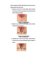
2.5 Teknik Operasi Pada umumnya, jika dilakukan secara berhati-hati perbaikan sederhana dua lapisan cukup untuk memastikan hasil fungsional dan kosmetik. Berikut beberpa tahapan teknik operasi kelopak mata: 1. Jahitan lapisan pertma adalah jahitan dengan pola jahitan terputus mattress, dilakukan di tengah- tengah Antara konjungtive dan kulit penutup stroma kecuali bila konjungtiva letaknya jauh dari penutup stroma, umumnya tidak memerlukan penjahitan. 2. Jahitan pertma lapisan ini adalah jahitan yang paling penting dari seluruh perbaikan dan harus dilakukan penutupan tepi jahitan yang sempurna pada permukaan laserasi menggunkaan absorbable chromic gut 3-0 atau 4-0 dan jarum khusus ophthalmic. Kesempurnaan perbaikan dapat mengurangi gangguan kontinuitas margin yang tertutup.
9
Perawatan yang baik harus diperhatikan terutama pada jahitan pertama, untuk memastikan keamanan pasca jahitan. 3. Selanjutnya, pada lapisan kedua juga dibutuhkan kehati-hatian untuk kesempurnaan menyelaraskan tepi yang tertutup dan dipastikan bahwa tidak ada bahan jahit yang kontak dengan kornea. Lapisan kedua berperan dalam aposisi kulit dan dilakukan sebuah pola jahitan simple interrupted dengan menggunkan nanabrorbable 3-0 atau 4-0 polypropylenen atau nilon. Dan lapisan kedua umumnya dilakukan perawatan selama 14 hari untuk memungkinkan kekuatan jaringan secara maksimal. 4. Setelah dilakukannya perbaikan kelopak mata, penting untuk mengevaluasi kembali abnormalitas mata. Jika terjadi trauma yang cukup parah sampai laserasi, maka penyebab yang paling berpotensi adalah uveitis sehingga diperlukan perawatan lebih lanjut. 5. Perawatan harus dilakukan dengan kortokosteroid topical karena tidak menggangu proses penyembuhan luka serta berpotensi untuk mengurangi resiko keratomycosis yang dapat menimbulkan cacat pada epitel kornea. 6. Melakukan evaluasi kornea secara cermat untuk mencegah adanya epitel. Penggunaan pewarnaan fluprescein dengan pencahayaan oleh sinar ultraviolet (Wood’s lamp) dapat sangat membantu dalam mengidentifikasi lesi pada kornea. Jika tidak tersedia lampu Wood untuk lebih ekonomis dapt lebih ekonomis dapt menggunakan “black-light”.
Figure 3. Ilustrasi Teknik Operasi Laserasi Kelopak Mata.
2.6 Pasca Operasi Pemberian antibiotik topikal dan antibiotik sistemik (trimetoprim-sulfa atau penicillin) umumnya diberikan selama lima sampai tujuh hari pasca operasi. Dalam penanganan juga diberikan obat anti inflamasi non-steroid (fenilbutazon atau flunixin meglumine selama tiga sampai lima hari pasca operasi). Tetanus toxoid dapat diberikan jika diperlukan. 10
Penanganan luka pasca operasi juga dapat dilakukan dengan membersihkan area sekitar luka jahitan. Membersihkan area sekitar luka dapat dilakukan dengan pemberian larutan garam, takarannya ¼ sendok teh garam meja dicampur dengan 1 cangkir air hangat. Pemberian larutan garam dapat diberikan sembari menunggu dokter hewan. Larutan ini sama seperti air mata. Pada kuda dapat diberikan masker terbang untuk mata, tujuannya untuk menghindari lalat hinggap pada area luka, menjaga lingkungan kuda agar terbebas dari debu dan benda-benda asing (UC Davis, 2009).
11
BAB III PENUTUP 3.1 Kesimpulan Laserasi kelopak mata yaitu suatu keadaan dimana terjadi kerobekan kulit bagian mata bagian mata yang disebabkan karena adanya yang disebabkan karena adanya trauma akibat trauma akibat benda tumpul maupun benda tajam. Operasi laserasi kelopak mata dianggap sebagai tindakan emergensi untuk mencegah terjadinya keparahan kasus, seperti terjadinya devitalisasi jaringan yang tidak diinginkan, infeksi, pembentukan jaringan parut, ulserasi dan kekeringan pada kornea. Sebelum operasi pemeriksaan klinis termasuk pemeriksaan luar dan refleks. Sebelum melakukan tindakan operasi, terlebih dahulu dilakukan persiapan operasi yaitu persiapan alat, bahan, obat, persiapan ruangan operasi, persiapan hewan kasus dan operator. Operasi dapat dilakukan setelah hewan diberikan injeksi antropin sulfat secara intra muscular yang bertujuan untuk tachycardia, untuk mencegah terjadinya aspirasi, setelah 10 menit kemudian suntikan xylazine HCl 2% dan ketamin HCl 10% sebagai obat untuk anestesi. Perawatan pasca operasi dapat diberikan antibiotik topikal dan antibiotik sistemik (trimetoprim-sulfa atau penicillin) umumnya diberikan selama lima sampai tujuh hari pasca operasi. Dalam penanganan juga diberikan obat anti inflamasi non-steroid (fenilbutazon atau flunixin meglumine selama tiga sampai lima hari pasca operasi). Tetanus toxoid dapat diberikan jika diperlukan.
3.2 Saran Dalam pelaksanaan operasi laserasi kelopak mata harus diperhatikan kondisi hewan. Setelah dilakukan tindakan operasi sebaiknya menghindari kontaminasi karena bisa terjadi infeksi sekunder.
12
DAFTAR PUSTAKA Fossum, T. W. 2013. Small Animal Surgery. College of Veterinary Medicine. Texas. Ferraro, Gregory L. 2009. The Equine Eye. A Publication of the Center for Equine Health, UC Davis School of veterinary Medicine. Vol. 27(1): 3-11. Hendrix, Diane V.H., DVM, Diplomate ACVO. 2013. How to Repair Eyelid Lacerations. Aaep Proceedings. Vol. 59: 149-154. Jena, Biswadeep, Abrar Ahmed, Nilesh Kumar Pagrut. 2015. Successful Management of Upper Eyelid Laceration in a Dromedary Camel (Camelus dromidarius). Advances in Animal and Veterinary Sciences. Vol 3(2): 133-135. Verma, Mahesh Kumar, Sangeeta Devi Khangembam and Anil Kumar Gangwar. 2018. Surgical Repair of Equine Eyelid Laceration. An International Refereed, Peer Reviewed & Indexed Quarterly Journal in Science, Agriculture & Engineering. Vol. 6: 353-354. Kangmaruf. 2017. Bedah Laserasi Kelopak Mata pada Hewan (Bedah Veteriner). Diakses pada tanggal 6 September 2021 melalui https://mydokterhewan.blogspot.com/2016/03/bedahlaserasi-kelopak-mata-bedah.html Kumar, P Ravi, V Devi Prasad, D Bhagya Raju, Makkena Sreenu and B Sailaja. 2016. Repair of Extensive Eyelid Laceration with Exposed Lacrimal Gland in Buffalo – A Report of Two Cases. International Journal of Science, Environment and Technology. Vol. 5(4): 19461951.
13
Click to edit Master title style
ILMU BEDAH KHUSUS VETERINER TEKNIK OPERASI LASERASI KELOPAK MATA Kelompok 3D N u r I n t a m W u l a n Yu n i t a
1 8 0 9 5 111 0 0
I Made Surya Meganugraha
1 8 0 9 5 111 0 1
Mohammad Gus Shofi
1 8 0 9 5 111 0 2
Putu Raditya Kurnia Putra
1 8 0 9 5 111 0 3 1
Click to edit Master title style
Terminologi
Laserasi kelopak mata yaitu suatu keadaan dimana terjadi kerobekan kulit bagian mata bagian mata yang disebabkan karena adanya yang disebabkan karena adanya trauma akibat trauma akibat benda tumpul maupun benda tajam. . Laserasi kelopak mata atas pada kuda
Laserasi kelopak mata dapat terjadi pada semua spesies hewan.
Laserasi kelopak mata bawah pada kuda.
2 2
Click to edit Master title style
Indikasi
Operasi ini dianggap sebagai tindakan emergensi untuk mencegah terjadinya keparahan kasus, seperti terjadinya devitalisasi jaringan yang tidak diinginkan, infeksi, pembentukan jaringan parut, ulserasi dan kekeringan pada kornea. Operasi ini diindikasikan untuk menangani kasus laserasi yang terjadi pada kelopak mata hewan.
3 3
Click to edit Master title style
Anestesi
Hewan yang akan di operasi diberikan premedikasi dengan menyuntikan antropin sulfat secara intra muscular. Setelah hewan teranestesi cukur rambut di bagian mata kemudian dicuci dengan menggunakan sabun dan dibilas hingga bersih selanjutnya diusap dengan kapas beralkohol 70% dan didesinfeksi dengan iodium tincture 3% dengan arah dari dalam keluar. Pada daerah mata disuntikan lignocaine HCl untuk memblok saraf sensorik pada kelopak mata. Pada hewan kecil, setelah hewan teranestesi letakan di meja operasi dengan posisi ventrodorsal/dorsal. Selanjutnya hewan siap di operasi.
4 4
Click to edit Master title style Persiapan Operasi Laserasi pada kelopak mata harus segera
ditutup. Penutupan harus dilakukan di bawah anestesi umum sebagai sedasi dan anestesi lokal. Sebelum melakukan, luka harus disterilkan dari semua nanah dan debris, bila perlu dilakukan disinfeksi permukaan permukaan kelopak kelopak mata serta menghindari debriment debriment luka yang berlebihan berlebihan karena akan menggangu proses aposisi yang benar.
Persiapan Kelopak Mata 1. Disarankan untuk menggunakan scrub bedah berbahan dasar iodin karena chlorhexidine telah terbukti menjadi racun bagi epitel kornea. 2. Meminimalisasi kontak scrub dengan kornea, kornea dapat dilindungi dengan menggunakan salep berbahan dasar petroleum.
3. Jika luka terjasi 12 – 24 jam harus segera lakukan penutupan primer. 4. Melakukan evaluasi menyeluruh pada mata jika tidak ada kelainan, maka akan mudah melakukan penutupan.
5 5
Click to edit Master title style Teknik Operasi 1. Jahitan lapisan pertma adalah jahitan dengan pola jahitan terputus mattress, dilakukan di tengahtengah antara konjungtive dan kulit penutup stroma kecuali bila konjungtiva letaknya jauh dari penutup stroma, umumnya tidak memerlukan penjahitan. 2. Jahitan pertama lapisan ini adalah jahitan yang paling penting dari seluruh perbaikan dan harus dilakukan penutupan tepi perbaikan jahitan yang sempurna pada permukaan laserasi. 3. Selanjutnya, pada lapisan kedua juga dibutuhkan kehati–hatian untuk kesempurnaan menyelasraskan tepi yang tertutup dan dipastikan bahwa tidak ada bahan jahit yang kontak dengan kornea. 4. Setelah dilakukannya perbaikan kelopak mata, penting untuk mengevaluasi kembali abnormalitas mata. 5. Perawatan harus dilakukan dengan kortokosteroid topical karena tidak menggangu proses penyembuhan luka serta berpotensi untuk mengurangi resiko keratomycosis yang dapat menimbulkan cacat pada epitel kornea. 6. Melakukan evaluasi kornea secara cermat untuk mencegahadanya epitelnya. Penggunaan pewarnaan fluprescein dengan pencahayaan oleh sinar ultraviolet (Wood’s lamp) dapat sangat membantu dalam mengidentifikasi lesi pada kornea.
6 6
Click to edit Master title style
Pasca Operasi
Pemberian antibiotik topikal dan antibiotik sistemik (trimetoprimsulfa atau penicillin) umumnya diberikan selama lima sampai tujuh hari pasca operasi. Dalam penanganan juga diberikan obat anti inflamasi non-steroid (fenilbutazon atau flunixin meglumine selama tiga sampai lima hari pasca operasi). Tetanus toxoid dapat diberikan jika diperlukan. Penanganan luka pasca operasi juga dapat dilakukan dengan membersihkan area sekitar luka jahitan. Membersihkan area sekitar luka dapat dilakukan dengan pemberian larutan garam, takarannya ¼ sendok teh garam meja dicampur dengan 1 cangkir air hangat.
Pada kuda dapat diberikan masker terbang untuk mata, tujuannya untuk menghindari lalat hinggap pada area luka, menjaga area mata agar terbebas dari debu dan benda-benda asing 7 7
Click to edit Master title style
Thank You 8
International Journal of Science, Environment and Technology, Vol. 5, No 4, 2016, 1946 – 1951
ISSN 2278-3687 (O) 2277-663X (P)
Case Report
REPAIR OF EXTENSIVE EYELID LACERATION WITH EXPOSED LACRIMAL GLAND IN BUFFALO – A REPORT OF TWO CASES P Ravi Kumar1*, V Devi Prasad2, D Bhagya Raju3, Makkena Sreenu4 and B Sailaja1 1 Assistant professor, Department of Veterinary Surgery and Radiology, 2 Associate professor, Department of Veterinary Surgery and Radiology, 3 Professor & Head, Department of Veterinary Surgery and Radiology, 4 Contract Teaching Faculty, Department of Veterinary Surgery and Radiology, NTR College of Veterinary Science, Gannavaram- 521 102(AP) E-mail: [email protected] (*Corresponding Author)
Abstract: Two buffaloes with lacerations on the left upper eyelid in one and right upper eyelid in the other were diagnosed with exposed lacrimal glands resulted from a horn gore. The lacerated wounds were managed surgically by three layered suture technique, under local analgesia which yielded a favorable outcome. No postoperative complications were recorded in both the cases. Keywords: Three layered suture technique, eyelid lacerations, Auriculopalpebral nerve block, Buffaloes. Introduction Eye lids form the outer protecting layer of the eye which protects the eye ball from trauma besides controlling the entry of light into the eye by its movement. They spread the tear film uniformly over the eyeball by blinking (Maggs, 2008; Siddiqui and Telfah, 2010). Wounds over the eye lids in animals, may happen during grazing at thorny bushes or trees, sometimes by contact of barbed wires (Bishnoi and Gahlot, 2004; Gahlot et al., 2007). These wounds may range from a simple laceration perpendicular to the margin of eyelid to an extensive one with a flap of eye lid hanging from a pedicle or laceration with complete loss of eyelid margin (Irby, 2004).
Usually these wound are edematous and bloody with mucoid to
mucopurulent discharges in the periocular areas. Jena et al., (2015), reported management of eyelid laceration in a camel whereas, only a very few reports were available on lacerated wound of eyelids in buffaloes. In the present paper, surgical management of extensive upper eyelid lacerations in buffaloes was reported. Case history and Observations: Two graded Murrah she buffaloes were presented to the clinics with a complaint of injury on the upper eyelids. The injuries were said to have happened during infighting. In Case 1, Received June 9, 2016 * Published Aug 2, 2016 * www.ijset.net
1947
P Ravi Kumar, V Devi Prasad, D Bhagya Raju, Makkena Sreenu and B Sailaja
Graded murrah she buffalo of 8 years showed a laceration from the lateral canthus
on right
upper eye lid and in case 2, Graded murrah she buffalo of 6 years showed laceration from the middle of left upper eye lid to the lateral canthus. In both the cases, the symptom, lesions etc were more or less similar except the difference of dexterity. On physical examination, swollen lacrimal glands were found exposed. Soiling was observed in both the cases but it was heavier in case 2 when compared to that in case 1. No discharges were noticed, as
they were presented immediately. Blepharitis along with
blepharedema was noticed. Mild conjunctivitis was noticed but without any apparent defects in vision. Hematological and serum biochemical parameters were found to be normal in both the cases. Based on clinical findings, the condition was diagnosed as extensive laceration of eyelid and decided to reconstruct. Treatment The buffaloes were sedated with xylazine hydrochloride IM at the dose rate of 0.03 mg per Kg body weight and regional analgesia was achieved by performing Auriculo palpebral, supra orbital, nerve blocks using 2% lignocaine hydrochloride. As the sensory innervation to the eye lids in buffaloes is received from nasociliary branches of ophthalmic nerves, besides supra orbital nerve, a line of analgesia was also created in eyelids through linear infiltration at a considerable distance from the margin of wounds so as to avoid distortion of wound edges besides causing analgesia. After ascertaining prompt analgesia, they were controlled in standing position. Antibiotic eye drops were instilled into eye before starting the surgical procedure. After aseptic preparations, the wound edges of the eyelids were freshened with the help of gauze instead of scalpel, to avoid loss of more tissue in the margins of wound. Suturing of margins was done in three layers with deep layer involving fibrous tarsal plate and orbicularis oculi layer of the eyelid margins with polyglactin 910 No 3-0 followed by subcutaneous sutures (intermediate layer). Then the cutaneous edges were sutured with interrupted sutured (superficial layer) using No 2-0 braided silk. The palpebral conjunctiva was left unsutured as suturing of the skin and orbicularis oculi layer enables its apposition. After surgical correction of eyelid, the eye was irrigated with normal saline. Postoperatively, both the animals were given Streptopencillin at dose rate of 500mg/50Kg Body weight IM once daily for 5 days, Meloxicam at the dose rate of 0.2 mg/Kg Body weight subcutaneously once daily for 3 days and ciprofloxacin eye drops were instilled in to the eye three times a day for 10 days.
Repair of Extensive Eyelid Laceration with Exposed ….
1948
Results and Discussion Complete healing of the wound was noticed by the end of 8th day in case 1 and 14th day case 2. No postoperative complications like ectropion or entropion, step formation in the margin of eyelids and wound dehiscence were noticed in a follow up period of six months. Eyelid lacerations observed in the present report were due to horn gore. As buffaloes are vicious and are known for their fighting nature, injuries due to butting among themselves are very frequently reported in large animal practice. Due to nosy posture, injuries to the head in general and eyes in particular can be thought to happen quite often. Irby, (2004) stated that, upper or lower eyelid injuries are resulted from hooks, nails or other pointed objects where as extensive wounds may also occur due to crushing of the tissue by blunt objects. As the eyelids are sensitive and most vascular, edema is usually seen immediately after the injury (Jena et al., 2015) as noticed in the present study. No discharges were noticed in the present study which could be attributed to their early presentation. Both case cases were operated immediately to prevent further discomfort to them by possible Keratoconjunctivitis due to continuous exposure of cornea to environment besides irritation by hanging flap of the injured eyelid. Irby, (2004) also opined that, the management of eyelid laceration in animals requires immediate treatment. In the present study surgical repair was done under sedation besides local analgesia using a combination of nerve blocks along with local infiltration of anesthetic solution at a considerable distance from the site of injury. Bedi (2015) stated that, injection of anesthetic mixture at the site of injury may distort the local anatomy causing difficulty in appropriate apposition. Hence, the linear infiltration was carried out well above the site of damage. The margins of the eyelids in the present study were sutured by traditional three layered technique as advised by Irby, (2004) to treat eyelid laceration with deep layer covering fibrous tarsal layer and orbicularis oculi layer, intermediate layer involving subcutaneous tissue and the superficial layer involving skin. Chawla et al (1993) recommended a two layered technique for suturing the major lacerations of eye lid with first layer involving the palpebral conjunctiva followed by second layer involving muscle layer and skin. In present study, palpebral conjunctiva was not sutured as the suturing of fibrous tarsal layer and orbicularis oculi layer causes apposition of palpebral conjunctiva at the same time there will not be any irritation to cornea due to suture material. Similar technique was also followed by Bedi (2010) in human eye lid lacerations keeping in view of the same principle. Proper
1949
P Ravi Kumar, V Devi Prasad, D Bhagya Raju, Makkena Sreenu and B Sailaja
reconstruction strict postoperative care ensured a good recovery in both cases with acceptable aesthetic appearance. Conclusion Lacerated wound of eyelid is a condition which should not be neglected as it may lead to other ocular disorders like Keratoconjunctivitis, corneal ulcers etc which may interfere with the vision of animal. A three layered suturing technique using an absorbable suture material is recommended to be carried out under regional analgesia in order to get encouraging results. References [1] Bedi KD (2010) Lid and Canalicular Injuries – Pearls in the Primary Repair. KEral J. of Ophthalmology, Vol. XXII, No.3 Page no: 133-135. [2] Bishnoi P, Gahlot TK (2004). Ophthalmic affections in camels (Camelus dromedarius), Vet. Pract. 5(2):89-93. [3] Chawla Sk, Panchbhal VS and Gahlot TK (1993). The special sense organs In: In: ruminant Surgery by RPS Tyagi and Jit Singh, CBS Publishers and Distributors Pvt Ltd., PP 398-400. [4] Gahlot TK, Dudi PR, Sharma CK, Bishnoi P, Purohit S (2007). Surgeries of head and neck region of dromedary camel in India. In: Proceedings of the International Camel Conference, Rajasthan, 2007 (Recent trends in Camelids research and Future strategies for saving Camels, India), 171-175. [5] Irby NL (2004), Surgical disease of the eye in farm animals In: In: Farm animal surgery by S. Fubini and N Ducharme, Elseiver publications; pp 444-45. [6] Jena B, Ahmed A, Pagrut NK (2015). Successful Management of Upper Eyelid Laceration in a Dromedary Camel (Camelus dromidarius). Advances in Animal and Veterinary Sciences. 3(4): 133-135. [7] Maggs DJ (2008). Eyelids In: Maggs, D.J., Miller, P., & Ofri, R. (eds), Slatter’s fundamentals of veterinary ophthalmology, 4th Edition, Elsevier Health Sciences, St. Louis, Missouri, USA: 107-134. [8] Siddiqui MI, Telfah MN (2010). Injury to the Eyelids. In: A Guide Book of Camel Surgery, 1st Edition, Abu Dhabi Food Control Authority, United Arab Emirates: 182-183.
Repair of Extensive Eyelid Laceration with Exposed ….
1950
Fig-1: Photograph showing extensive laceration of right upper eyelid with exposed lacrimal gland (shown by an arrow) in a buffalo
Fig-2: Photograph showing extensive laceration of left upper eyelid with exposed lacrimal (shown by arrow) gland in a buffalo
1951
P Ravi Kumar, V Devi Prasad, D Bhagya Raju, Makkena Sreenu and B Sailaja
Fig-3: photograph showing reconstructed right upper eye lid in a buffalo
Fig-4: Photograph showing reconstructed left upper eye lid in a buffalo
See discussions, stats, and author profiles for this publication at: https://www.researchgate.net/publication/344385787
SURGICAL REPAIR OF EQUINE EYELID LACERATION Article · January 2018
CITATIONS
READS
0
83
2 authors: Anil Gangwar
Sangeeta Khangembam
Narendra Deva University of Agriculture and Technology
Narendra Deva University of Agriculture and Technology
91 PUBLICATIONS 395 CITATIONS
25 PUBLICATIONS 42 CITATIONS
SEE PROFILE
Some of the authors of this publication are also working on these related projects:
Tissue engineering and Intravenous Regional Anesthesia (IVRA) in large animals View project
Intravenous regional anesthesia View project
All content following this page was uploaded by Anil Gangwar on 26 September 2020. The user has requested enhancement of the downloaded file.
SEE PROFILE
VOL. VIII, SPECIAL ISSUE, RKVY NOV-2018 SEMINAR NDUAT AYODHYA
MULTILOGIC IN SCIENCE
ISSN 2277-7601
An International Refereed, Peer Reviewed & Indexed Quarterly Journal in Science, Agriculture & Engineering SURGICAL REPAIR OF EQUINE EYELID LACERATION Mahesh Kumar Verma, Sangeeta Devi Khangembam and Anil Kumar Gangwar Department of Veterinary Surgery and Radiology, College of Veterinary Science and Animal Husbandry, Narendra Deva University of Agriculture and Technology, Kumarganj-224 229, Faizabad (UP) Abstract Eyelid lacerations are common in horses and can occur in isolation or in conjunction with injury to the globe. A horse aged 3 years was presented with the history of eye injury by an iron hook in the stable since last 3 days. The horse was anesthetized by using xylazine (@1.1 mg/kg b.wt.)- ketamine (@2.2 mg/kg b.wt.) protocol. The nerves were desensitized by infiltrating local anesthetic laterally or medially along the rim of the orbit. The lower lid was desensitized by infiltrating local anaesthetic along the ventral rim of the orbit to block the zygomatic nerve. The lacerated lid margins were freshened by B.P. blade no. 23 sutured using PGA no. 2-0. Post operative care included a topical antimicrobial (Tobramycin eye drop©) instilled in the eye thrice a day for 7 days. Ointment Neosporin was applied locally over the suture site. After the repair was complete, the eye was protected by applying the clean cotton cloth over the affected eye. Systemic Intamox (2.5g) and Meloxicam (@ 0.6 mg/kg b.wt.) were administered for 5 and 3 days, respectively. Sutures were removed on 10th post-operative day. The eyelid healed completely without any complication. Key words: Eyelid laceration, Horse, Reconstruction Introduction: The most frequent eye related problems in horses are corneal ulcers (abrasions to the surface of the eye), eyelid lacerations and uveitis (Plumer, 2005). Eyelid lacerations are common in horses and can occur in isolation or in conjunction with injury to the globe. Eyelid lacerations are mainly caused when the eyelid simply gets snagged on a piece of stall hardware, an overlooked raised nail or piece of wire. In most species, the eyelids are important in protecting the eye. A functional eyelid is essential to maintain the health of the globe itself. If the eyelid is incomplete or immobile, it is unable to physically protect the eye and is unable to spread the tear film over the ocular surface. The upper lid is the more commonly damaged and is also the more important, as it plays a greater role in distributing tear film and preventing exposure keratitis (Brooks and Matthews, 2007). The eyelid integrity of the eyelid margins is vital for maintaining a proper tear film on the surface of the eye. The eyelid injuries are much easier to diagnose than most other eye emergencies but are equally important to seek rapid veterinary attention. Most lacerations involving the margin of the eyelid require repair with suture. Surgical repair should be undertaken promptly before edema and inflammation and before (further) corneal damage occurs. Delay in treatment distorts the affected tissue and hampers effective repair. All full-thickness eyelid lacerations must be repaired surgically, either by primary closure or by grafting. An irregular eyelid margin can cause recurring and chronic eye irritation. There are no retrospective studies discussing equine eyelid laceration repair in the peer-reviewed literature. However, we repaired the eyelid laceration successfully in a horse. Materials and methods: A horse aged 3 years was presented with the history of eye injury by an iron hook in the stable since last 3 days (Fig. 1). The animal was quite alert. First of all, the whole eye was examined clinically without sedation and nerve blocks to check ocular reflexes and to determine the injury's extent. Clinical examination revealed
Fig.1: Lower eyelid laceration in a horse
pupillary light response and no corneal injury were noted. Once the initial examination completed, the horse was sedated and auriculo-palpebral nerve was blocked (which temporarily prevents eyelid movement) to complete a thorough ophthalmic examination. The lower eyelid was lacerated from the lateral canthus up to the middle of the lid margin. The lacerated part of eyelid was slightly contracted. The eyelid mucosa was exposed with slight granulation tissue over the lacerated site. Tetanus toxoid (5 ml) was administered one day before the surgical repair. The site was prepared to repair the lacerated eyelid. The area around the eye was disinfected by using a dilute povidoneiodine (2%) solution and saline (both Betadine scrub and alcohol are toxic to the corneal epithelium and can cause ulcers). The horse was anesthetized by using xylazine (@1.1 mg/kg b.wt.)- ketamine (@2.2 mg/kg b.wt.) protocol. The nerves were desensitized by infiltrating local anesthetic laterally or medially along the rim of the orbit. The lower lid was desensitized by infiltrating local anaesthetic along the ventral rim of the orbit to block the zygomatic nerve. The lacerated lid margins were freshened by B.P. blade no. 23 sutured using PGA no. 2-0. The eyelid edges were sutured in two layers. The eyelid margins were aligned first and the tip of the lacerated part was sutured at its original position on the lateral canthus of the eye. Later, the first (deep) layer of sutures was placed. Absorbable sutures were placed in such a way that the suture material does not penetrate the conjunctiva. The skin was closed with interrupted silk sutures. Post operative care included a topical antimicrobial (Tobramycin eye drop©) instilled in the eye thrice a day for 7 days. Ointment Neosporin was applied locally over the suture site. After the repair was complete, the eye was protected by applying the clean cotton cloth over the affected eye. Systemic Intamox® (2.5g) and Meloxicam® (@ 0.6 mg/kg b.wt.) were administered for 5 and 3 days, respectively. Sutures were removed on 10th post-operative day. The eyelid healed completely without any complication (Fig.2).
Fig.2: Complete recovery after 10 days
®Intas Pharmaceuticals Ltd, Ahmdabad-380054, Gujrat, India, © Taj Pharmaceuticals amenable to regional analgesia, enabling virtually all repairs to be Results and Discussion: Although eyelid lacerations are common but this condition performed standing under sedation (Henriksen and Brooks, 2014). should be considered as surgical emergency. The eyelids are The surgical repair of eyelid lacerations differs somewhat from that
VOL. VIII, SPECIAL ISSUE, RKVY NOV-2018 SEMINAR NDUAT AYODHYA
MULTILOGIC IN SCIENCE
ISSN 2277-7601
An International Refereed, Peer Reviewed & Indexed Quarterly Journal in Science, Agriculture & Engineering of most skin lacerations. The reasons for this include the need to lid must be protected from self-trauma by hard eye cup in horses. maintain a functional eyelid and ensure a cosmetic outcome as well as Because the blink response is often impaired by the swollen lid, a the fact that the eyelid is more vascularized than many other skin temporary tarsorrhaphy is necessary to protect the cornea. Avoid regions. Repair is necessary to maintain a horse's ocular health and using cautery (burning tissue) when repairing eyelid lacerations to often can be accomplished in a standing, sedated horse. Most eyelid prevent further damage to the lacerated tissue. Use extreme caution lacerations can be successfully repaired (Labelle and Clark-Price, when placing sutures, and avoid burying them. The delicate cornea 2013). can be damaged if it comes in contact with sutures, and can lead to Eyelid lacerations are extremely important to repair and complications. In present case the wound healed without any should be reapposed as soon as possible. Use care when repairing complication. Brooks (2006) reported that some dehiscence of the lacerations of the eyelid to ensure that functionality of the eye and eyelid margin nearly always occurs postoperatively. This margin cosmetic appeal are maintained for the patient. The eyelids have breakdown can cause varying amounts of lid misalignment with excellent blood supply, so with prompt and accurate repair, eyelids resulting entropion or ectropion. Gaping of the conjunctival side of have a tremendous capacity to heal and good outcomes can be the incision due to poor suture placement can slow healing and allow achieved, even if tears are large or the lids are virtually avulsed a suture to rub on the cornea to cause painful corneal ulcers. Such (Schaer, 2007). Eyelid lacerations can bleed profusely, and sutures would need to be replaced or removed. Skin sutures can also haemorrhage can be a nuisance during repair the defects. Lacerations become infected. involving the lid margin require exact apposition to prevent long term References: v-shape defects and an impaired lid function. Most lacerations heal Brooks, D.E. (2006). Ocular Emergencies and Trauma. In Equine and maintain an intact eyelid margin. Even large eyelid flaps will Surgery, Auer, J.A., Stick, J.A. (eds), 3rd ed, W.B. Saunders, St heal well with minimal debridement if they are repaired promptly Louis. pp 767-774. before there is extensive swelling. Due to the highly vascularity of the Brooks, D.E. and Matthews, A.G. (2007). Equine Ophthalmology. eyelids, the tissue that appears devitalised will often survive. If a In Gelatt, KN (ed.), Veterinary Ophthalmology, 4th ed., Blackwell portion of the eyelid margin is destroyed, the surface of the eye is at Pub, Ames, I.A.,p p 1165-1274. risk for future disease due to lack of protection from the eyelids and Henriksen, M. de Linde and Brooks, D. E. (2014). Standing lack of tear film covering the eye. In order to maintain eyelid ophthalmic surgeries in horses. Veterinary Clinics of North function, debridement of wounds must be minimal. Avoid simply America: Equine Practice, 30(1): 91-110 excising any potentially viable tissue as serious complications can Labelle, A. L. and Clark-Price, S. C. (2013). Anesthesia for ensue. Even if you think its dead, try to replace it ( Lavach et al., ophthalmic procedures in the standing horse. Veterinary Clinics of 1984). Precise apposition of the eyelid margin is essential if future North America: Equine Practice, 29(1):179-191. keratitis is to be prevented. Debridement at the eyelid margin should Lavach, D., Severen, G.A. and Roberts, S.M. (1984). Lacerations be avoided if at all possible, as this will distort the normal eyelid of the equine eye: A review of 48 cases. J. Vet. Med. Assoc., margin. Temporary tarsorrhaphy may be beneficial to reduce the risk 184:1243–1248. of corneal damage while eyelid lacerations heal and normal function Plummer, C.E. (2005). Equine Eyelid Disease. Current Techniques returns. in Equine Practice, 4(1): 95-105. Horses generally require double-layer closure. A two-layer Schaer, B. (2007). Ophthalmic emergencies in horses, Veterinary closure is typically preferred with a third subcutaneous layer being an Clinics of North America Equine Practice 23(1): 49-65. option if there is marked oedema. When skin sutures are in place, the
www.ycjournal.net
View publication stats
NAAS Rating- 5.20
UGC Approved journal
Impact factor-1.137
354
�����������
��� �� 27 No 1, April 2009 Volume
A Publication of the Center for Equine Health, UC Davis School of Veterinary Medicine
The Equine Eye An animal will always look for a person’s intentions by looking them right in the eyes.
R
abbits run, possums play dead, chameleons change color and skunks spray. Other animals have horns or claws to fight off predators. Mobbing behavior is common in birds and is usually done to protect the young in social colonies. While survival techniques vary among animals, what many have in common are highly developed senses of sight, smell, or hearing to detect danger and escape. Unlike other large prey species, horses do not have antlers, horns or cloven hooves with which to defend themselves. Instead, they have an exceptional ability to see and spot the movement of any potential
INSIDE THIS ISSUE… Directorʼs Message.................. 2 Eye Problems in Horses ........... 4 Traumatic Injuries ................... 5 Corneal Disorders .................. 5 Uveitis/Moon Blindness............ 6 Cancer .................................. 7 Cataracts ................................ 7 Case Study from the VMTH .. ... 8 Handling Eye Injuries ............... 9 Ophthalmology Service .. ...... 10 Tribute to Dr. Wheat .. ........... 10 Cataracts in Foals .. ............... 11 Dr. OʼBrien Honored .. .......... 11 Help for Unwanted Horses .. .. 12
predator. Horses have survived by using a standard defense mechanism for animals in open grasslands and prairies: they frequently scan and monitor their surroundings to avoid attack by spotting a predator before it reaches a critical distance. While most horses today are domesticated and do not have a constant need to safeguard their survival, they are expected to perform in ways that require an unnatural demand on their visual system. It seems only fair then that we become partners in the effective use of their eyes and learn how their visual system can affect behavior. For example, how does the lateral placement of a horse’s eyes affect its vision? Visual perspective/field of view. The lateral placement of the eyes, more to the side of the head than to the front, combined with a horizontally elongated, roughly rectangular pupil, provides the horse with the ability to see a very broad field of landscape, much like a camera’s wide-angle lens. In fact, horses are capable of seeing almost 360 degrees around with
monocular vision, where each eye is used separately. This extensive visual field makes it difficult for a predator or a human handler to sneak up on a horse. The wide range, however, has two “blind spots” or areas where the horse cannot see. The first is directly in front of the face, in a cone-shaped area that comes to a point about 3 feet in front of the horse. The second area is right behind the head, in an area that extends over the horse’s back and behind the —Continued on page 3
Volume 27, Number 1 -April 2009
2 - The Horse Report
DIRECTOR’S MESSAGE
Best Results Through Early Intervention
Dr. Gregory L. Ferraro
F
ew would argue the importance of a pair of healthy, well-functioning eyes to the well being and performance of their horse. Indeed, unimpaired sight is as important to the horse as it is to any other animal, including human. Yet few people have a good understanding of how the horse’s eye functions or how their vision differs from that of our own. Infections of or injuries to the horse’s eye always have the potential to pose serious risk to their visual capacity. Severe insults can result in permanent damage very quickly, and minor incidents left unattended can progress swiftly into serious threats. Yet again, few horse owners have the ability to recognize and properly respond to those minor problems. Of all the illnesses and injuries that can befall our beloved horses, we are generally the least prepared to deal with those related to the eye. While we can recognize the signs of colic,
we are likely to not notice the development of a corneal ulcer in the eye until the condition has progressed to the stage of significant pain and corneal damage. Many experienced horse owners can treat and care for minor cuts and abrasions themselves and have a good idea of when those acute injuries are severe enough to require the attention of a veterinarian, but too many will have trouble in assessing both the severity of an eye injury and when veterinary intervention is required. For that reason, this issue of The Horse Report provides some information intended to help you assess equine ophthalmologic problems. It provides a description of the basic anatomy and function of the horse’s eye as well as some common but potentially serious problems. We hope this will help you more quickly recognize any abnormalities that may appear in the eye of your horse and provide guidance as to what type of condition you may be looking at. You will notice that we are providing very little information about how to treat these conditions. There is a very good reason for that. Basically, it is because you are not qualified to treat them! As a long-time equine practitioner, I can tell you in all honesty that if your horse’s eye has a problem that you are able to recognize with your own naked and untrained eyes, then you should call your veterinarian immediately. If you follow this one bit of advice, you will have UC Davis Center for Equine Health
taken the correct course of action 99% of the time. If your horse has a painful and watering eye, don’t wait a couple of days to see if it gets better on its own, and definitely do not treat it with the contents of some tube of medication left over from the treatment of another horse, five years previously. If you see something growing on the globe of your horse’s eye or some foreign object sticking out of the region of his eye socket, don’t be a hero. Call your veterinarian. Remember, discretion is the better part of valor. Don’t do something you will regret later. In all conditions related to the eye and its surrounding structures, the best results are always obtained through early intervention by trained and experienced equine veterinarians. Sunday-morning ophthalmologists need not apply! Trust me on this one, read what we have written here but use it only as a means to know when you are in trouble, not as a license to practice. We want you to be informed and educated in all forms of equine health care, but part of that learning experience is to recognize your limitations. There is no more appropriate arena for the teachings of “Dirty Harry” than medical ophthalmology.
The Horse Report - 3
Volume 27, Number 1 - April 2009
The Equine Eye
—Continued from page 1
tail. Thus, as a horse jumps an obstacle, the obstacle briefly disappears from sight just before the horse takes off. Depth perception. The tradeoff to having monocular vision is that a horse’s depth perception is somewhat more limited than a human’s. The placement of the horse’s eyes decreases the possible range of binocular vision (using both eyes at the same time). Horses use their binocular vision by looking straight at an object, raising the head when looking at a distant predator or focusing on an obstacle to jump. To use binocular vision on a closer object near the ground, such as a snake or threat to its feet, a horse will drop its nose and look downward with the neck somewhat arched. Thus, a horse will raise or lower its head to increase its range of binocular vision. Riders of jumpers allow their horse to raise its head a few strides before a jump so it can assess the jump and the proper take-off spot. Sensitivity to light. Horses, like humans, must adapt to different light intensities varying from the dimmest star to bright sunlight on snow. One mechanism for adjusting to this wide range is to switch back and forth between two different types of photoreceptors: the rods and cones located in the retina of the eye, each of which has been optimized to perform best at different ends of the intensity spectrum. Rod receptors are the primary receptors used when the light levels range from virtually complete darkness to those
found at dawn and twilight. Horses are well endowed with a high proportion of rod to cone photoreceptors—about 20 million rods to 1 million cones, which makes them sensitive to light and gives them extremely good night vision. Moreover, a layer of tissue in the eye of many animals, called the tapetum lucidum (see Anatomy of the Eye
Practically speaking, horses tend to have better vision on slightly cloudy days than on bright, sunny days. On the other hand, they are less able to adjust to sudden changes of light, such as when moving from a bright day into a dark barn. This should be taken into consideration during training, because certain tasks such as loading into a trailer may frighten a horse simply because he cannot see. It is also important in riding, because quickly moving from light to dark or vice versa will temporarily blind the horse and make it difficult for him to judge what is in front of him. Sensitivity to motion. Horses are very sensitive to motion, because motion is usually the first alert that a predator is approaching. The horse’s retina—the light-sensitive tissue at the back of the eye—is richly endowed with motiondetecting cells known as rods, evidence that the horse’s eyes are optimally constructed and positioned for its survival.
below), is thought to function to reflect light back into the retina, thereby enhancing vision in lowlight conditions such as at night. In addition to these photoreceptors and other mechanisms for adjusting to light, the horse has one of the largest eyes among land mammals, which allows more light to enter the eye. Admission of light to the eye is further improved by the horizontal elongation of the cornea and pupil and by the pupil’s ability to dilate to an area six times larger than that of a human pupil.
Anatomy of the Eye The equine eye is similar to the eye of most mammalian species. Essentially a piece of brain tissue—the retina—has been brought “outside” the brain and placed inside a tough, protective structure known as the globe (the eyeball). The globe is constructed in such a way that an optically clear and focusable pathway is provided, whereby light rays from the outside world pass into the globe and reach the light-sensitive retina. In turn, the retina transmits its perceived —Continued on page 4
UC Davis Center for Equine Health
Volume 27, Number 1 -April 2009
4 - The Horse Report
The Equine Eye
—Continued from page 3
information via the optic nerve to the final processing unit, the brain, where visual perceptions are converted into images. The eyeball is protected by eyelids, comprised of two folds of skin and muscle just like the human eye. The skin has many blood vessels so that abrasions and lacerations that are properly treated heal well and are resistant to infection. The eyelid muscles are quite strong and can be opened or closed at will. Hence, examination of the equine eye often requires sedation and nerve blocks. Like most other mammalian species, the horse has a third eyelid, which is a T-shaped cartilage covered with a transparent mucous membrane known as the conjunctiva.
Choroid Cornea Corpora nigra
Retina Tapetal area Optic nerve
Lens
Optic disk
Iris Ciliary body
Sclera
As shown in the above illustration, the cornea is part of the fibrous outer layer of the eye, which serves to protect the intraocular contents. The cornea is transparent and is involved in transmitting and refracting (bending) light into the eye. Although it is made up of living cells, the cornea has few or no blood vessels. The surface cornea receives its nutrition from tears, while the back portion is nourished by a clear fluid called the aqueous humor, which is normally present in the front chamber of the eye. The sclera, like the cornea, is made up primarily of collagen fibers. It is part of the outer fibrous layer of the eye and functions to protect the interior of the eye. The iris and ciliary body, part of the middle vascular layer of the eye, perform a number of functions. The iris contains muscles that control the size of the pupil, which in turn controls the
amount of light that enters the eye. The ciliary body produces the clear aqueous humor that nourishes the front chamber of the eye. It also contains the muscles that allow for focus on both distant and close objects. This function in horses is relatively poorly developed. The lens sits directly behind the iris and functions to refract light in order to produce a focused image on the retina. Like the cornea, it has no blood supply but is nourished by the aqueous humor. The choroid is part of the middle vascular layer of the eye and helps provide nutrition to the retina. In the innermost portion of the choroid is the tapetum lucidum, a layer of tissue that is thought to function to enhance low-light vision by reflecting light back into the retina. The retina is made up of nervous tissue and functions to transform light into a neurologic impulse. Axons that originate from the retina— axons are a long fiber of a nerve cell (a neuron) that act somewhat like fiber-optic cables carrying an outgoing message—come together at the optic disk, where they leave the eye as the optic nerve. The optic nerve takes the neurologic message from the retina to the brain.
Eye Problems in Horses Horses respond to eye discomfort much the same as people. The eyelids may become swollen, the eyes may become reddened, and there may be excessive tearing. The horse may also squint or blink constantly. Excessive rubbing of the eye on a post or foreleg may also indicate an underlying problem. If any of these signs occur, a veterinarian should be consulted for diagnosis and treatment. One feature of eye disease is that relatively minor problems initially present with the same signs as more severe problems. Therefore, all cases of suspected eye problems should receive the attention of a veterinarian. As with virtually all other disease processes, the chances of successful treatment of these problems rely heavily on early detection. This principle is even more critical with eye disease. The five most common eye problems in horses seen at UC Davis are:
UC Davis Center for Equine Health
The Horse Report - 5
Volume 27, Number 1 - April 2009
• Traumatic injuries • Corneal disorders • Inflammation inside the eye (uveitis) • Cancer on or around the eye • Cataracts
Traumatic Injuries Horses can sustain a wide range of traumatic injuries to their eyes and related structures. These can vary in severity from simple abrasions of the eyelids that may be treated in a “first aid” manner to full thickness lacerations of the eyelids or cornea. This latter category usually presents with sudden squinting and discharge in the form of excess tearing, mucous or blood. All of these should be considered an emergency and require the immediate attention of a veterinarian. The longterm prognosis depends on the structures injured. Eyelid lacerations are frequently obvious because the affected lid is often hanging loose and there is a moderate amount of blood on the face around the eyelid. Prompt and meticulous repair by a veterinarian is essential in restoring eyelid function (including tear film distribution) and protecting the underlying cornea from exposure. It is recommended that horses receive tetanus prophylaxis and topical and systemic antibiotics to prevent infection. Corneal lacerations are often less apparent because the only clinical sign may be intense squinting. Horses with corneal lacerations will usually vigorously resist any attempt to determine the cause of the pain. A veterinarian should assess the
is another key element for saving an injured eye, and researchers at UC Davis have developed effective ways to administer eye medications and antibiotics every few hours using indwelling medicinal delivery systems. This photo shows a severe laceration of the upper eyelid. Eyelid lacerations usually heal well if they are repaired soon after injury.
extent of the damage by use of sedation and nerve blocks of the eyelid muscles. Frequently such injuries can be repaired by a veterinarian, although he/she may advise referral to a veterinary ophthalmologist. Foreign objects such as plant material, dust, sand or ash may become lodged under a horse’s eyelids. If the horse will allow it, flushing the debris with water or saline solution may be helpful, but a veterinarian should still examine the eye for any further damage since the debris can scratch the cornea and cause a corneal ulcer. If a foreign object such as a piece of wood has pierced an eyelid and become embedded, you can remove it but follow up with a full eye examination with a veterinarian to determine whether further damage has occurred from splinters. If the eyeball itself has a foreign object embedded in it, DO NOT REMOVE IT. Seek immediate veterinary attention as microsurgery may be required to remove the object and save the eye. Ophthalmologists at UC Davis have successfully removed foreign objects from eyes using very fine microsurgical instruments. Preventing infection UC Davis Center for Equine Health
Corneal Disorders By far the most common corneal problem in horses is corneal ulceration. This is a condition where the most superficial cells of the cornea are abraded away. Like most eye problems, this will initially manifest as a suddenly painful eye with excessive tearing. Corneal ulcers require immediate care by a veterinarian as they have the potential to worsen. Because the cornea does not have blood vessels running through it to help clear an infection, the eye is more susceptible to infection from bacteria and fungus. These secondary infections can cause serious problems and complicate healing, which may result in a “melting” ulcer. Signs associated with an infected corneal ulcer include a creamy color or
Superficial corneal ulcer. If caught early and treated correctly, these will usually heal quickly.
—Continued on page 6
Volume 27, Number 1 -April 2009
6 - The Horse Report
The Equine Eye
—Continued from page 5
melting appearance to the surface of the eye. Melting ulcers are a serious threat to eye survival and require intensive treatment by a veterinarian.
are many theories about the possible causes of this type of inflammation, including autoimmune conditions and herpesvirus infection. UC Davis researchers are currently studying herpesvirus as a potential cause of this type of unexplained corneal inflammation.
that results in blindness, but the cumulative effects of many attacks.
Inflammation Inside the Eye (Uveitis)
Infected corneal ulcer. Note accumulation of white blood cells within the cornea.
Melting corneal ulcer. Note degraded area. This ulcer could perforate at any time and is a true emergency.
The most common cause of blindness in horses is a disease known to veterinarians as equine recurrent uveitis (ERU). Other common names for this condition include moon blindness and periodic ophthalmia. As the name suggests, ERU is characterized by recurrent episodes of intraocular inflammation. Signs of an acute ERU attack include squinting, redness, cloudiness and tearing. The severity of the signs and the frequency of the attacks vary among individual horses. Blindness results from cataracts, glaucoma and retinal degeneration. It is not one attack
A minor wound to the cornea may occur and appear to have healed, but if an infection is introduced into the deeper layers of the cornea, a corneal abscess may develop. Again, this is a serious condition that requires immediate treatment by a veterinarian. Less frequently, horses can develop corneal inflammation, which tends to be intermittent and of variable severity. There
Active episode of equine recurrent uveitis. Note the redness to the conjunctiva, excess tearing, and hazy appearance. UC Davis Center for Equine Health
End stage equine recurrent uveitis. Note extensive corneal scarring.
The causes associated with ERU are not completely understood. The disease itself is an autoimmune condition, but various other factors may be involved, such as leptospirosis, an infectious disease caused by a particular type of bacteria called a spirochete transmitted by rats as well as by skunks, opossums, raccoons, foxes, and other vermin. The therapeutic approach to managing ERU is to aggressively treat acute attacks and also minimize the frequency and severity of recurrences. As mentioned above, any horse with a suddenly painful, teary eye should receive immediate attention from a veterinarian who can then make a definitive diagnosis and initiate appropriate treatment. New methods used by veterinary ophthalmologist to treat ERU involve the implantation of medicated “wafers” or injections of medication into the back of the eye. These anti-inflammatory drugs are then absorbed slowly over the course of years.
The Horse Report - 7
Volume 27, Number 1 - April 2009
Cancer By far the most common ocular/periocular cancer seen in horses is squamous cell carcinoma. These tumors typically occur in one of three locations: on the surface of the eye, on the third eyelid, or within the eyelid. These masses are often readily visible and have a “wart-like” appearance.
Other types of cancer such as sarcoids and melanomas, which are common throughout the body, can also affect the eyes and interfere with eye function. In most instances, a tumor may begin as a small area of roughened eyelid or a small bump. If diagnosed early, a number of treatment methods may be used. Note that when eyelid tumors are found, it is important to examine the rest of the body for the presence of similar tumors.
Cataracts
Squamous cell carcinoma on the eye. Note the pink growth on the left side of the photo.
Treatment of squamous cell carcinoma on the surface of the eye usually involves referral to an ophthalmologist. In addition to removing the mass, some type of freezing (cryotherapy) or radiation treatment is usually performed. If the carcinoma is on the third eyelid, it is usually treated by surgical removal of the entire third eyelid. Many general practitioners perform this procedure. Squamous cell carcinoma within the eyelid itself is often the most challenging location because the tumors are often not noticed in this location until they are large. Treatment consists of surgical removal and some type of added treatment such as chemotherapy.
The lens is a somewhat flattened, transparent, flexible disc consisting of layers of lens fibers. The lens sits behind the iris and helps focus the images onto the retina. A diseased lens usually responds by becoming opaque; the opacity (or cataract) may be localized or diffuse. Cataracts are often progressive in
nature and result in serious visual loss over time. The affected lens can be removed. Cataracts can form secondary to equine recurrent uveitis (ERU) as described above. They can be treated with surgery, but because the cataract has formed as a result of months or years of intraocular inflammation, the success rates are usually not high. Horses can be born with cataracts due to developmental or heritable causes. Congenital cataracts in newborn foals are especially amenable to surgical removal. Veterinary attention should be sought as soon as possible. Signs include a milkyappearing pupil or evidence of impaired vision (the foal stays very close to the mare, stumbles, or is reluctant to move). Once the lens is removed, the horse will be far-sighted (not able to see things up close), but many go on to live productive lives. v
The eye is an intricate and delicate organ. In all conditions related to the eye and its surrounding structures, the best results are always obtained through a cooperative effort of observant horse owner and experienced equine veterinarian. UC Davis Center for Equine Health
Volume 27, Number 1 -April 2009
8 - The Horse Report
Early Recognition of a Problem Saves Teekla’s Eye A Case Study from the UC Davis Veterinary Medical Teaching Hospital
O
bservant owners and quick-acting referring veterinarians are vital elements to providing a successful outcome to a horse with a serious problem affecting the eye. This principle was once again demonstrated one morning last summer when the owner of Teekla, a 9-year-old Friesian gelding, removed the horse’s fly mask and noticed he was squinting and tearing out of his left eye. She immediately called Teekla’s veterinarian who realized the seriousness of Teekla’s eye problem and referred him at once to the UC Davis Veterinary Medical Teaching Hospital. At UC Davis, Teekla was examined by ophthalmologist Dr. Steven Hollingsworth, who determined that the horse had an infected corneal ulcer characterized by a “melting” component. This “melting” appearance is a particularly worrisome clinical sign because it means that the cornea could rupture at any time, allowing the infectious organisms to invade the inside of the eye. While it is possible to save an eye that has ruptured, the prognosis is significantly more grave. Teekla was scheduled for emergency surgery and, within 5 or 6 hours of his owner’s first noticing a problem, was on the operating table. The specific surgical procedure performed on Teekla’s melting corneal ulcer was a pedicle conjunctival graft. This technique involves dissecting a fingershaped strip of the thin membrane that covers the conjunctiva (the white part of the eye). The degraded area of cornea is then trimmed off and the graft is sutured directly onto the corneal defect. This procedure provides immediate structural support to the weak area of the cornea as well as a blood supply to help fight the infection.
Melting ulcer on Teekla’s left eye immediately before surgery.
Teekla’s conjunctival pedicle graft immediately after surgery.
Before Teekla awoke from his anesthesia, a small tube was placed under his lower eyelid. This is called a subpalpebral lavage and allows for topical medications to be applied without having to constantly pry the eyelids open. Samples taken from Teekla’s left eye just before surgery revealed that the ulcer was infected with a fungus called Aspergillus. Teekla remained in the hospital for 4 days after surgery to ensure that everything was healing well. He then returned at 2 weeks, at 1 month, and at 2 months after the surgery to check for progress. During this time, the medications he was receiving to Teekla’s conjunctival pedicle graft one month after surfight the infection were slowly tapered down. gery. Note how the graft At his last recheck, the connecting portion of the graft was trimmed away leaving only a small scar where the original ulcer had been. Teekla recovered completely with no loss to his eye or vision because of the coordinated efforts of his owner, veterinarian, and ophthalmologist.
UC Davis Center for Equine Health
has shrunken to allow for a relatively large area of clear cornea.
The Horse Report - 9
Volume 27, Number 1 - April 2009
Preventing and Handling Eye Injuries
E
ye injuries and infections are not uncommon in horses, but minor problems left untreated can quickly become serious and result in blindness if unattended. Here are some recommendations for preventing problems and some guidelines to follow if your horse should sustain an eye injury. • One way to prevent problems is to ensure that your horse’s environment is safe. Make sure that sharp edges on water troughs, metal buildings, pipes, hooks or other objects are covered, padded, or are inaccessible. Pound in or pull any old nails that may be protruding from fences and other structures. • Keep the horse’s environment as dust-free as possible. • If your horse sustains an eye injury or develops an eye infection, contact your veterinarian immediately. (Read the section on Traumatic Injuries on page 5 of this Horse Report). • If the injury is a laceration to the eyelid, clean the area very gently with a saline solution while waiting for the veterinarian. If you don’t have one on hand (like the saline solution for contact lenses), you can make some. The ratio is about ¼ teaspoon of table salt to 1 cup of lukewarm water. It should taste like tears. • If the injury consists of a foreign object such as a piece of wood that has pierced the eyelid and become embedded, you can remove it but follow up with a full eye examination with a veterinarian to determine the extent of damage. Clean the area very gently with a saline solution while waiting for the veterinarian. • If the eyeball itself has a foreign object embedded in it, DO NOT REMOVE IT. Seek immediate veterinary attention as microsurgery may be required to remove the object and save the eye. • Put a fly mask on the horse to keep flies off the eye area. • If possible, keep your horse in subdued light, such as his stall, until the veterinarian arrives. • With all medications prescribed by your veterinarian, make sure that you follow the instructions to the letter, including medicating your horse through the full course of treatment. Do not stop medication because you see marked improvement. This can result in an infection flaring up again. Discard all medications at the end of the treatment course. • When working with a horse with an eye problem, be aware that he may have obscured vision and be a little more spooky than usual. Talk gently so you don’t surprise him if you walk up on a “blind side”.
UC Davis Center for Equine Health
Volume 27, Number 1 -April 2009
10 - The Horse Report
Ophthalmology Service UC Davis Veterinary Medical Teaching Hospital
T
he UC Davis Veterinary Medical Teaching Hospital has a complete Ophthalmology Service for all species large and small, with an emphasis on horses. The service comprises one of the largest veterinary ophthalmology centers in the United States. It is completely equipped with sophisticated equipment for the diagnosis and treatment of all eye diseases in horses, including an operating microscope for microsurgery and a phacoemulsification unit for cataract surgery (a system that uses ultrasound to break up, flush, and remove the affected lens material). The service also uses cryotherapy and beta irradiation for eye tumors.
Dr. Hollingsworth
Dr. Steven Hollingsworth is Chief of the Ophthalmology Service at UC Davis and Assistant Professor of Veterinary Ophthalmology in the UC Davis School of Veterinary Medicine. He obtained a DVM from Purdue University and was a veterinary ophthalmologist in private practice for nine years before coming to Davis. He completed a residency in comparative ophthalmology at UC Davis in 1992 and then joined the faculty in 1994. In addition to his clinical duties, Dr. Hollingsworth teaches clinical ophthalmology to third- and fourth-year veterinary students. He also serves as Chair of the Examination Committee of the American College of Veterinary Ophthalmologists and has been a reviewer for a number of professional journals, including the American Journal of Veterinary Research, Veterinary Anaesthesia and Analgesia, Veterinary Surgery, and Veterinary Ophthalmology. The ophthalmology department is open to both referrals and the general public. To make an appointment, call the UC Davis Veterinary Medical Teaching Hospital at (530)752-0290.
Dr. J. D. Wheat
April 10, 1923 – March 4, 2009 Dr. Don (J.D.) Wheat, world renowned equine surgeon and clinician and an integral part of the UC Davis School of Veterinary Medicine, passed away on March 4, 2009. Dr. Wheat dedicated his career to the care and welfare of the equine athlete and to the training of several generations of equine practitioners. He was a role model for many students, interns, residents and graduate students, stimulating them to envision and explore new ideas for improving the care of horses. Dr. Wheat also left his legacy in the establishment of the J.D. Wheat Dr. Wheat Veterinary Orthopedic Research Laboratory in 1988. The laboratory conducts research on orthopedic diseases of animals, with an emphasis on musculoskeletal disorders of racehorses. The laboratory provides an environment in which complex problems presented by most musculoskeletal diseases can be addressed by using the collective expertise of investigators from several different disciplines in the basic and clinical sciences of the School of Veterinary Medicine. Dr. Wheat’s interest, encouragement and continued curiosity to understand and discover better ways to treat musculoskeletal diseases provided the laboratory with a firm foundation on which to build the success it enjoys today. Dr. Wheat was a charter diplomate of the American college of Veterinary Surgeons; recipient of the American Association of Equine Pracititioners teaching award; member of the National Academies of Practice; and an inductee in the International Equine Veterinary Hall of Fame. He will be sorely missed.
UC Davis Center for Equine Health
The Horse Report - 11
Volume 27, Number 1 - April 2009
Cataracts in Foals
H
orses can be born with cataracts due to developmental or heritable causes—known as congenital cataracts. Foals with cataracts often present very early in life, usually at 1 to 2 months of age. Typically, the owners will notice the whiteness in the pupil of one eye almost immediately after birth. This change is often followed closely by the other eye. Visual problems experienced by the foal are noticed through behaviors such as hesitancy to go anywhere without the mare or outright bumping into things. Otherwise, the foals are usually systemically healthy, and the eyes are not painful.
Complete cataract in a foal less than 2 months old, immediately before surgery.
One week after cataract surgery. Note the incision sutures on the right and the gree glow from the tapetum at the back of the eye.
Congenital cataracts in newborn foals are especially amenable to surgical removal. Veterinary attention should be sought as soon as possible. Signs include a milky-appearing pupil or evidence of impaired vision as mentioned above. Immediately before and after surgery, the foal is given a number of medications including topical antibiotics, topical anti-inflammatories (steroidal and nonsteroidal), and systemic anti-inflammatories (usually Banamine). The surgery is performed using a technology known as phacoemulsification. This is the exact same technology and equipment used in human cataract surgery. Most of the foals spend about 5 to 7 days in the hospital after surgery. Rechecks are usually required at approximately 2 weeks and 2 months after surgery, although the interval and frequency is determined by the presence or absence of complications.
The most common post-operative problem is glaucoma. During the post-surgical period, the medications are usually reduced. Cataract surgery will usually result in the horse being far-sighted (not able to see things up close), but many go on to live productive lives.
Typical healing two months after cataract surgery.
Dr. Timothy O’Brien Honored at AAEP Convention Professor emeritus Timothy O’Brien of the UC Davis School of Veterinary Medicine was recently honored for significantly advancing the development and training of equine veterinarians. The distinguished educator award was presented to Dr. O’Brien by the American Association of Equine Practitioners during the organization’s annual meeting in San Diego. He was recognized for his efforts as an educator and mentor to the many students and colleagues with whom he has been affiliated. O’Brien, a veterinary radiologist, specialized in characterizing bone and joint problems of horses and spent much of his career at UC Davis. He has trained approximately 120 large-animal and equine surgery residents.
UC Davis Center for Equine Health
Dr. O’Brien
Volume 27, Number 1 - April 2009
12 - The Horse Report
Urgent Need to Help Unwanted Horses in California Before 1998, California’s unwanted horses often ended up at the slaughterhouse, but with legislation prohibiting slaughter and processing plants now closed in the U.S., the problem of unwanted horses has risen to a new level. Of an estimated 9 million horses in the U.S., as many as 80,000 to 100,000 are reported each year as unwanted animals. The current economic downturn has only made a problem that has been building over several years worse. The need for a solution is now extremely urgent. In response to this crisis, a group of experts in animal control, veterinary practice and the humane community throughout the state have come together to form the International Animal Welfare Training Institute (IAWTI) whose goal is to create science-based solutions to welfare issues on the farm, in animal shelters and in other areas where animals and humans intersect. Public education and outreach programs are being planned to train members of the horse community about options for the care of unwanted horses. Those interested in helping financially toward solving this problem may contact Mr. Kelly Nimtz at (530)752-7024 ([email protected]), Dr. John Madigan at (530)752-6513 ([email protected]), or Dr. Gregory Ferraro at (530)752-6433.
VISIT OUR WEB SITE . . . If you are accessing The Horse Report from our website and no longer want a paper copy, PLEASE LET US KNOW .... save us the postage; the horses will benefit!
www.vetmed.ucdavis.edu/ceh
CEH
HORSEREPORT Mail ID#1415 Center for Equine Health School of Veterinary Medicine University of California One Shields Avenue Davis, CA 95616-8589 RETURN SERVICE REQUESTED
Send an e-mail request to [email protected]
CEH
HORSEREPORT ©The Regents of the University of California April 2009
Center for Equine Health (530) 752-6433 www.vetmed.ucdavis.edu/ceh Director: Dr. Gregory L. Ferraro e-mail: [email protected] Writer/Editor: Barbara Meierhenry e-mail: [email protected] Management Services Officer: Katie Glide e-mail: [email protected] Dean, School of Veterinary Medicine: Dr. Bennie I. Osburn The Center for Equine Health is supported with funds provided by the State of California Pari-Mutuel Fund and contributions by private donors. The University of California does not discriminate in any of its policies, procedures or practices. The University is an affirmative action/ equal opportunity employer. The information you provide will be used for University business and will not be released unless required by law. To review your record, contact Advancement Services, 1480 Drew Avenue, Ste. 130, Davis, CA 95616. A portion of all gifts is used to defray the costs of administering the funds. All gifts are taxdeductible as prescribed by law.
Nonprofit Org. U.S. POSTAGE PAID UC Davis