![Tsapaki Effective Dose in CT [PDF]](https://pdfs.asia/img/200x200/tsapaki-effective-dose-in-ct.jpg)
6 0 1 MB
Practical exercise: Effective dose estimate in CT TRAINING COURCE PROGRAM 19 – 20 May 2011, Sofia, Bulgaria
Virginia Tsapaki Medical Physics Dpt Konstantopoulio General Hospital email: [email protected]
Dose DataMed1 project “In order to assess population exposures from medical radiology in terms of the collective or per caput effective dose it is necessary to estimate representative mean effective doses (E), for each type of x-ray examination that makes a significant contribution to the collective dose in a country”
2
CT exams in the TOP20 list of the report 154 CT CT CT CT CT CT CT
head neck chest spine abdomen pelvis trunk 3
4
focused to organ of interest
5
Summary of Dose Metrics
Air dose
CTDI – CT Dose Index
DLP
Organ Dose
Effective Dose
Slice Plane
Rotation Axis
6
CT Dose Index in multislice CT
CTDIvol = CTDIw / pitch CTDIvol was introduced to allow for variations in exposure in the z direction when the pitch was not equal to one 7
Dose Length Product (DLP) DLP = CTDI vol × ( Exposure _ length ) 100 mGyCm = 10 mGy CTDI × 10 cm
300 mGyCm = 10 mGy CTDI × 30 cm
Dose in one rotation x exposure length (mGycm) DLP : A convenient index for total dose 8
Effective dose (E) E=
Σ
wT.HT
E : effective dose wT : weighting factor for organ or tissue T HT : equivalent dose in organ or tissue T The E, is defined in ICRP 60 [ICRP, 1991], ICRP 103 [ICRP, 2007b] and ICRU 51 [ICRU, 1993]. It is the sum over all the organs and tissues of the body of the product of the equivalent dose, HT, to the organ or tissue and a tissue weighting factor, wT, for that organ or tissue
9
Effective dose
Calculation from organ doses
Calculation from DLP using conversion factors. (European Guidelines) 10
CTDI pencil chamber method
A specially designed ion chamber with a shape of a pencil.
11
12
Dose Length Product (DLP) DLP = CTDI vol × ( Exposure _ length ) 100 mGyCm = 10 mGy CTDI × 10 cm
300 mGyCm = 10 mGy CTDI × 30 cm
Dose in one rotation x exposure length (mGycm) DLP : A convenient index for total dose 13
A first order effective dose estimate (for mean population dose purposes)
E = E DLP × DLP Region of Body Head Neck Chest Abdomen Pelvis
E/DLP Conversion Factor mSv.mGy-1.cm-1 0.0023 0.0054 0.017 0.015 0.019
EUR16262 Guidelines on Quality Criteria for CT (http://www.drs.dk/guidelines/ct/quality) 14
European Guidelines for Multislice Computed Tomography ttp://www.msct.info/CT_Quality_Criteria.htm .
The document includes both adult and paediatric conversions coefficients 15
Important for paediatric exams
When using conversion coefficients for children one must be aware that these coefficients have been obtained for a 16 cm CT dose phantom, whereas the CT console indicator will provide DLP or CTDI assuming the use of the 32-cm diameter body phantom. In pediatric examinations, the figures displayed in the CT console should be multiplied by a factor of 2 for children and of 3 for infants in order to give a realistic estimate of the patient's dose.
16
Organ doses
NRPB
Jones DG and Shrimpton P Nomalised Organ Doses for x-ray computed tomography calculated GSF using Monte Carlo techniques NRPB SR 250 NRPB Chilton Oxon 1993
Zankl M, Panzer W and Drexler G The Calculation of Dose from External Photon Exposures using Reference Human Phantoms and Monte Carlo Methods Part IV: Organ Doses from CT Examinations GSF-Bericht 30/91 GSFForschungszentrum fur Umwelt und Gesundheit, Institut fur Strahlenschutz, Neuherberg, Germany 1991 17
Organ doses Anthropomorphic phantoms (Rando phantom):TLDs placed in specific positions in the phantom for organ dose measurement
18
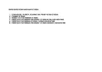
Software methods
CT-Expo [www.sascrad.com]
ImPACT CT Patient Dosimetry Calculator (www.impactscan.org)
19
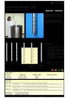
I mPACT CT Patient Dosimetry Calculator Ve rsion 1.0.3 24/08/2010 Introduction This spreadsheet is a tool for calculating patient organ and effective dos es from CT scanner examinations. It mak es use of the NRPB Monte Carlo dose data s ets produced in report SR250 (link at bottom of page). SR250 provides normalised organ dose data for irradiation of a mathematical phantom by a range of CT scanners . As SR250 was produced in 1993, it does not include data for more modern scanners. To overc ome this problem, the ImPACT CT scanner dose survey was carried out by physicists in the UK and Europe. This work , provides a method for 'matching' the dose dis tribution of newer scanners to s canners included in SR250. The matching results are included in this spreads heet. As new sc anners are introduc ed, their matches will be included in updates to this s preadsheet. More details can be found on the dose survey page on the ImPACT website (link below) The results produced by the CTDosimetry spreadsheets have been checked against those produced by CTDOSE, produced by John Le Heron, the standard software used to calculate doses from the NRPB SR250 datasets. The two methods produce identic al results for a range of s cans and sc anners, with the exception of small differences between the doses calculated for muscle and for the 'remainder' organs. These differences are present despite an apparent similarity in calc ulation method, and are typically 1-2%. Insta lla tion The system should work on any PC with Microsoft Excel 2000 or above. Apple computer, but it is anticipated that it should work on a Mac.
It has not yet been tested on a
Ins tallation is fairly simple, and only requires the SR250 data sets (MCSET01.DAT to MCSET23.DAT) to be present in the same directory as this spreadsheet. (SR250 is sold by the NRPB - see link below) Macros are used on this spreadsheet for a variety of purposes. Depending on your version of Ex cel , and macro options , the sec urity level may have to be switched to 'medium' (selec t 'Tools' -> 'Options' -> 'Security' -> 'Macro Sec urity'), and/or ImPACT added to your trusted Macro sources. This spreadsheet has been checked for macro viruses, and the logic and c alculations been tested extensively, however ImPACT accept no responsibility for loss or damage inc urred as a result of its use. Workshe e ts CTDosimetry.xls consists of 12 worksheets Introduction
Provides an introduc tion and instructions for use
Scan Calc ulation
The data entry and results sheet
Paediatric
Information on relative dos es to adult and paediatric patients
Phantom
Allows interactive s election of the scan range used for dos e calculation us ing a diagram of the phantom us ed to generate SR250
Scanners
Provides data on CT scanner models , including CTDI in air and phantom, as well as the s canner matching data
MatchData
Gives data required to perform the scanner matchings in the Scanners work sheet
Collimation
Lists relative CTDI values at different collimations for a range of CT s canners. These values are more useful for multi-slice sc anners, as the CTDI can vary considerably over the range of available collimations
MonteCarloData
Contains the unformatted SR250 data set.
Doses
Contains the formatted dose data from the SR250 data s et that is currently loaded.
DoseCalculations
Performs the organ dose c alculations, and calc ulation of remainder organ doses etc. 20
Selections Version
Provides data for the drop down selection boxes in the ScanCalculation worksheet, and performs calculations for 'remainder' organ doses Details changes made in each version, from vers ion 0.99e onwards
Using CTDosimetry.xls To calculate doses using CTDosimetry.xls, the user must enter a number of parameters relating to the scanner and the scan series The following four selections, made in the top left box on the ScanCalculations worksheet define the Monte Carlo data set that is used: Manufacturer
Select the scanner manufacturer from the drop down list
Scanner
Select the scanner model or scanner model group for the drop down list.
kV
Choose the appropriate scan kV.
Scan Region
Choose head or body.
The Monte Carlo data set that is used for this combination of scanner, kV and body part is displayed in the cell marked 'Data Set'. The data set that is currently loaded is displayed below. If these do not match, no dose is calculated. To load the appropriate data set, and enable dose calculation, press the 'Update Data Set' button. Scan and patient data is entered in the box on the top right of the ScanCalculations worksheet. Tube current
The x-ray tube current. Note that this should be the actual scanner mA, and not the 'effective mAs' displayed on some multi-slice scanners
Rotation time
The scanner tube rotation time
Spiral pitch
The scanning pitch (table travel per rotation/total collimated slice width). For axial scanning, (couch increment)/(collimated slice width) should be used
mAs/rotation
The total mAs per gantry rotation. Do not enter data in this box - it is calculated automatically. The mAs/per rotation divided by the spiral pitch. This is a calculated value that provides a basis for comparison of spiral protocols with different pitches The total nominal x-ray beam width along the z-axis, selected from a range of possible 21 values in the drop down box. This determines the relative CTDI compared to the reference (usually 10 mm) collimaiton.
Effective mAs Collimation
Zoom In Zoom Out
42,5
-1
-10
64
-1
-10
90
80
70
60
50
40
30
20
10
0
-10
22
I mPACT CT Patient Dosimetry Calculator version 0.99m, 1/07/2002 Scanner Model: Manufacturer: GE Scanner: GE QX/i, LightSpeed, LightSpeed Plus kV: 120 Scan Region: Body Data Set MCSET19 Update Data Set Current Data MCSET19 Scan range Start Position -5 cm Get From Phantom Diagram End Position 45 cm Patient Sex: f
Acquisition Parameters: mA 340 Rotation time 0.8 mAs / Rotation 272 Collimation 5 Slice Width 10 Pitch 1.35 Rel. CTDI Look up 1.26 CTDI (air) Look up 34.5 CTDI (soft tissue) 36.9 12.8 nCTDIw
mA s mAs mm mm
at selected collimatio mGy/100mAs mGy/100mAs mGy/100mAs
Organ Gonads Bone Marrow (red) Colon Lung Stomach Bladder Breast Liver Oesophagus (Thymus) Thyroid Skin
wT 0.2 0.12 0.12 0.12 0.12 0.05 0.05 0.05 0.05 0.05 0.01
HT 33.434 15.854 32.619 7.010 35.678 39.058 1.498 33.378 1.209 0.100 13.381
wT.HT 6.687 1.902 3.914 0.841 4.281 1.953 0.075 1.669 0.060 0.005 0.134
Remainder Organs Adrenals Brain Upper Large Intestine Small Intestine Kidney Pancreas Spleen Thymus Uterus Muscle
Bone Surface
0.01
22.972
0.230
CTDIw (mGy)
0.443 0.443 22.637
CDTIv ol (mGy) DLP (mGy.cm)
Remainder1 Remainder 2
0.025 17.709 0.025 17.709 Total Effective Dose (mSv)
HT 30.370 0.006 35.786 34.884 38.987 30.545 33.313 1.209 35.652 17.640 34.7 25.7 1284.5 23
2. Scan Range
Calculate 1.
Scan Range Data (Slice Positions)
Age Group
Get Values
Gender male
Adult
3.
Scanner
4. Select mode
L [cm]
0
female
Scanner Model Manufacturer
Scan Range z from zto z+
Scanner Data for Scan Region "Body" nCTDIw
Uref
[mGy/mAs]
[kV]
0,150
130
-0,0070
3,35
Demo Spiral Scanner
Body mode for head/neck region
P B,H
kCT
kOB
∆L [cm]
0,42
0,80
1,00
0,0
0,00
0,00
p
Ser.
Spiral mode
5. Scan Parameters
Please Enter Actual Settings:
U
I
t
Qel
Q
N * hcol
TF
hrec
[kV]
[mA]
[s]
[mAs]
[mAs]
[mm]
[mm]
[mm]
0
1,0
Dose Values per Scan or per Series*
6. Results
*
Tissue or HT per Series Remainder HT per Series [mSv]
Organs
[mSv]
CTDIvol
[mGy]
[mGy]
[mGy*cm]
[mSv]
[mSv]
Thyroid
0,0
Brain
0,0
0,0
0,0
hrec?
hrec?
n.a.
Breasts
0,0
Thymus
0,0
Oesophagus Lungs Liver
0,0 0,0 0,0 0,0
Spleen P ancreas Adrenals
0,0 0,0 0,0 0,0
E
* Duterus
Organ
CTDIw
* DLPw
Child/Baby: all CTDI and DLP values refer to 16cm head phantom!
Dose Values per Examination
DLPw
E
Duterus
[mGy*cm]
[mSv]
[mSv]
Stomach Colon
Ser?
Ser?
n.a.
Effective dose E refers to ICRP 60
Please note: All organ doses HT are based on conversion coeff icients f or standard patients (ADAM, EVA, CHILD, BABY) and serve f or inf ormation purposes only (in particular organs outside the scan range) !
0,0
Kidneys Small intest.
Testicles
0,0
Upp. large int.
0,0
Ovaries Bladder
0,0 0,0 0,0
Uterus
0,0
Bone marrow Bone surface Skin
0,0 0,0
Misc.
0,0
HT per Series
24[mSv] Eye lenses
0,0
Dose Reference Level (DRL) values are reported by various national or international organizations for abdomen CT DRL
CTDI
DLP
Abdomen EUR 16262 SC 1999
35
900
Abdomen EC MDCT 2004
25
524
Abdomen UK SC 2003
20
460
Abdomen UK MDCT 2003
20
470
Abdomen Germany MDCT 2003
24
1500
Abdomen IAEA 2006
10.9
696
Abdomen USA NEXT Survey
30*
740
* Data from USA are not DRL but 75th percent of data distribution. They are put in this table for comparison purposes.
25
Table 1. Effective dose in various exams. Examination
Effective dose (mSv)
Abdomen radiography
0.7
Chest or Abdomen CT
8.0
3 phase CT liver study
15.0
CT guided biopsy
23.0
CT guided RF ablation
35.0
Repeated CT guided RF ablation
112.0
CT coronary angiography
10.0
Coronary angiography
8
Thallium heart scan
35
CT urography
14.8
CT colonography
5.1
Neonatal abdomen CT
13.1
1 year old abdomen CT
11.1
Annual natural background radiation
2.0 26
Thank you for your attention
27